Publicado
Modelo Experimental de la Respuesta Ósea a Xenoinjertos de Origen Bovino. Estudio Radiográfico e Histomorfométrico.
Experimental models of bone response to bovine xenograft radiographic and histomorphometric
Palabras clave:
Biomateriales, Sustitutos Oseos, Xenoinjertos, Histología, Radiografía digital (es)Biomaterials, Bone substitutes, Xenografts, Histology, Digital Radiography (en)
Descargas
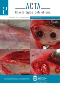
Objetivos: Evaluar la respuesta ósea a injertos de origen bovino insertados en tibia de conejos mediante técnicas radiográficas e histomorfométricas. Materiales y Métodos: Estudio experimental en el que se emplearon veinte conejos de Nueva Zelanda con un peso entre 3900-4500 g. Veinte injertos de hueso bovino en polvo, con un tamaño de partícula de 500-1000 mm fueron insertados en defectos óseos de 4mm de diámetro en la metáfisis proximal de la tibia derecha, y veinte defectos con las mismas dimensiones fueron realizados como control en la metáfisis proximal de la tibia izquierda. Los animales fueron sacrificados en grupos de cinco al cabo de 1, 2, 3, y 4 meses. Se tomaron radiografías anteroposteriores y laterales. Las muestras fueron seccionadas y teñidas con hematoxilina-eosina y tricrómico de Masson. Resultados: Al cabo de 4 meses, las imágenes radiográficas mostraron la reparación completa de los defectos óseos sin alteraciones atribuibles a la presencia del injerto. El análisis histomorfométrico a los 4 meses mostró valores de 22.8±1.5% para el hueso nuevo, 39.4±2.3% para el material de injerto residual y 37.7±2.5% para el tejido conectivo no calcificado. No hubo diferencias significativas en el cierre cortical con hueso bovino 98.8±1.1% comparado con los controles 99.1±0.7% al final del período de tiempo estudiado. Conclusiones: El biomaterial utilizado en este estudio mostro ser biocompatible, osteoconductivo, poco reabsorbible y puede ser considerado como un sustituto óseo que no interfiere con el proceso normal de reparación del hueso.
Referencias
McAllister B, Haghighat K. Bone augmentation techniques. Journal of Periodontology 2007; 78: 377–396.
Esposito M, Grusovin M, Rees J, Karasoulos D, FeliceP, Alissa R, Et Al. Effectiveness of sinus lift procedures for dental implant rehabilitation: a Cochrane systematic review. European Journal of Oral Implantology 2010;3: 7–26.
Ersanli S, Olgac V, Leblebicioglu B. Histologic analysis of alveolar bone following guided bone regeneration. Journal of Periodontology 2004;75: 750–756.
Esposito M, Grusovin M, Coulthard P, Worthington H. The efficacy of various bone augmentation procedures for dental implants: a Cochrane systematic review of randomized controlled clinical trials. The International Journal of Oral & Maxillofacial Implants 2006; 21: 696–710.
Aghaloo T, Moy P. Which hard tissue augmentation techniques are the most successful in furnishing bony support for the implant placement?. The International Journal of Oral & Maxillofacial Implants 2007 ;22 (Suppl. 1): 49–70.
Schlegel K, Fichtner G, Schultze-Mosgau S, Wiltfang J. Histologic findings in sinus augmentation with autogenous bone chips versus a bovine bone substitute. The International Journal of Oral & Maxillofacial Implants 2003; 18: 53–58.
Barone A, Covani U. Maxillary alveolar ridge reconstruction with non-vascularized autogenous block bone: clinical results. The International Journal of Oral and Maxillofacial Surgery 2007; 65: 2039–2046.
Johansson L, Isaksson S, Lindh C, Becktor J, Sennerby L. Maxillary sinus floor augmentation and simultaneous implant placement using locally harvested autogenous bone chips and bone debris: a prospective clinical study. The International Journal of Oral and Maxillofacial Surgery 2010;68: 837–844.
Nkenke E, Radespiel-Tröger M, Wiltfang J. Morbidity of harvesting of retromolar bone grafts: a prospective study. Clinical Oral Implants Research 2002;13: 514–520.
Cricchio G, Lundgren S. Donor site morbidity in two different approaches to anterior iliac crest bone harvesting. Clinical Implant Dentistry & Related Research 2003; 5: 161–169.
Sasso R, Lehuec J, Shaffrey C. Illiac crest bone graft donor site pain after anterior lumbar interbody fusion: a prospective patient satisfaction outcome assessment. Journal of Spinal Disorders and Techniques 2005;18: 77–89.
Arcuri C, Cecchetti F, Germano F, Motta A, Santacroce C. Clinical and histological study of a xenogenic bone substitute used as a filler in postextractive alveolus. Minerva Stomatologica 2005; 54:351–362.
Piatelli M, Favero G, Scarano A, Orsini G, Piatelli A. Bone reactions to anorganic bovinebone used in sinus augmentation procedures: a histologic long- term report of 20 cases in humans. The International Journal of Oral & Maxillofacial Implants 1999; 14: 835–840.
Norton M, Odell E, Thompson I, Cook R. Efficacy of bovine mineral for alveolar augmentation: a human histologic study. Clinical Oral Implants Research 2003; 14: 775–783.
Worth A, Mucalo M, Home G, Bruce, W, Burbidge H. The evaluation of processed cancellous bovine bone as a bone graft substitute. Clinical Oral Implants Research 2005; 16: 379–386.
Barone A, Crespi R, Aldini N, Fini M, Giardino R, Covani U. Maxillary sinus augmentation:histologic and histomorphometric analysis. The International Journal of Oral and Maxillofacial Implants 2005; 20: 519–525.
Orsini G, Scarano A, Piatelli M, Piccirilli M, Caputi S, Piattelli A. Histologic and ultrastructural analysis of the regenerated bone in maxillary sinus augmentation using a porcine bone derived biomaterial. Journal of Periodontology 2006; 77: 1984–1990.
Nannmark U, Sennerby L. The bone tissue responses to prehydrated and collagenated corticocancellous porcine bone grafts: a study in rabbit maxillary defects. Clinical Implant Dentistry & Related Research 2008;10: 264–270.
Di Stefano D, Artese L, Lezzi G, Piattelli A, Pagnutti S, Piccirilli M, Et Al. Alveolar ridge regeneration with equine spongy bone: a clinical, histological, and inmunohistochemical case series. Clinical Implant Dentistry and Related Research 2009;11: 90–100.
Hing K, Best S, Tanner K, Bonfield W, Revell P. Biomechanical assessment of bone ingrowth in porous hydroxyapatite. Journal of Materials Science: Materials in Medicine 1997;8: 731–736.
Briem D, Linhart W, Lehmann W, Meenen N, Rueger, J. Long-term outcomes after using porous hydroxyapatite ceramics (Endobon®) for surgical management of fractures of the head of the tibia. Der Unfallchirurg 2002;105: 128–133.
Kehr P, Gosset F. Endobons as a bone substitute in spine surgery. Preliminary study in 11 patients. European Journal of Orthopaedic Surgery and Traumatology 2000;10: 217–221.
Werber K, Brauer R, Weiss W, Becker K. Osseous integration of bovine hydroxyapatite ceramic in metaphyseal bone defects of the distal radius. Journal of Hand Surgery 2000; 25: 833–841.
Grimm J, Mueller-Huelsbeck S, Mueller M, Egbers H, Brinkmann G, Heller, M. Evaluation of hydroxyapatite implants in vertebral bodies and extremities by contrast-enhanced magnetic resonance imaging. Archives of Orthopaedic and Trauma Surgery 2001;121: 158–161.
Motomiya M, Ito M, Takahata K, Irie K, Abumi K, Minami A. Effect of hydroxylapatite porous characteristics on healing outcomes in rabbit posterolateral spinal fusion model. European Spine Journal 2007;16: 2215–2224.
Wiltfang J, Merten H, Wiltfang J. Ectopic bone formation with the help of growth factor bFGF. Journal of Cranio-Maxillofacial Surgery 1996; 24: 300–304.
Hing K, Best S, Tanner K, Bonfield W, Revell P. Mediation of bone ingrowth in porous hydroxyapatite bone graft substitutes. Journal of Biomedical Materials Research. Part A 2004;68: 187–200.
Gierse H, Donath K. Reactions and complications after the implantation of Endobon including morphological examination of explants. Archives of Orthopaedic and Trauma Surgery 1999;119: 349–355.
Baer W, Schaller P, Carl H. Spongy hydroxyapatite in hand-surgery: a five year follow-up. Journal of Hand Surgery Br. 2002;27:101-3
TamaiN, Myoui A, Tomita T, Nakase T, Takana J, OchiT, Yoshikawa H. Novel hydroxyapatite ceramics with an interconnective porous structure exhibit superior osteoconduction in vivo. Journal of Biomedical Materials Research 2002; 59: 110–117.
Tadic D, Epple M. A thorough physicochemical characterization of 14 calcium phosphate-based bone substitution materials in comparison to natural bone. Biomaterials 2004; 25: 987–994.
Yamamichi N, Itose T, Neiva R, Wang H. Long-term evaluation of implant survival in augmented sinuses: a case series. The International Journal of Periodontics and Restorative Dentistry 2008; 28: 163–169.
Cutter C, Mehrara B. Bone grafts and substitutes. Journal of Long-Term Effects of Medical Implants 2006; 16: 607–618.
Van Steenberghe D, Callens A, Geers L, Jacobs R. The clinical use of deproteinized bovine bone mineral on bone regeneration in conjunction with immediate implant installation. Clinical Oral Implants Research 2000; 11: 210–216.
Hising P, Bolin A, Branting C. Reconstruction of severely resorbed alveolar ridge crests with dental implants using a bovine bone mineral for augmentation. The International Journal of Oral & Maxillofacial Implants 2001; 16: 90–97.
Lang N, Hämmerle C, Oesch B, Schenk R. Risk of transmission of agents associated with Creutzfeldt-Jakob disease and bovine spongiform encephalopathy. Plastic and Reconstructive Surgery 2000;105: 2273–2275.
Wenz B, Oesch B, Horts M. Analysis of the risk of transmitting bovine spongiform encephalopathy through bone grafts derived from bovine bone. Biomaterials 2001; 22: 1599–1606.
Liebendörfer A, Tröster S. Hydroxyapatite ceramics in clinical application. Histological findings in 23 patients. Unfallchirurgie 1997;23: 60–68.
Santos F, Pochaspski M, Martins M, Zenobio E, Spolidoro L, Marcantonio E. Comparison of biomaterial implants in the dental socket: histological analysis in dogs. Clinical Implant Dentistry & Related Research 2010; 12: 18–25.
Khodadadyan-Klostermann C, Liebi T, Melcher I, Raschke M, Haas N. Osseous integration of hydroxyapatite grafts in metaphyseal bone defects of the proximal tibia (CT-study). Acta Chirurgiae Orthopaedicae et Traumatologiae Cechoslovaca 2002;69: 16–21.
Schnettler R, Dingeldein E, Herr G. Defect reconstruction using demineralized bone matrix. Experimental studies on piglets. Orthopadie 1998; 27: 80–88.
Jensen S, Aaboe M, Pinholt E, Hj_rting-Hansen E, Melsen F, Ruyter I. Tissue reaction and material characteristics of four bone substitutes. The International Journal of Oral & Maxillofacial Implants 1996;11: 55–66.
Liljensten E, Adolfsson E, Strid K, Thomsen P. Resorbable and non-resorbable hydroxyapatite granules as bone graft substitutes in rabbit cortical defects. Clinical Implant Dentistry & Related Research 2003;5: 95–101.
Cómo citar
APA
ACM
ACS
ABNT
Chicago
Harvard
IEEE
MLA
Turabian
Vancouver
Descargar cita
Visitas a la página del resumen del artículo
Descargas
Licencia
Derechos de autor 2011 Rafael Arcesio Delgado Ruíz, José Luis Calvo Guirado, María Piedad Ramírez Fernández, José Eduardo Maté Sanchez del Val, Gerardo Goméz Moreno, Javier Guardia

Esta obra está bajo una licencia internacional Creative Commons Atribución-NoComercial-SinDerivadas 4.0.
Aquellos autores/as que tengan publicaciones con esta revista, aceptan los términos siguientes:
- Los autores/as conservarán sus derechos de autor y garantizarán a la revista el derecho de primera publicación de su obra, el cuál estará simultáneamente sujeto a la licencia Reconocimiento-NoComercial-SinObraDerivada 4.0 Internacional que permite a terceros compartir la obra siempre que se indique su autor y su primera publicación esta revista.
- Los autores/as podrán adoptar otros acuerdos de licencia no exclusiva de distribución de la versión de la obra publicada (p. ej.: depositarla en un archivo telemático institucional o publicarla en un volumen monográfico) siempre que se indique la publicación inicial en esta revista.
- Se permite y recomienda a los autores/as difundir su obra a través de Internet (p. ej.: en archivos telemáticos institucionales o en su página web) antes y durante el proceso de envío, lo cual puede producir intercambios interesantes y aumentar las citas de la obra publicada. (Véase El efecto del acceso abierto).
- Una vez sometido el artículo no se aceptaran cambios respecto a la incorporación o retiro de autores.