IDENTIFICATION PROCESS OF THE CAUSAL ELEMENT USING CUTMARK ANALYSIS IN BONE: CASE STUDY
National Institute of Legal Medicine and Forensic Sciences, Special Edition
Isla Y. Campos-Varela
Anthropologist.
National Institute of Legal
Medicine and Forensic Sciences.
Anthropology Laboratory.
- Bogotá Office -
Bogotá, D.C. - Colombia.
Correspondence to:
Isla Y. Campos-Varela.
Laboratorio de Antropología,
National Institute of Legal Medicine and Forensic Sciences.
Email: icampos@medicinalegal.gov.co
SUMMARY
An anthropological trauma analysis was conducted on an individual killed by multiple injuries from a combined sharp and blunt mechanism using a cold weapon (machete) to the head, trunk and upper extremities. After conducting the autopsy, the cranial fragments were submitted to the anthropology laboratory for cleaning, restoration and study; later, plaster reproductions of the cutmarks with clear striations were made and a gel was developed to take impressions of the marks left by the machete recovered at the scene and submitted for study; finally, a visual comparison of the striations observed on the bone and those left by the machete in the gel was conducted.
Keywords: Tool mark analysis; Forensic anthropology; Bone trauma; chop wound; striation.
The striation pattern left by the element in the gel was found to be random. In other words, it did not correspond to the irregular edge of the construction, but rather to a unique pattern resulting from the deformity on the cutting edge caused by the use of the tool. Considering that the same pattern of striations was observed in the cranial cutmarks, the conclusion was made that the causal element of the cuts was the same machete submitted for study.
The development of the study involved exploring odontological techniques using alginate impressions, which facilitated the analysis along with plaster molds. The development of a highly effective alternative gel for taking cutmark impressions is also reported on.
INTRODUCTION
Fragments of the cranial vault belonging to a 38-year-old adult male who died in a public establishment and presented multiple injuries from a combine sharp and blunt mechanism to the head, trunk and upper extremities. A machete covered in blood was recovered at the scene and submitted to the Institute of Legal Medicine for a fingerprints search.
The anthropological approach was initiated during the autopsy procedure with documentation of bone injuries and their relationship to soft-tissue injuries, recovering bone fragments for further analysis (Figure 1). The presence of striations in the cutmarks was noticeable from the start, so authorization was requested from the physician responsible for the case to analyze the tool marks from the machete once lophoscopy and biological studies were completed.
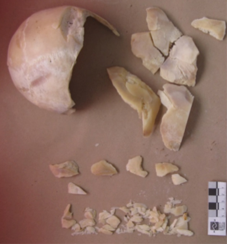
Fig 1. Bone fragments extracted from the cadaver after the cleaning procedure.
Source: I. Campos
METHODOLOGY
The first challenge of the case was removing the fat from the bone fragments because even though the soft tissue was removed, the bones maintained adiposity, which prevented the gluing of the fragments for the analysis, making it necessary to test various cleaning techniques.
This process required submerging the bones in boiling water. Later, water with conventional detergents was used, then calcium oxide (lime) was added. Finally, hydrogen peroxide (oxygenated water) was added until the bones were in suitable condition for restoration using a synthetic adhesive.
To carry out the analysis, the methodology proposed by Steve Symes in the short course on bone trauma he conducted at the National Institute of Legal Medicine and Forensic Sciences was applied (1,2). Symes proposes matching the striations left by the blade of a tool in question in order to determine if said tool could have been used to make the cuts in the bone.
For the analysis of the machete, a high density gel with the capacity of accurately capturing the smallest irregularities of the blade was used. While these industrial gels are widely known and used throughout the world, they are not available in Colombia, so various recipes were prepared from edible gelatin in an attempt to replace it. The first recipes had flaws such as rapid shrinking due to dehydration, the appearance of fungi and lack of consistency for the cuts. After many trials, an adequate preparation was obtained by mixing ingredients such as gelatin without flavour, sodium benzoate (food preservative) and acetic acid (commercial vinegar).
Impressions of the bone and machete cuts were taken as outlined by Buitrago (3), using alginate and type 4 plaster to create the molds and final models.
After cleaning, the fragments were restored (Figure 2) paying special attention to the location of marks that presented striation, avoiding applying adhesive to those areas. After assembling the majority of fragments, the three largest surfaces with the most notable striations and the easiest to reproduce were selected; alginate molds were created and then cast with dental plaster (Figures 3 and 4).
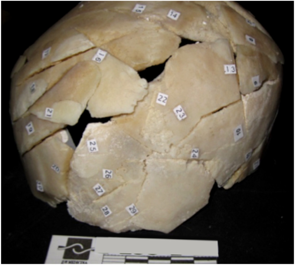
Fig 2. Bone fragments extracted from the cadaver after the cleaning procedure and restoration. The numbers denote the events caused by a combined sharp and blunt mechanism.
Source: I. Campos
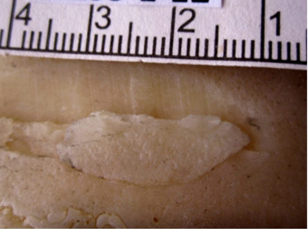
Fig 3. Cranial injury caused by a combined sharp and blunt mechanism showing striations.
Source: I. Campos
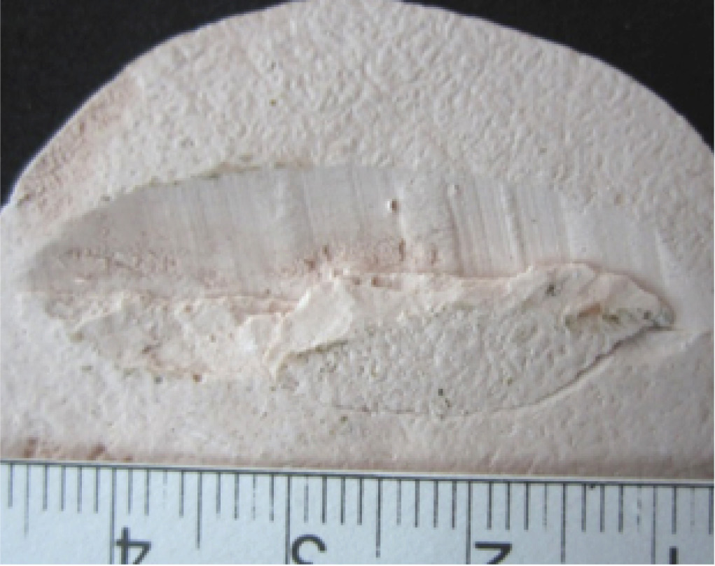
Fig 4. Plaster reproduction of skull injury due to combined sharp and blunt mechanism.
Source: I. Campos
The striations left by the blade of the machete were analyzed by dividing the blade into 12 cm-long segments and later making cuts with each one in the gelatin, taking care to number each segment and identify the location of the tip and which side of the edge left marks on the gel. After making the cuts, and faced with the difficulty of undertaking a photographic comparison due to lack of contrast with the color of the gelatin, the decision was made to make the molds out of alginate and to then take the impressions using dental plaster (Figure 5).
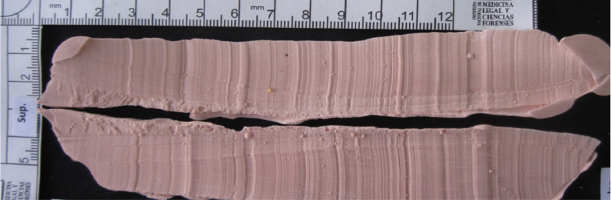
Fig 5. Model of striations left by segment of machete blade corresponding to segment 1; above, impression of the right edge and below, the left edge.
Source: I. Campos
With the molds of the striated cutmarks of the skull and those left by the blade of the machete, a visual comparison was conducted to identify if any matches could be identified among these patterns. The comparison consisted of observing the width of the grooves and elevations as well as the side on which they were cast, considering that each striation was produced by a small crease in the edge of the element.
To facilitate the comparison, photographs were taken with metric scales, which were uploaded onto a software to edit photos and both photographs were amplified to scale.
Results
Over 30 cutmarks were found on the skull which caused its fragmentation. Although the cutmarks were all in the region of the cranial vault, they were largely concentrated in the posterior region. Three of the cuts observed presented clearly defined striations since they compromised the bone in a tangential way, only injuring the external surface.
Oxidation was found on the metal of the machete blade. The blade had irregularities on the edges - small creases towards both sides - (Figure 6), which, when making the incision, produced striations on the affected surface. Studying the impressions left by the machete demonstrated that the striations had different widths and depths that were not repeated along the entire edge.
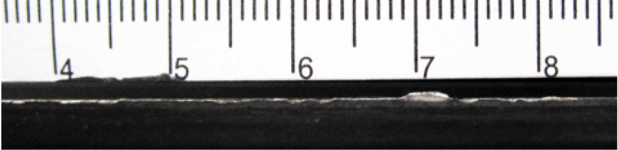
Fig 6. Detail of the blade of the machete - mid segment - which highlights the biggest irregularities.
Source: I. Campos
Various segments in the bone and gel impressions can be identified that present coinciding striations in the sequence of width, depth and inclination (Figure 7, 8 and 9), which leads to the conclusion that the machete submitted for laboratory analysis is the causal element of the cutmarks, since the striation pattern is random and produced by the deterioration of the blade. It also does not correspond to a deliberate sequence used in the mass fabrication of this element.
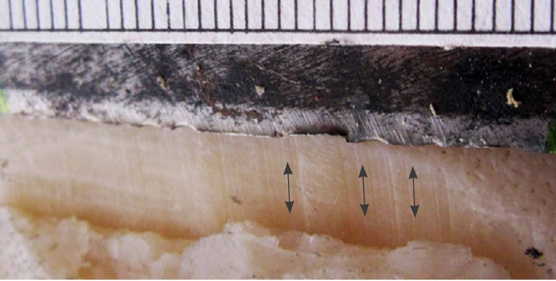
Fig 7. Detail comparison of the surface of the cut with the corresponding segment of the machete.
Source: I. Campos
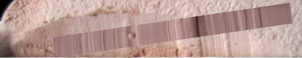
Fig 8. Detail assembly to scale of the cut in the frontal bone with the superimposed photograph of the cutmarks of the right edge of the machete in gel (corresponding to 200mm to 240mm of the blade measured from the tip), which shows an almost complete match of striations.
Source: I. Campos
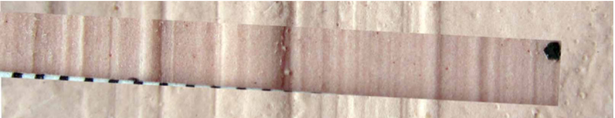
Fig 9. Detail assembly to scale of the photograph of the chop wound located in the right parietal, with superimposed photograph of the cutmarks of the right edge of the machete in gel (corresponding to 94mm to 132 mm of the blade measured from the tip), which shows an almost complete match of striations.
Source: I. Campos
In Figure 7, the arrows highlight the areas in the bone that have more noticeable grooves and elevations that correspond to small creases in the machete.
Upon observation, not all segments corresponded to a 100% match, which has a few different explanations. Firstly, if a gel is softer, it captures more characteristics of the element than cortical tissue, which is rigid, can. Furthermore, given that the striations were left by defects produced by the use of the machete, we deduce that the blade was deformed with each successive blow to the hard cranial surface.
DISCUSSION
When comparing tool marks in bone with suspicious elements, it is worth noting that the ideal moment for anthropologists to approach the case is in the autopsy room, since a complete observation of the cadaver —reading of the bone injury in relation to findings of soft tissues— allows them to understand the trauma that occurred and take the samples that they deem most appropriate.
To adequately interpret bone trauma, it is necessary to know the anatomical location of the injury, which can be achieved with the exact reconstruction of the structure. Therefore, the cleaning procedure should focus on the removal of fat to enable the adhesion of fragments and the reconstruction of bone. Cleaning itself carries the risk of eroding the edges of the fragments, which should be avoided at all costs in order to keep the characteristics of the cutmarks intact.
The restoration process is long and can take several days. It consists of not only unifying all possible fragments but also of recording characteristics such as striations or inclinations of the edge on the fractures, among others, before gluing the pieces together.
The gel for taking impressions must have mechanical properties that allow for highly accurate representations of all the small details and irregularities of the cutting elements and the striations it can form on the bone. It must also be a homogenous material that does not clump. It should also be rigid enough not to deform when making the cut to prevent distorting the pattern of striations. The volume of the gel should be kept constant for the longest time possible under different environmental conditions and preservatives should be added to avoid the spread of fungi.
Creating reproductions, not only of cutmarks but also impressions in gel, significantly facilitates the photographic documentation and subsequent visual comparison of striation patterns.
Studies such as these require the multidisciplinary and collective effort of physicians, anthropologists, assistants and forensic experts, and criminal investigators, since without the recovery of elements at the scene, the request for anthropological analysis or careful handling during the extraction and processing of samples, anthropological analysis would not have been able to take place.
ACKNOWLEDGEMENTS
The study of this case would not have been possible without the decision of Dr. Mabel Zurbarán, the forensic doctor who requested the anthropological analysis in an unprecedented exploration at the Institute; the dedication of Álvaro Gutiérrez, assistant to the Pathology Group, who took great pains to recover even the smallest cranial fragment during the autopsy; the patience of Edwin Cardona, who constantly tries new techniques to improve the effectiveness of cleaning bones while conserving their characteristics; the collaboration of Sergio González, now anthropologist at the National University of Colombia, with whom a final formula for the gel was created to take impressions; finally, I am especially grateful for the work of odontologist Edna Buitrago Suárez, who created the models used in the analysis of this case.
REFERENCES
1. Symes S. Curso corto de trauma y pseudo-trauma óseo. Bogotá, D.C.: Instituto Nacional de Medicina Legal y Ciencias Forenses; 2010.
2. Symes S, Chapman EN, Rainwater CW, Cabo LL, Myster S. Technical Report: Knife and Saw Mark Analysis on Bone: A manual designed for the examination of criminal mutilation and dismemberment. Washington, D.C.: Department of Justice; 2010.
3. Buitrago, E. Uso de métodos de impresión dental y exploración de materiales para el análisis de marcas de corte para identificación de elemento causal. Case Report. 2015;1(Suppl 1).
4. Cardona E, Campos I. Limpieza de tejido óseo. In: Téllez N. Editor. Consideraciones para el análisis de fracturas óseas: una visión desde la antropología en Texto de Patología Forense; 2015.