Case report:
Acute generalized exanthematous pustulosis related to phenytoin administration
Palabras clave: Erupciones por medicamentos; Pustulosis exantematica aguda generalizada; hidantoínas.
Keywords: Drug eruptions, Acute Generalized Exanthematous Pustulosis, hydantoins.
Juan R Castro–Ayarza
Dermatologist
Specialist in university teaching
Faculty of Medicine
Universidad Nacional de Colombia
Bogotá, D.C. – Colombia
Eduardo Fierro
Dermatologist. Oncologist
Faculty of Medicine
Universidad Nacional de Colombia
Bogotá, D.C. – Colombia.
Corresponding author:
Juan Raul Castro Ayarza
Calle 91 #19c 62,
office 206
Phone number: 031 530 0048
Email: juanraulcastro@yahoo.com
SUMMARY
The occurrence of acute generalized exanthematous pustulosis adverse reactions to medication administration is becoming more frequent. This article reports the case of a 78-year-old woman who attended the clinic with generalized papules and pustules on the scalp, trunk and limbs, with a concordant histology study and who was diagnosed with acute generalized exanthematous pustulosis (AGEP) associated with the use of phenytoin, a medication that may cause different skin reactions and that has been previously related to this disease. The patient was treated with systemic steroids and the disease had a satisfactory outcome.
INTRODUCTIoN
Adverse drug reactions are common in in-patients and can happen in several ways, for example as the emergence of inflammatory lesions on the skin (1). Sometimes the cause of the disease can be determined from the morphology of primary lesions. Presence of pustules, in particular, may help to guide the diagnosis of some reactions to medications (1).
From the presence of pustules it is possible to make an acute generalized exanthematous pustulosis (AGEP) diagnosis, a condition related to the administration of some medications such as anticonvulsants, antimalarials and antibiotics (2). This paper presents a case of AGEP associated with the use of phenytoin.
Case Report
A 78-year-old woman from Bogotá, Colombia, with seven days of course of the disease and who had pruritic erythematous lesions in her trunk and scalp. She was experiencing fever from the fifth day, which is why she was hospitalized.
The patient had a history of a two-month-old stroke associated with a seizure she suffered two weeks prior to her hospital admission. Due to this seizure episode, the patient was administered oral phenytoin 200mg/day. She had been previously under pharmacological management for dyslipidemia and dyspepsia with acetylsalicylic acid 100mg/day, lovastatin 40mg/day and omeprazole 20mg/day.
Physical examination of the patient revealed erythematous plaques with fine scale, as well as superficial pustules on the scalp, chin, trunk, and thighs (see Figure 1). In addition, there were no signs of lymphadenopathy and lesions were easy to detach by using curettage. Likewise, a 38.1°C temperature was found in one of her records during hospitalization.
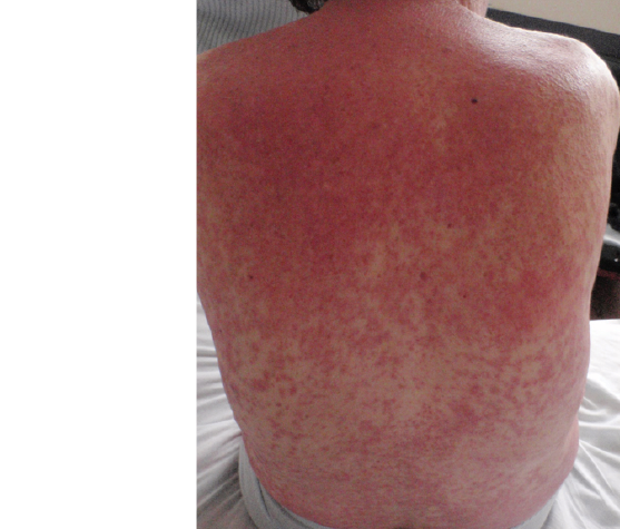

Fig 1. Dorsum. An erythematous base plaque and small pustules are observed when looking closer.
Source: Images obtained from the data collected in the study.
Blood tests showed a complete blood count with hemoglobin 9.7g/dl, hematocrit 30.0%, leukocytes 13950 mm3/dl, neutrophils 81%, lymphocytes 15%, eosinophils 1% and platelets 107000 mm3/dl, whereas transaminases and creatinine studies were normal. The haematoxylin and eosin histopathology report of a skin biopsy allowed identifying epidermis with basal layer vacuolation and subcorneal pustules, as well as dermal edema and dense superficial perivascular lymphocytic infiltrate with presence of eosinophils (see Figure 2). By relating findings made on the pustular skin to its presence in subcorneal histology with measured fever and leukocytosis an AGEP diagnosis was made, thus phenytoin administration was suspended, since it was considered as a possible cause of the reaction, instead a prednisolone dose of 0.5mg/kg administered orally during seven days was added to the patient’s treatment. Once phenytoin was suspended, pustules and fever disappeared at the second day, while the erythema lasted 10 days, then skin symptoms started to decrease until they disappeared after a month, as evidenced in the dermatology outpatient visit.

Fig 2. 10x magnified haematoxylin and eosin stain histopathology. Epidermis with presence of orthokeratosis and foci of spongiosis and superficial epidermal and superficial perivascular lymphoid infiltrate with eosinophils are observed.
Source: Image obtained from the data collected in the study.
Discussion
AGEP type adverse drug reaction was first described in 1968 when studying patients suffering psoriasis with a clinical suspicion of pustular disease. Its name was established from the French translation made in 1980, distinguishing it from acute generalized pustulosis, a postreptococcal infection (2-3).
Prevalence of AGEP by age or sex has not been determined and there are few reports describing it in children (3-5). Despite this, it is thought that it has a prevalence of 1 to 5 per million people annually, although it is likely this disease is underdiagnosed (2). In up to 90% of the cases of AGEP that have been reported the disease has been associated with a medication. In addition, there seems to be a predisposition to this condition in haplotypes like HLA-B51, HLA-DR11 and HLA-DQ3, although there is no a specificity according to the type of drug (5). On the other hand, the increase of the expression of Fas, p53 and bcl-2, which leads to keratinocyte apoptosis, could cause AGEP, although a delayed hypersensitivity reaction has also been involved (5).
Additionally, in the literature on this pathology there are studies describing some antibacterial agents, mainly macrolides and penicillins, as well as several antimycotics (6-9); likewise, a large variety of medications, including some antihypertensives (calcium antagonists, angiotensin-converting enzyme inhibitors), antiarrhythmics, anticonvulsants, antidepressants and anxiolytics, even acetaminophen, have been reported (6). The clinical profile of this disease occurs from one to three weeks after the medication has been administered, however in the case of antibiotics the occurrence average time is 2.5 days, while for other medications is 18 days (6). It is important to note that, although less frequently, AGEP has been related to viral and bacterial infections and ultraviolet light exposure (2,5,6).
Symptoms usually found in this clinical profile include fever, asthenia and adynamia. The main cutaneous manifestation of the disease is the presence of pustules, however AGEP clinical picture starts with edematous alike erythematous macules that extend mainly in intertriginous areas (2,6). Generally, pustules emerge in these areas, they do not have a follicular pattern and, sometimes, they can come together, which causes a false Nikolsky sign (2,9). Another characteristic of AGEP is the presence of lymphadenopathies (2).
Systemic involvement is reflected in the appearance of neutrophilic leukocytosis, although eosinophilia may occur in one third of cases. Aminotransferases elevation is mild, less than the double of the normal value, whereas in the creatinine clearance process a 30% reduction occurs (2,6).
However, analyzing AGEP through histopathology does not allow the medical doctor to make a diagnosis at the disease acute moment, which delays initial treatment; but histopathology findings such as the appearance of subcorneal and intraepidermal pustules with peripheral spongiosis may help in making the diagnosis. The dermis may also be affected by a superficial perivascular infiltrate containing lymphocytes, neutrophils and, to a lesser extent, eosinophils. Some mild vasculitis changes, as well as necrotic keratinocytes may also happen (2,6).
Sidoroff et al. (2) suggest five diagnostic criteria for this disease:
1.Multiple pustules, from tens to hundreds, on an erythematous base.
2.Compatible histopathological changes.
3.Fever higher than 38 °C.
4.Neutrophil count higher than 7000 mm3.
5.Spontaneous resolution in 15 days.
In their research, in order to determine the diagnosis, Sidoroff et al. (2) created a table that includes the following items: morphology (pustules, erythema, distribution and scaling); course of the disease (acute onset, fever, spontaneous resolution, mucosal involvement and neutrophil count increase higher than 7000 mm3), and histological findings (neutrophil exocytosis, papillary edema and spongious changes). Nevertheless these items are complemented by an unpractical score in terms of daily clinical practice (1,2).
Patch tests and lymphocyte transformation tests have proved useful to determine the agent involved in the disease, achieving a positivity rate of up to 80%; besides they make clear the process in the pathogenesis of T cells (3).
The course of the disease implies that its clinical scenario should disappear between 4 and 10 days, with fever and lymphadenopathy being the first symptoms to do so. On the other hand, pustules usually heal spontaneously at approximately nine days resulting in scaling after their resolution (2,6).
AGEP is a disease difficult to diagnose in its initial stage and can be confused with an infectious process. Similarly, its initial development may resemble that of a DRESS (drug rash with eosinophilia and systemic symptoms) hypersensitivity reaction to medications, where there is low or null presence of pustules. Furthermore, clinical presentation of pustular psoriasis is difficult to differentiate, but usually this is a long course disease that has frequent relapses, while subcorneal pustular dermatosis has a less acute development (1,6). Likewise, Reiter’s disease differs in joint involvement, which is not found in AGEP, and acneiform eruptions, which also have pustules, generally triggered by previous use of corticosteroids, which is a situation that constitutes the treatment of AGEP, rather than its cause (3).
Despite there are vasculitis features in AGEP, it should be histologically differentiated from vasculitis purpura. Really severe cases may look like toxic epidermal necrolysis when pustules coincide with skin scaling (1).
As it happened in the case reported here, the use of phenytoin has also been described in other AGEP cases where idiosyncratic adverse skin reactions to medications are reported, including DRESS-type hypersensitivity, maculopapular exanthema, Steven Jonhson syndrome and necrolysis toxic epidermal (10,11). In addition, there are cases reporting cross-reactivity between phenytoin and carbamazepine and between phenytoin and phenobarbital with skin manifestations (12). Nonetheless, there is only one case in the literature that reports the association of AGEP with this medication (13).
Conclusion
Quitting the use of the medication is fundamental in the treatment for AGEP. In addition, using systemic corticosteroids may be appropriate in cases with hepatic or systemic involvement. The administration of antipyretics is adequate based on the symptomatology of the patient (1).
AGEP is an adverse drugs reaction that usually finds its resolution after stopping the use of the medication causing it. There are different drugs related to its onset, including phenytoin, as seen in this case.
References
1.Mockenhaupt M. Severe drug-induced skin reactions: clinical pattern, diagnostics and therapy. J Dtsch Dermatol Ges. 2009;7(2):142-60. http://doi.org/bkps7n.
2.Sidoroff A, Halevy S, Bavinck JN, Vaillant L, Roujeau JC. Acute generalized exanthematous pustulosis (AGEP) - a clinical reaction pattern. J Cutan Pathol. 2001;28(3):113-119. http://doi.org/dbgwpb.
3.Auer-Grumbach P, Pfaffenthaler E, Soyer HP. Pustulosis acuta generalisata is a post-streptococcal disease and is distinct from acute generalized exanthematous pustulosis. Br J Dermatol. 1995;133(1):135-9. http://doi.org/bwrdpr.
4.Mengesha YM, Bennett ML. Pustular skin disorders: diagnosis and treatment. Am J Clin Dermatol. 2002;3(6):389-400. http://doi.org/dbszx9.
5.Meadows KP, Egan CA, Vanderhooft S. Acute generalizated exanthematous pustulosis (AGEP), an uncommon condition in children: case report and review of the literature. Pediatr Dermatol. 2000;17(5):399-402. http://doi.org/fkkrdk.
6.Beylot C, Doutre MS, Beylot-Barry M. Acute Generalized Exanthematous Pustulosis. Semin Cutan Med Surg. 1996;15(4):244-9.
7.Beltraminelli HS, Lerch M, Arnold A, Bircher AJ, Haeusermann P. Acute generalized exanthematous pustulosis induced by the antifungal terbinafine: case report and review of the literature. Br J Dermatol. 2005;152(4):780-3. http://doi.org/cgg5hd.
8.Heymann WR, Manders SM. Itraconazole-induced acute generalized exanthemic pustulosis. J Am Acad Dermatol. 1995;33(1):130-1. http://doi.org/c4m3dw.
9.Cuchía HJ, Arévalo NF, Castellanos HJ. Pustulosis aguda exantemática generalizada inducida por terbinafina: reporte de un caso. Rev Asoc Col Dermatol. 2008 [cited 2016 Sep 22];16(3):214-6. Available from: https://goo.gl/wGNpmf.
10.Sánchez X, Merlano C, Cruz CM. Síndrome de hipersensibilidad a medicamentos con eosinofilia y síntomas sistémicos (DRESS). Rev Asoc Col Dermatol. 2008 [cited 2016 Sep 22];16(3);208-10. Available from: https://goo.gl/ZI4DLq.
11.Maoz KB, Brenner S. Drug rash with eosinophilia and systemic symptoms syndrome: sex and the causative agent. Skinmed. 2007;6(6):271-3. http://doi.org/bw2kzk.
12.Mendiratta V, Bhushan P. Phenytoin-induced DRESS with cross-reactivity to carbamazepine in a 10-year-old Indian child. Clin Exp Dermatol. 2006;31(5):720-1. http://doi.org/dj5sxd.
13.Mallo S, Fernández E, Cardeñoso E, Ingelmo JM, Pascual AM. Pustulosis aguda exantemática generalizada: aportación de dos casos. Med Cutan Iber Lat Am. 2003 [cited 2016 Sep 22];31(4):246-51. Available from: https://goo.gl/kTkQah.