Letter to the editor
A three-dimensional approach to learning coronary anatomy
Un enfoque tridimensional para el aprendizaje de la anatomía coronaria
Rafael Eddison Tacuri-Sauñe1,2![]()
1 Universidad Nacional de San Cristóbal de Huamanga - Professional School of Human Medicine - Ayacucho - Peru.
2 Sociedad Científica Medico Estudiantil San Cristóbal - Ayacucho - Peru.
Open access
Received: 15/04/2023
Accepted: 19/07/2023
Corresponding author: Rafael Eddison Tacuri Sauñe. Escuela Profesional de Medicina Humana, Universidad Nacional de San Cristóbal de Huamanga. Ayacucho. Perú. Email: all.anatomia@gmail.com.
Keywords: Coronary Vessels; Heart; Students, Medical; Education (MeSH).
Palabras clave: Vasos coronarios; Corazón; Estudiantes de medicina; Educación (DeCS).
How to cite: Tacuri-Sauñe RE. A three-dimensional approach to learning coronary anatomy. Rev. Fac. Med. 2024;72(1):e108354. English. doi: https://doi.org/10.15446/revfacmed.v72n1.108354.
Cómo citar: Tacuri-Sauñe RE. Un enfoque tridimensional para el aprendizaje de la anatomía coronaria. Rev. Fac. Med. 2024;72(1):e108354. English. doi: https://doi.org/10.15446/revfacmed.v72n1.108354.
Copyright: Copyright: ©2024 Universidad Nacional de Colombia. This is an open access article distributed under the terms of the Creative Commons Attribution License, which permits unrestricted use, distribution, and reproduction in any medium, as long as the original author and source are credited.
Dear Editor:
In medical programs, the anatomy of the heart is learned through a theoretical-practical methodology in the classrooms and dissection halls of the faculties of medicine and health sciences. However, there are still some challenges for the adequate transfer of knowledge on this subject from professors to students.1,2 For this reason, the objective of this paper is to present a comprehensible and educational method for teaching coronary anatomy to students of medicine and other health sciences. We propose an update of the three-dimensional method developed by Sos & Sniderman,3 in which we include the arteriovenous trigone of the heart, also known as the triangle of Brocq & Mouchet, and the region of the interventricular septum (IVS), which is irrigated by the anterior interventricular (AI) artery and the posterior interventricular (PI) artery.
In this method, the right hand is used to represent the interventricular groove and the left hand to represent the atrioventricular groove.3 The margin of the right hand represents the interventricular groove, while the thumb and index fingers of the left hand, which encircle the right wrist (RW), represent the coronary sulcus. In addition, the palmar and dorsal region of the right hand mark the position of the left and right ventricle, respectively (Figure 1 A).
The right coronary artery (RCA) and left coronary artery (LCA) originate in the first portion of the aorta. In the model, they are represented by the index finger and thumb of the left hand, respectively, where the index finger reaches the lower margin of the RW, corresponding in the model to the crux of the heart, and from there it continues on as the PI artery, which irrigates the diaphragmatic face and the posterior third of the interventricular septum in 85% of the cases.4 In the remaining 15% of the cases, the thumb does not border the inferior margin of the RW due to the dominance of the LCA of the individuals (15%) (Figure 1A).
On the other hand, the common trunk or left main trunk is represented by the first metacarpal of the right hand, which expands in a posture similar to that of a right-handed baseball pitcher. The anterior interventricular branch and circumflex artery, originating from the bifurcation of the left coronary artery, are represented by the right index finger and thumb, respectively (Figure 1B). Finally, the triangular region created between the first and second metacarpal of the right hand reflects the limits of the triangle of Brocq & Mouchet (Figure 1C), an important region because it contains smaller vessels that can pose a greater surgical risk in case of damage.
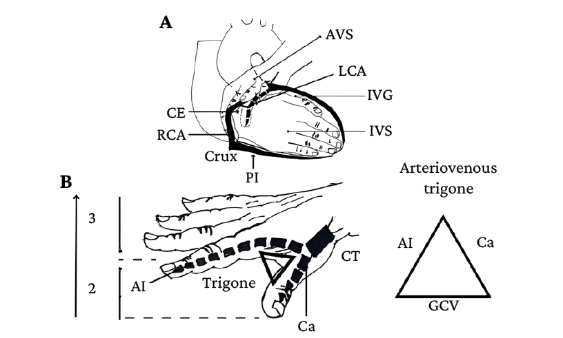
Figure 1. Three-dimensional representation of the anatomy of the heart. Updated Sos & Sniderman’s method. A) The right hand represents the interventricular septum and the left hand represents the atrioventricular septum; B) The right thumb and index finger represent the anterior interventricular and circumflex arteries; C) Arteriovenous trigone of the heart (Triangle of Brocq & Mouchet).
IVS: interventricular septum; AVS: atrioventricular septum; CS: coronary sulcus; IVG: interventricular groove; RCA: right coronary artery; LCA: left coronary artery; PI: posterior interventricular artery; AI: anterior interventricular artery; CT: common trunk; CA: circumflex artery; GCV: great cardiac vein.
Source: Elaborated based on Sos & Sniderman.3
Conflicts of interest
None stated by the authors.
Funding
None stated by the authors.
Acknowledgments
None stated by the authors.
References
1.Ruiz-Cerrillo S. Enseñanza de la anatomía y la fisiología a través de las realidades aumentada y virtual. Innovación Educativa. 2019;19(79):57-76.
2.Alfalah SFM, Falah JFM, Alfalah T, Elfalah M, Muhaidat N, Falah O. A comparative study between a virtual reality heart anatomy system and traditional medical teaching modalities. Virtual Reality. 2019;23(3):229-34. https://doi.org/mvcr.
3.Sos TA, Sniderman KW. A simple method of teaching three-dimensional coronary artery anatomy. Radiology. 1980;134(3):605-6. https://doi.org/mvcq.
4.Marroquín C, Duque J, Rivera-Cardona GA. Patologías asociadas a las variaciones anatómicas encontradas en el origen aórtico de las arterias coronarias. Salutem Scientia Spiritus. 2022;8(2):42-9.