Publicado
Comparación del grosor óseo palatino bilateral para inserción de miniimplantes
Comparison of bilateral palatal bone thickness for insertion of mini implants
DOI:
https://doi.org/10.15446/aoc.v9n2.78843Palabras clave:
Métodos de anclaje en ortodoncia, hueso cortical, paladar, tomografía de haz cónico, mini-implantes (es)Orthodontic anchorage procedures, cortical bone, palate, Cone Beam Computed Tomography, mini implants (en)
Objetivo: comparar el grosor óseo palatino bilateral e identificar zonas seguras para inserción de miniimplantes. Métodos: se llevó a cabo un estudio transversal que incluyó 100 tomografías de haz cónico (CBCT) pretratamiento de pacientes que asistieron a la Especialidad de Ortodoncia en la Universidad Autónoma de Nayarit (UAN). Las imágenes fueron orientadas utilizando el mismo protocolo. En el corte sagital, las mediciones se realizaron de los 0 a los 24mm, con intervalos de 3mm entre cada una, con la primera medición en el borde posterior del foramen incisivo. Las mediciones del grosor palatino se realizaron en el corte coronal a los 2, 4, 6 y 8mm de la sutura media palatina hacia ambos lados. Resultados: se encontró mayor grosor en la zona anterior en las medidas sagitales de 0 mm y 3 mm, a diferencia del resto del paladar. Se hallaron diferencias estadísticamente significativas al comparar los valores tanto sagitales como transversales. Con la prueba Post Hoc de Tukey no se encontraron diferencias estadísticamente significativas al comparar el lado derecho con el izquierdo en cada una de las medidas transversales. Conclusiones: no se encontraron diferencias significativas entre los dos lados, se determinó como zona segura la región anterior del paladar de 0mm a 6mm y es primordial la CBCT en todos los pacientes con indicación de miniimplantes en paladar.
Objective: Bilateral comparison of palatal bone thickness and identification of safe areas for insertion of mini implants. Methods: Cross-sectional Study that included 100 Cone beam computed tomography (CBCT) pretreatment, of patients who attended the Orthodontic Specialty at the Autonomous University of Nayarit (UAN). The images were oriented using the same protocol. In the sagittal plane, measurements were made from 0 to 24mm, with intervals of 3mm between each, being the first measurement at the posterior rim of the incisive foramen. Measurements of the Palatine thickness were made on the coronal plane at 2, 4, 6 and 8mm of the mid palatal suture on both sides. Results: Greater thickness was found in the anterior area in the sagittal measurements of 0mm and 3mm, unlike the rest of the palate. Statistically significant differences were found when comparing both sagittal and transverse values. With Tukey's post hoc test no statistically significant differences were found when comparing the right and left sides in each of the cross measures. Conclusions: No significant differences were found between the two sides, the anterior palatal region of 0mm to 6mm was determined to be safe and CBCT is paramount in all patients in whom mini-implants are to be placed on the palate.
Comparación del grosor óseo palatino bilateral para inserción de miniimplantes
Comparison of bilateral palatal bone thickness for insertion of mini implants
Citación sugerida
Tirado Núñez A, Castrejón Jaime S, Silva Zatarain A, Gutiérrez Rojo J. Comparación del grosor óseo palatino bilateral para inserción de miniimplantes. Acta Odont Col [en línea] 2019 [fecha de consulta: dd/mm/aaaa]; 9(2): - 35. Disponible en: https://revistas.unal.edu.co/index.php/actaodontocol/article/view/78843
|
Recibido 03/04/2019 |
Aprobado 8/06/2019 |
Publicado 15/07/2019 |
Resumen
Objetivo: comparar el grosor óseo palatino bilateral e identificar zonas seguras para inserción de miniimplantes. Métodos: se llevó a cabo un estudio transversal que incluyó 100 tomografías de haz cónico (CBCT) pretratamiento de pacientes que asistieron a la Especialidad de Ortodoncia en la Universidad Autónoma de Nayarit (UAN). Las imágenes fueron orientadas utilizando el mismo protocolo. En el corte sagital, las mediciones se realizaron de los 0 a los 24mm, con intervalos de 3mm entre cada una, con la primera medición en el borde posterior del foramen incisivo. Las mediciones del grosor palatino se realizaron en el corte coronal a los 2, 4, 6 y 8mm de la sutura media palatina hacia ambos lados. Resultados: se encontró mayor grosor en la zona anterior en las medidas sagitales de 0 mm y 3 mm, a diferencia del resto del paladar. Se hallaron diferencias estadísticamente significativas al comparar los valores tanto sagitales como transversales. Con la prueba Post Hoc de Tukey no se encontraron diferencias estadísticamente significativas al comparar el lado derecho con el izquierdo en cada una de las medidas transversales. Conclusiones: no se encontraron diferencias significativas entre los dos lados, se determinó como zona segura la región anterior del paladar de 0mm a 6mm y es primordial la CBCT en todos los pacientes con indicación de miniimplantes en paladar.
Palabras clave: Métodos de anclaje en ortodoncia; hueso cortical; paladar; tomografía de haz cónico; miniimplantes.
Abstract
Objective: Bilateral comparison of palatal bone thickness and identification of safe areas for insertion of mini implants. Methods: Cross-sectional Study that included 100 Cone beam computed tomography (CBCT) pretreatment, of patients who attended the Orthodontic Specialty at the Autonomous University of Nayarit (UAN). The images were oriented using the same protocol. In the sagittal plane, measurements were made from 0 to 24mm, with intervals of 3mm between each, being the first measurement at the posterior rim of the incisive foramen. Measurements of the Palatine thickness were made on the coronal plane at 2, 4, 6 and 8mm of the mid palatal suture on both sides. Results: Greater thickness was found in the anterior area in the sagittal measurements of 0mm and 3mm, unlike the rest of the palate. Statistically significant differences were found when comparing both sagittal and transverse values. With Tukey’s post hoc test no statistically significant differences were found when comparing the right and left sides in each of the cross measures. Conclusions: No significant differences were found between the two sides, the anterior palatal region of 0mm to 6mm was determined to be safe and CBCT is paramount in all patients in whom mini-implants are to be placed on the palate.
Key words: Orthodontic anchorage procedures; cortical bone; palate; Cone Beam Computed Tomography; mini implants.
Introducción
Los miniimplantes han revolucionado los tratamientos de ortodoncia debido a que, por tratarse de sistemas de anclaje absoluto, disminuyen los movimientos secundarios o adversos de los órganos dentarios (1-3).
El lugar de inserción y las características del miniimplante dependen de los siguientes factores: grosor del hueso cortical, densidad ósea, cantidad de hueso disponible, estructuras anatómicas adyacentes, y grosor del tejido blando (4).
Los miniimplantes se insertan comúnmente en la cortical bucal en maxilar y mandíbula, en la zona palatina del proceso alveolar maxilar, en el paladar duro del maxilar o en el área retromolar en el cuerpo de la mandíbula (5, 6).
El paladar duro tiene importantes ventajas sobre otros lugares de inserción de miniimplantes: en primer lugar, en virtud de la ausencia de estructuras anatómicas críticas (vasos sanguíneos, nervios y raíces dentales), su zona paramedial ha sido identificada como el mejor sitio de anclaje para el maxilar superior; en segundo lugar, es quirúrgicamente accesible; en tercer lugar, es menos susceptible a la inflamación; y, por último, presenta una adecuada calidad de hueso cortical. Por ello, los miniimplantes en el paladar presentan alta estabilidad y alto porcentaje de éxito (4, 7-10).
Las principales desventajas de estos sistemas son la posible falta de espesor óseo vertical y su variabilidad anatómica entre los pacientes. Otras preocupaciones referidas son: perforación en la cavidad nasal, interferencia con el canal incisivo, posible efecto sobre la sutura medialpalatina en pacientes en crecimiento, y necesidad de una anestesia más profunda (5). Por lo tanto, es primordial conocer el grosor del hueso palatino ya que de este modo es posible evitar perforaciones contralaterales o perforaciones en el seno maxilar durante la inserción de miniimplantes.
El objetivo de este estudio fue comparar el grosor óseo palatino bilateral e identificar zonas seguras del paladar para inserción de miniimplantes mediante tomografías de haz cónico (CBCT).
Métodos
Se llevó a cabo un estudio descriptivo de tipo transversal. El universo de estudio comprendió 1159 tomografías de haz cónico pretratamiento, del tomógrafo VATECH PAX-I3D. La totalidad de estas tomografías corresponde a pacientes que asistieron al Posgrado de Ortodoncia de la Universidad Autónoma de Nayarit en el período comprendido entre agosto de 2014 y diciembre de 2018 y, entre ellas, se seleccionaron de forma aleatoria 100 tomografías que cumplieron con los criterios de inclusión y exclusión.
Las tomografías de haz cónico se tomaron de los pacientes que acudieron a diagnóstico en la Clínica de Especialidad en Ortodoncia. Todos los pacientes fueron posicionados con el plano de Frankfurt paralelo al piso. Los participantes fueron informados acerca del procedimiento del estudio, y el consentimiento informado otorgado por estos siguió las directrices propuestas por la Declaración de Helsinki de la AMM, Principios éticos para las investigaciones médicas en seres humanos (2013).
Los criterios de inclusión de este estudio fueron:
- pacientes entre 12 y 40 años;
- sin tratamiento de ortodoncia previo;
- dentición completa y permanente erupcionada;
- salud periodontal, según la nueva clasificación de enfermedad periodontal (11);
- ausencia de apiñamiento anterior o posterior.
Los criterios de exclusión observados en esta investigación fueron:
- presencia de lesiones periapicales o de patologías óseas;
- presencia de enfermedades sistémicas que podrían influenciar la calidad y cantidad ósea;
- presencia de torus palatino;
- órganos dentarios incluidos.
Las imágenes fueron orientadas utilizando el mismo protocolo. En el corte sagital, la intersección entre el plano sagital y coronal se coloca sobre el borde posterior del foramen incisivo, de manera que el plano axial coincida con la espina nasal posterior (Figura 1).
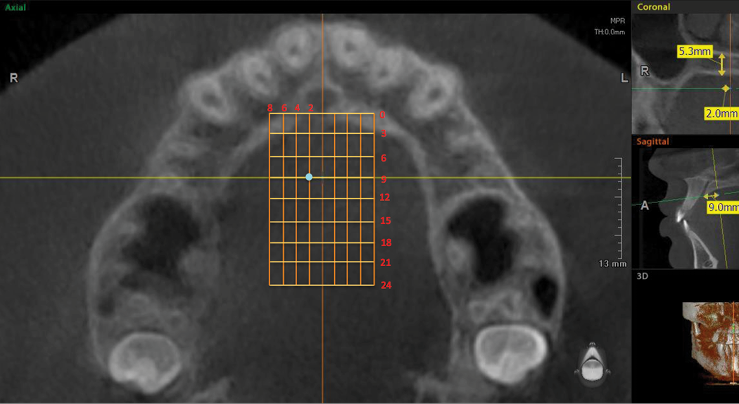
En el corte sagital, las mediciones se realizaron de los 0 a los 24mm, con intervalos de 3 mm entre cada una, con la primera medición en el borde posterior del foramen incisivo (Figura 2). Las mediciones del grosor palatino se realizaron en el corte coronal a los 2, 4, 6 y 8mm, de la sutura media palatina hacia ambos lados (Figura 3).
Posteriormente, en el corte coronal (Figura 3), perpendicular al plano axial se tomaron las medidas del grosor palatino en cada uno de los puntos de intersección de las líneas de referencia antes mencionadas, formando una cuadrícula de 72 sitios (Figura 4). Así, en las Figuras 3 y 4 se observa el grosor palatino de 5.3mm a los 2mm (sagital) y 9mm (coronal) del lado derecho.
Los datos fueron recopilados en una hoja clínica y vaciados a un formato del programa Microsoft Excel. Posteriormente, los mismos fueron trasladados a un paquete estadístico SPSS versión 2.0 para su análisis. Se realizó la prueba estadística para variables con estadística descriptiva. La prueba de Anova se utilizó debido a que en este estudio se compararon más de dos variables, se comparó el lado derecho con el lado izquierdo en cada una de sus medidas transversales 0, 2, 4, 6 y 8mm tratando de encontrar similitudes y diferencias entre todas las medidas y se complementó con la prueba Post Hoc de Tukey para saber qué medias difieren entre sí. El valor de significancia para todas las pruebas fue de p<0.05.
Figura 1. Plano axial posicionado en el borde posterior del foramen inci sivo y espina nasal posterior.
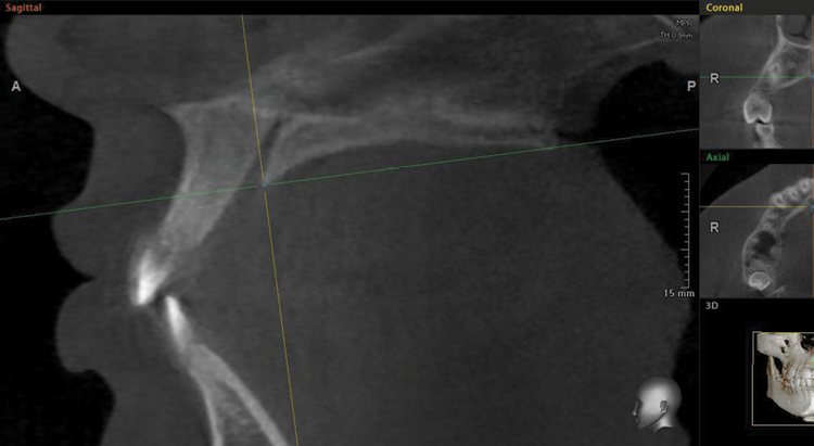
Figura 2. Intersección de plano axial y coronal a los 0mm (a) y 24mm (b).

Figura 3. Grosor palatino medido a los 9mm del borde posterior del foramen incisivo.

Figura 4. Cuadrícula del paladar. Grosor palatino de 5.3mm a los 2mm (sagital) y 9mm (coronal) del lado derecho.
Resultados
El promedio de edad de la población estudiada fue de 22 años ± 7. El mayor grosor palatino se encontró en el corte sagital en 0mm y 3mm; y los valores menores se encontraron a 24 mm para el corte sagital, y a 8mm para el corte transversal. La estadística descriptiva se encuentra en las Tablas 1 y 2.
La comparación de los valores de forma sagital arrojó diferencias estadísticamente significativas p<0.05 (Tablas 3 y 4). En las pruebas Post Hoc de Tukey, en el corte transversal a los 2mm del lado derecho se encontraron diferencias estadísticas significativas (p<0.05) a nivel del corte sagital de 0, 3 y 6mm con todos los valores. Sin embargo, en el corte sagital entre 9, 12 y 15 mm no se encontraron diferencias estadísticas significativas (p<0.05) entre ellos, ya que la diferencia fue de 1mm. Adicionalmente, a partir de 15 a 24mm la diferencia de las medidas fue de 0.7mm, por lo que no existieron diferencias estadísticas significativas entre ellos.
En el corte transversal a 2mm del lado izquierdo se encontraron diferencias estadísticamente significativas (p<0.05) en el corte sagital de 0 a 6mm con el resto de las medidas, en las medidas sagitales de 9 a 24mm la diferencia fue de 1.41mm por lo que no se encontraron diferencias estadísticas entre todos estos valores (p<0.05).
Al comparar las medidas transversales de 4, 6 y 8mm del lado derecho e izquierdo se encontraron diferencias estadísticas significativas (p<0.05) en el corte sagital de 0 a 9mm con todos los valores. A partir de 12 a 24mm no existen diferencias estadísticas significativas entre los valores encontrados del lado derecho. Del lado izquierdo no se encontraron diferencias significativas en el corte sagital de 15 a 24mm.
Al comparar los valores de forma sagital se encontraron diferencias estadísticas significativas (p<0.05) en cada uno de ellos (tabla 5). En las pruebas Post Hoc de Tukey en el corte sagital a 21 y 24mm no se encontraron diferencias (p<0.05) entre los valores encontrados en 2, 4, 6 y 8mm del corte transversal. Con excepción de los valores 2 y 8 mm en los cuales si se encontraron diferencias estadísticas significativas.
En el corte sagital a 3mm no se encontraron diferencias (p<0.05) entre los valores encontrados en los 2, 4, y 6 mm del corte transversal. A los 8mm del corte transversal no se encontraron diferencias estadísticas significativas entre los valores derechos e izquierdos, pero con los valores de 2, 4 y 6mm si se encontraron diferencias estadísticas (p<0.05). En el corte sagital a 6mm no se encontraron diferencias (p<0.05) entre los valores encontrados en los 2, 4, 6 y 8mm del corte transversal. Con excepción de los valores 2 y 8mm en los cuales si se encontraron diferencias estadísticas significativas.
En el corte sagital a 9 mm no existieron diferencias estadísticas significativas (p<0.05) entre los valores. En el corte sagital a 12 y 18mm no se encontraron diferencias estadísticas (p<0.05) entre los valores de 2 y 8mm del corte transversal, pero si con los valores de 4 y 6mm. En el corte sagital a 15mm se encontraron diferencias estadísticas significativas (p<0.05) con los valores de 2mm en el corte transversal con todos los grupos. En el corte transversal entre los grupos de 4, 6 y 8mm no existen diferencias estadísticas significativas. En el Post Hoc de Tukey en las medidas transversales no se encontraron diferencias estadísticas (p<0.05) al comparar los valores del lado derecho con los del lado izquierdo de cada uno.
|
Tabla 1. Estadísticos descriptivos del lado derecho |
|||||||
|
Corte sagital |
Corte Transversal |
Media |
Desviación |
Máximo |
Mínimo |
Rango |
Varianza |
|
N válido (según lista) |
|||||||
|
0 |
2 mm |
10.2450 |
4.01943 |
19.60 |
.00 |
19.60 |
16.156 |
|
4 mm |
11.0260 |
2.60457 |
18.20 |
4.50 |
13.70 |
6.784 |
|
|
6 mm |
11.3020 |
2.55987 |
17.60 |
5.50 |
12.10 |
6.553 |
|
|
8 mm |
12.2250 |
2.91396 |
19.20 |
5.70 |
13.50 |
8.491 |
|
|
3 |
2 mm |
7.7530 |
3.18730 |
17.10 |
1.70 |
15.40 |
10.159 |
|
4 mm |
8.3530 |
2.86538 |
17.20 |
.00 |
17.20 |
8.210 |
|
|
6 mm |
9.1060 |
2.64081 |
15.50 |
3.40 |
12.10 |
6.974 |
|
|
8 mm |
10.5400 |
2.80141 |
17.20 |
4.10 |
13.10 |
7.848 |
|
|
6 |
2 mm |
5.8670 |
2.17790 |
14.00 |
2.10 |
11.90 |
4.743 |
|
4 mm |
5.9200 |
2.20459 |
13.80 |
1.80 |
12.00 |
4.860 |
|
|
6 mm |
6.3380 |
2.38178 |
12.80 |
2.00 |
10.80 |
5.673 |
|
|
8 mm |
7.4770 |
2.72674 |
14.70 |
1.60 |
13.10 |
7.435 |
|
|
9 |
2 mm |
4.6400 |
1.62822 |
10.10 |
1.30 |
8.80 |
2.651 |
|
4 mm |
4.1610 |
1.70228 |
9.20 |
.90 |
8.30 |
2.898 |
|
|
6 mm |
4.1700 |
1.79446 |
8.20 |
.50 |
7.70 |
3.220 |
|
|
8 mm |
4.8630 |
2.18376 |
11.50 |
.80 |
10.70 |
4.769 |
|
|
12 |
2 mm |
3.9670 |
1.45074 |
7.20 |
1.00 |
6.20 |
2.105 |
|
4 mm |
3.1700 |
1.27505 |
6.60 |
.40 |
6.20 |
1.626 |
|
|
6 mm |
2.9600 |
1.41607 |
7.70 |
.50 |
7.20 |
2.005 |
|
|
8 mm |
3.3240 |
1.81687 |
10.70 |
.60 |
10.10 |
3.301 |
|
|
15 |
2 mm |
3.6440 |
1.55019 |
7.90 |
.00 |
7.90 |
2.403 |
|
4 mm |
2.8160 |
1.25591 |
6.40 |
.50 |
5.90 |
1.577 |
|
|
6 mm |
2.4790 |
1.26278 |
7.10 |
.50 |
6.60 |
1.595 |
|
|
8 mm |
2.6060 |
1.54180 |
9.60 |
.40 |
9.20 |
2.377 |
|
|
18 |
2 mm |
3.4930 |
1.57892 |
9.60 |
.60 |
9.00 |
2.493 |
|
4 mm |
2.5890 |
1.26426 |
6.10 |
.40 |
5.70 |
1.598 |
|
|
6 mm |
2.0680 |
1.15241 |
5.60 |
.00 |
5.60 |
1.328 |
|
|
8 mm |
2.1390 |
1.41485 |
9.40 |
.20 |
9.20 |
2.002 |
|
|
21 |
2 mm |
3.2540 |
1.81873 |
10.40 |
.20 |
10.20 |
3.308 |
|
4 mm |
2.2650 |
1.22156 |
5.10 |
.20 |
4.90 |
1.492 |
|
|
6 mm |
1.8050 |
1.10109 |
6.30 |
.00 |
6.30 |
1.212 |
|
|
8 mm |
1.7730 |
1.28675 |
8.40 |
.00 |
8.40 |
1.656 |
|
|
24 |
2 mm |
2.9420 |
1.68026 |
7.30 |
.20 |
7.10 |
2.823 |
|
4 mm |
2.0260 |
1.27300 |
4.80 |
.20 |
4.60 |
1.621 |
|
|
6 mm |
1.5170 |
1.06752 |
5.30 |
.00 |
5.30 |
1.140 |
|
|
8 mm |
1.5000 |
1.14636 |
7.20 |
.00 |
7.20 |
1.314 |
|
|
Tabla 2. Estadísticos descriptivos de lado izquierdo |
|||||||
|
Corte sagital |
Corte Transversal |
Media |
Desviación |
Máximo |
Mínimo |
Rango |
Varianza |
|
N válido (según lista) |
|||||||
|
0 |
2 mm |
10.2760 |
3.53631 |
17.20 |
.00 |
17.20 |
12.505 |
|
4 mm |
10.9940 |
2.46244 |
17.70 |
5.40 |
12.30 |
6.064 |
|
|
6 mm |
11.1420 |
2.47669 |
17.30 |
2.90 |
14.40 |
6.134 |
|
|
8 mm |
12.0110 |
2.91610 |
21.50 |
6.80 |
14.70 |
8.504 |
|
|
3 |
2 mm |
7.7570 |
2.94794 |
15.30 |
2.90 |
12.40 |
8.690 |
|
4 mm |
8.2930 |
2.46578 |
14.90 |
3.10 |
11.80 |
6.080 |
|
|
6 mm |
8.9910 |
2.43074 |
14.20 |
3.60 |
10.60 |
5.909 |
|
|
8 mm |
10.4220 |
2.57221 |
17.10 |
4.20 |
12.90 |
6.616 |
|
|
6 |
2 mm |
5.7630 |
2.35598 |
13.60 |
1.50 |
12.10 |
5.551 |
|
4 mm |
5.8890 |
2.26577 |
12.30 |
1.20 |
11.10 |
5.134 |
|
|
6 mm |
6.2270 |
2.35389 |
12.20 |
1.10 |
11.10 |
5.541 |
|
|
8 mm |
7.2840 |
2.65744 |
14.60 |
1.20 |
13.40 |
7.062 |
|
|
9 |
2 mm |
4.5110 |
1.66235 |
10.00 |
1.50 |
8.50 |
2.763 |
|
4 mm |
4.1000 |
1.69533 |
8.90 |
.90 |
8.00 |
2.874 |
|
|
6 mm |
3.9960 |
1.82939 |
10.20 |
.90 |
9.30 |
3.347 |
|
|
8 mm |
4.6330 |
2.21469 |
12.30 |
.70 |
11.60 |
4.905 |
|
|
12 |
2 mm |
3.8110 |
1.47470 |
7.20 |
1.00 |
6.20 |
2.175 |
|
4 mm |
3.1280 |
1.38980 |
8.00 |
.60 |
7.40 |
1.932 |
|
|
6 mm |
2.9480 |
1.52855 |
9.00 |
.40 |
8.60 |
2.336 |
|
|
8 mm |
3.2750 |
1.83377 |
11.30 |
.70 |
10.60 |
3.363 |
|
|
15 |
2 mm |
3.5680 |
1.40187 |
7.30 |
1.10 |
6.20 |
1.965 |
|
4 mm |
2.7240 |
1.24642 |
7.20 |
.80 |
6.40 |
1.554 |
|
|
6 mm |
2.3400 |
1.24600 |
7.30 |
.40 |
6.90 |
1.553 |
|
|
8 mm |
2.5510 |
1.44198 |
8.70 |
.40 |
8.30 |
2.079 |
|
|
18 |
2 mm |
3.4700 |
1.51947 |
7.60 |
.90 |
6.70 |
2.309 |
|
4 mm |
2.4960 |
1.17773 |
6.50 |
.60 |
5.90 |
1.387 |
|
|
6 mm |
2.0100 |
1.12524 |
6.40 |
.20 |
6.20 |
1.266 |
|
|
8 mm |
2.0060 |
1.18653 |
6.70 |
.20 |
6.50 |
1.408 |
|
|
21 |
2 mm |
4.0330 |
8.03969 |
82.00 |
.40 |
81.60 |
64.637 |
|
4 mm |
2.2130 |
1.22809 |
6.00 |
.30 |
5.70 |
1.508 |
|
|
6 mm |
1.6880 |
.98599 |
5.40 |
.10 |
5.30 |
.972 |
|
|
8 mm |
1.5880 |
1.03204 |
5.60 |
.10 |
5.50 |
1.065 |
|
|
24 |
2 mm |
3.0940 |
1.78189 |
9.30 |
.00 |
9.30 |
3.175 |
|
4 mm |
1.9750 |
1.21401 |
6.00 |
.10 |
5.90 |
1.474 |
|
|
6 mm |
1.4130 |
.98017 |
4.50 |
.00 |
4.50 |
.961 |
|
|
8 mm |
1.3820 |
.93715 |
4.80 |
.00 |
4.80 |
.878 |
|
|
Tabla 3. Tabla Anova medidas de corte transversal derecha |
||
|
Mm |
F |
Sig. |
|
2 |
116.146 |
.000 |
|
4 |
288.228 |
.000 |
|
6 |
376.251 |
.000 |
|
8 |
367.997 |
.000 |
|
* La diferencia de medias es significativa al nivel 0.05. |
||
|
Tabla 4. Tabla Anova medidas de corte transversal izquierda |
||
|
Mm |
F |
Sig. |
|
2 |
50.356 |
.000 |
|
4 |
317.810 |
.000 |
|
6 |
394.404 |
.000 |
|
8 |
396.855 |
.000 |
|
* La diferencia de medias es significativa al nivel 0.05. |
||
|
Tabla 5. Tabla Anova medidas de corte sagital |
||
|
Corte transversal |
F |
Sig. |
|
0 |
5.687 |
.000 |
|
3 |
15.770 |
.000 |
|
6 |
7.784 |
.000 |
|
9 |
2.916 |
.005 |
|
12 |
5.999 |
.000 |
|
15 |
12.933 |
.000 |
|
18 |
22.597 |
.000 |
|
21 |
8.016 |
.000 |
|
24 |
28.111 |
.000 |
Discusión
En el presente estudio se determinó el grosor óseo palatino y se delimitaron las zonas seguras del paladar para observar la variabilidad que existe en el grosor óseo entre cada individuo. Es fundamental enfatizar que no se deben colocar miniimplantes de la misma longitud en las diferentes zonas del paladar duro. Se registraron diferencias significativas en el corte transversal a los 2mm del lado derecho y se encontraron diferencias estadísticamente significativas (p<0.05) en el corte sagital a 0, 3 y 6mm con todos los valores.
En el corte transversal a 2mm del lado izquierdo se encontraron diferencias estadísticamente significativas (p<0.05) en el corte sagital de 0 a 6mm con el resto de las medidas, en las medidas sagitales de 9 a 24mm la diferencia fue de 1.41mm por lo que no se encontraron diferencias estadísticas entre todos estos valores (p<0.05).
Por lo anterior, se recomienda tomar tomografía de haz cónico a todos los usuarios futuros de miniimplantes, para determinar la seguridad de un posible sitio de inserción de miniimplante, así como conocer el espesor de hueso en el paladar (12).
La zona del paladar se ha utilizado como una alternativa para el anclaje ortodóntico ya que, según la clasificación de Misch, aquella ofrece la calidad y cantidad de hueso ideal para la colocación de miniimplantes, pues presenta hueso tipo D1 (>1250 HU) y D2 (850-1250 HU) (13, 14, 15).
Existen diversos estudios acerca del grosor óseo palatino. Los estudios realizados por Wang et al., Poorsattar et al. y Baumgaertel reportaron que la región anterior del paladar proporciona el mayor grosor óseo para la inserción de miniimplantes; lo que concuerda con los resultados del presente estudio, donde se encontró mayor grosor en la zona anterior en el corte sagital en 0 y 3mm posterior al foramen incisivo. Esta evidencia coincide con la investigación de Sung Hee Moon et al. donde se encontró que los miniimplantes en ortodoncia pueden ser colocados con éxito 3mm posterior al agujero incisivo. Por su parte, Marquezan et al. mencionan que las áreas adecuadas para la colocación de miniimplantes en el paladar es 4mm posterior al foramen incisivo, medida que está dentro del rango encontrado en el presente estudio (10, 16, 17, 18, 19).
Estudios realizados por Holm et al. y Baumgaertel coinciden en que en la región posterior del paladar el grosor óseo disminuye. Este planteamiento coincide con el de Poorsattar et al. quienes reportaron que el grosor óseo del paladar disminuye desde la parte anterior a la parte posterior; lo que es consistente con los resultados de este estudio (12, 18, 19).
Adicionalmente, en el presente estudio, la prueba de Post Hoc de Tukey no reveló diferencias estadísticamente significativas al comparar el lado derecho con el izquierdo en cada una de las medidas transversales, lo que concuerda con los estudios realizados por Sung He Moon et al. y Kang et al. (10, 20).
En contraste, Kang et al. encontraron diferencias estadísticamente significativas al comparar el lado derecho con el izquierdo, lo que difiere de los hallazgos del presente estudio (19). Por su parte, Huaynate menciona una disminución del grosor óseo palatino desde la zona medial hacia el proceso alveolar maxilar; lo que difiere del presente estudio, donde se encontró un aumento de la zona medial del paladar hacia el proceso alveolar maxilar (21).
Contribuciones de los autores
Ana Paulina Tirado Núñez realizó las mediciones de las 100 CBCT y colaboró en la redacción del artículo. Saraí Castrejón Jaime seleccionó las 100 CBCT de acuerdo con los criterios de inclusión y exclusión. Aida Nabile Silva Zatarain supervisó y colaboró en la redacción del artículo de investigación. Jaime Fabián Gutiérrez Rojo realizó la estadística y extrajo los resultados.
Conflictos de interés
Los autores declaran no tener conflictos de interés.
Referencias
- Pérez García LM, Márquez Rodríguez O, Mursulí Sosa M, González Calzada N. Utilización de mini implante en paciente con requerimiento de anclaje asimétrico. Presentación de caso. Gac méd espirit 2013; 15(3): 306-312.
- Chaves A, Grageda E, Uribe E. Zonas seguras de mayor cantidad ósea para colocación de mini-implantes interradiculares en cortical vestibular de maxilares superiores en pacientes periodontalmente comprometidos. Revista Mexicana de Ortodoncia 2015; 3 (3): 148-153. https://doi.org/10.1016/j.rmo.2016.03.039
- Arismendi JA, Ocampo ZM, González FJ, Morales M. Miniimplantes como anclaje en ortodoncia. Rev Fac Odontol Univ Antioq 2006; 18(1): 82-94.
- Ntolou P, Tagkli A, Pepelassi E. Factors related to the clinical application of orthodontic mini-implants. Journal of International Oral Health 2018; 10(3): 103-110. https://doi.org/10.4103/jioh.jioh_28_18
- Bourasssa C, Hosein Y, Pollmann S, Galil K, Bohany R, Holdsworth D, Tassi A. In-vitro comparison of different palatal sites for orthodontic miniscrew insertion: Effect of bone quality and quantity on primary stability. Am J Orthod Dentofacial Orthop 2018; 154(6): 809-819. https://doi.org/10.1016/j.ajodo.2018.02.010.
- Huertas A, Grageda E. Expansión ortopédica del maxilar con miniimplantes ortodóncicos: Reporte de un caso. Revista Mexicana de Ortodoncia 2014; 2(1): 47-56. https://doi.org/10.1016/S2395-9215(16)30017-4
- Lee J, Kim D, Park Y, Kyung S, Kim T. The efficent use of Midpalatal miniscrew implants. Angle Orthodontist 2004; 74(5): 711-714.
- Su-Jung M, Ji-Eun K, Eun J, Jong-Hyun N, Ji-Young K, Yoon-Goo K. Analysis of midpalatal miniscrew-assisted maxillary molar distalization patterns with simultaneous use of fixed appliances: A preliminary study. Korean J Orthod 2016; 46 (1): 55-61. https://dx.doi.org/10.4041%2Fkjod.2016.46.1.55
- Wang M, Sun Y, Yu Y, Ding X. Evaluation of Palatal Bone thickness for Insertion of Orthodontic Mini-Implants in Adults and Adolescents. The Journal of Craniofacial Surgery 2017; 28(6): 1468-1471. https://doi.org/10.1097/SCS.0000000000003906
- Moon S, Park S, Lim W, Chun Y. Palatal Bone Density in Adult Subjects: Implications for Mini-Implant Placement. Angle Orthodontist 2010; 80(1): 137-144. https://doi.org/10.2319/011909-40.1
- Herrera D, Figuero E, Shapira L, Jin L, Sanz M. La nueva clasificación de las enfermedades periodontales y periimplantarias. Revista Científica de la Sociedad Española de Periodoncia 2018; 1 (9): 94-110.
- Holm M, Jost Brinkmann P, Mah J, Bumann A. Bone thickness of the anterior palate for orthodontic miniscrews. Angle Orthodontist 2016; 86(5): 826-831. https://doi.org/10.2319/091515-622.1
- Summer A, Caliskan A, Uzun C, Karoz T, Summer M, Cankaya S. The evaluation of palatal bone thickness for implant insertion with cone beam computed. Int J Oral Maxillofac Surg 2016; 45(2): 216-220. https://doi.org/10.1016/j.ijom.2015.09.012.
- King K, Lam E, Faulkner M, Heo G, Major P. Vertical bone volumen in paramedian palate of adolescents: A computed tomography study. American Journal of Orthodontics and Dentofacial Orthopedics 2007; 132(6): 783-788. https://doi.org/10.1016/j.ajodo.2005.11.042
- Kravitz N, Kusnoto B. Risks and complications of orthodontic miniscrews. American Journal of Orthodontics and Dentofacial Orthopedics 2007; 131 Suppl 1: S43-51. https://doi.org/10.1016/j.ajodo.2006.04.027
- Marquezan M, Nojima L, Freitas A, Baratieri C, Alves Júnior M, Nojima M, Araujo M. Tomographic mapping of the hard palate and overlying mucosa. Braz Oral Res 2012; 26(1): 36-42. https://doi.org/10.1590/S1806-83242012000100007
- Wang Y, Qiu Y, Liu H, He J, Fan X. Quantitative evaluation of palatal bone thickness for the placement of orthodontic miniscrews in adults with different facial types. Saudi Medical Journal 2017; 38(10): 1051-1057. https://doi.org/10.15537/smj.2017.10.20967
- Baumgaertel S. Quantitative investigation of palatal bone depth and cortical bone thickness for mini-implant placement in adults. American Journal of Orthodontics and Dentofacial Orthopedics 2009; 136(1): 104-107. https://doi.org/10.1016/j.ajodo.2008.11.020.
- Poorsattar-Bejeh A, Haghanifar S, Poorsathar-Bejeh M, Rahmati-Kamel. Individual scoring and mapping of hard and soft tissues of the anterior hard palate for orthodontic miniscrew insertion. Journal of Investigative and Clinical Dentistry 2015; 8(1), 1-12. https://doi.org/10.1111/jicd.12186.
- Kang S, Lee S, Ahn S, Heo M, Kim T. Bone thickness of the palate for orthodontic mini-implant anchorage in adults. American Journal of Orthodontics and Dentofacial Orthopedics 2007; 131(Suppl 1): S75-81. https://doi.org/10.1016/j.ajodo.2005.09.029
- Huaynate S. Evaluación del espesor óseo en el plano coronal del paladar duro en tomografías computarizadas Cone-Beam. [Trabajo de grado para optar al título de Odontóloga]. Lima: Universidad Nacional Mayor de San Marcos; 2017.
Referencias
Pérez García LM, Márquez Rodríguez O, Mursulí Sosa M, González Calzada N. Utilización de mini implante en paciente con requerimiento de anclaje asimétrico. Presentación de caso. Gac méd espirit 2013; 15(3): 306-312.
Chaves A, Grageda E, Uribe E. Zonas seguras de mayor cantidad ósea para colocación de mini-implantes interradiculares en cortical vestibular de maxilares superiores en pacientes periodontalmente comprometidos. Revista Mexicana de Ortodoncia 2015; 3 (3): 148-153. https://doi.org/10.1016/j.rmo.2016.03.039
Arismendi JA, Ocampo ZM, González FJ, Morales M. Miniimplantes como anclaje en ortodoncia. Rev Fac Odontol Univ Antioq 2006; 18(1): 82-94.
Ntolou P, Tagkli A, Pepelassi E. Factors related to the clinical application of orthodontic mini-implants. Journal of International Oral Health 2018; 10(3): 103-110. https://doi.org/10.4103/jioh.jioh_28_18
Bourasssa C, Hosein Y, Pollmann S, Galil K, Bohany R, Holdsworth D, Tassi A. In-vitro comparison of different palatal sites for orthodontic miniscrew insertion: Effect of bone quality and quantity on primary stability. Am J Orthod Dentofacial Orthop 2018; 154(6): 809-819. https://doi.org/10.1016/j.ajodo.2018.02.010.
Huertas A, Grageda E. Expansión ortopédica del maxilar con miniimplantes ortodóncicos: Reporte de un caso. Revista Mexicana de Ortodoncia 2014; 2(1): 47-56. https://doi.org/10.1016/S2395-9215(16)30017-4
Lee J, Kim D, Park Y, Kyung S, Kim T. The efficent use of Midpalatal miniscrew implants. Angle Orthodontist 2004; 74(5): 711-714.
Su-Jung M, Ji-Eun K, Eun J, Jong-Hyun N, Ji-Young K, Yoon-Goo K. Analysis of midpalatal miniscrew-assisted maxillary molar distalization patterns with simultaneous use of fixed appliances: A preliminary study. Korean J Orthod 2016; 46 (1): 55-61. https://doi.org/10.4041/kjod.2016.46.1.55
Wang M, Sun Y, Yu Y, Ding X. Evaluation of Palatal Bone thickness for Insertion of Orthodontic Mini-Implants in Adults and Adolescents. The Journal of Craniofacial Surgery 2017; 28(6): 1468-1471. https://doi.org/10.1097/SCS.0000000000003906
Moon S, Park S, Lim W, Chun Y. Palatal Bone Density in Adult Subjects: Implications for Mini-Implant Placement. Angle Orthodontist 2010; 80(1): 137-144. https://doi.org/10.2319/011909-40.1
Herrera D, Figuero E, Shapira L, Jin L, Sanz M. La nueva clasificación de las enfermedades periodontales y periimplantarias. Revista Científica de la Sociedad Española de Periodoncia 2018; 1 (9): 94-110.
Holm M, Jost Brinkmann P, Mah J, Bumann A. Bone thickness of the anterior palate for orthodontic miniscrews. Angle Orthodontist 2016; 86(5): 826-831. https://doi.org/10.2319/091515-622.1
Summer A, Caliskan A, Uzun C, Karoz T, Summer M, Cankaya S. The evaluation of palatal bone thickness for implant insertion with cone beam computed. Int J Oral Maxillofac Surg 2016; 45(2): 216-220. https://doi.org/10.1016/j.ijom.2015.09.012.
King K, Lam E, Faulkner M, Heo G, Major P. Vertical bone volumen in paramedian palate of adolescents: A computed tomography study. American Journal of Orthodontics and Dentofacial Orthopedics 2007; 132(6): 783-788. https://doi.org/10.1016/j.ajodo.2005.11.042
Kravitz N, Kusnoto B. Risks and complications of orthodontic miniscrews. American Journal of Orthodontics and Dentofacial Orthopedics 2007; 131 Suppl 1: S43-51. https://doi.org/10.1016/j.ajodo.2006.04.027
Marquezan M, Nojima L, Freitas A, Baratieri C, Alves Júnior M, Nojima M, Araujo M. Tomographic mapping of the hard palate and overlying mucosa. Braz Oral Res 2012; 26(1): 36-42. https://doi.org/10.1590/S1806-83242012000100007
Wang Y, Qiu Y, Liu H, He J, Fan X. Quantitative evaluation of palatal bone thickness for the placement of orthodontic miniscrews in adults with different facial types. Saudi Medical Journal 2017; 38(10): 1051-1057. https://doi.org/10.15537/smj.2017.10.20967
Baumgaertel S. Quantitative investigation of palatal bone depth and cortical bone thickness for mini-implant placement in adults. American Journal of Orthodontics and Dentofacial Orthopedics 2009; 136(1): 104-107. https://doi.org/10.1016/j.ajodo.2008.11.020.
Poorsattar-Bejeh A, Haghanifar S, Poorsathar-Bejeh M, Rahmati-Kamel. Individual scoring and mapping of hard and soft tissues of the anterior hard palate for orthodontic miniscrew insertion. Journal of Investigative and Clinical Dentistry 2015; 8(1), 1-12. https://doi.org/10.1111/jicd.12186.
Kang S, Lee S, Ahn S, Heo M, Kim T. Bone thickness of the palate for orthodontic mini-implant anchorage in adults. American Journal of Orthodontics and Dentofacial Orthopedics 2007; 131(Suppl 1): S75-81. https://doi.org/10.1016/j.ajodo.2005.09.029
Huaynate S. Evaluación del espesor óseo en el plano coronal del paladar duro en tomografías computarizadas Cone-Beam. [Trabajo de grado para optar al título de Odontóloga]. Lima: Universidad Nacional Mayor de San Marcos; 2017.
Cómo citar
APA
ACM
ACS
ABNT
Chicago
Harvard
IEEE
MLA
Turabian
Vancouver
Descargar cita
Licencia
Derechos de autor 2019 Ana Paulina Tirado Núñez

Esta obra está bajo una licencia internacional Creative Commons Atribución-NoComercial-SinDerivadas 4.0.
Aquellos autores/as que tengan publicaciones con esta revista, aceptan los términos siguientes:
- Los autores/as conservarán sus derechos de autor y garantizarán a la revista el derecho de primera publicación de su obra, el cuál estará simultáneamente sujeto a la licencia Reconocimiento-NoComercial-SinObraDerivada 4.0 Internacional que permite a terceros compartir la obra siempre que se indique su autor y su primera publicación esta revista.
- Los autores/as podrán adoptar otros acuerdos de licencia no exclusiva de distribución de la versión de la obra publicada (p. ej.: depositarla en un archivo telemático institucional o publicarla en un volumen monográfico) siempre que se indique la publicación inicial en esta revista.
- Se permite y recomienda a los autores/as difundir su obra a través de Internet (p. ej.: en archivos telemáticos institucionales o en su página web) antes y durante el proceso de envío, lo cual puede producir intercambios interesantes y aumentar las citas de la obra publicada. (Véase El efecto del acceso abierto).
- Una vez sometido el artículo no se aceptaran cambios respecto a la incorporación o retiro de autores.