Preferential Protection of Domains II and III of Bacillus thuringiensis Cry1Aa Toxin by Brush Border Membrane Vesicles
Palabras clave:
Bacillus thuringiensis, site directed mutagenesis, -endotoxin, mutagénesis sitio dirigida, - endotoxina (es)Preferential Protection of Domains II and III of Bacillus thuringiensis Cry1Aa Toxin by Brush Border Membrane Vesicles
Protección preferencial de los dominios II y III de la toxina Cry1Aa de Bacillus thuringiensis en Vesículas de Membrana de Borde de Cepillo
Syed-Rehan A. Hussain1 , Álvaro M. Flórez2 , Donald H. Dean3 , Óscar Alzate4
1Molecular, Cellular and Developmental Biology (MCDB) Program, the Ohio State University, Columbus, OH 43210, USA
2Laboratorio de Biología Molecular y Biotecnología, Facultad de Medicina, Universidad de Santander, UDES, Campus Universitario, lagos del Cacique, Bloque Arahuaco piso 2. Bucaramanga, Colombia
3Molecular, Cellular and Developmental Biology (MCDB) Program, the Ohio State University, Columbus. Department of Biochemistry, The Ohio State University, Columbus, OH, USA.
4Department of Cell and Developmental Biology, University of North Carolina, Chapel Hill, USA.alzate@med.unc.edu
Recibido: agosto 20 de 2010 Aprobado: noviembre 25 de 2010
Abstract
The surface exposed Leucine 371 on loop 2 of domain II, in Cry1Aa toxin, was mutated to Lysine to generate the trypsin-sensitive mutant, L371K. Upon trypsin digestion L371K is cleaved into approximately 37 and 26 kDa fragments. These are separable on SDS-PAGE, but remain as a single molecule of 65 kDa upon purification by liquid chromatography. The larger fragment is domain I and a portion of domain II (amino acid residues 1 to 371). The smaller 26-kDa polypeptide is the remainder of domain II and domain III (amino acids 372 to 609). When the mutant toxin was treated with high dose of M. sexta gut juice both fragments were degraded. However, when incubated with M. sexta BBMV, the 26 kDa fragment (domains II and III) was preferentially protected from gut juice proteases. As previously reported, wild type Cry1Aa toxin was also protected against degradation by gut juice proteases when incubated with M. sexta BBMV. On the contrary, when mouse BBMV was added to the reaction mixture neither Cry1Aa nor L371K toxins showed resistance to M. sexta gut juice proteases and were degraded. Since the whole Cry1Aa toxin and most of the domain II and domain III of L371K are protected from proteases in the presence of BBMV of the target insect, we suggest that the insertion of the toxin into the membrane is complex and involves all three domains.
Key words: Bacillus thuringiensis, site directed mutagenesis, δ -endotoxin.
Resumen
La superficie de la toxina Cry1Aa, en el asa 2 del dominio II contiene expuesta la leucina 371, la cual fue modificada a lisina produciendo una mutante sensible a la tripsina, L371K. Esta mutante produce dos fragmentos de 37 y 26 kDa por acción de la tripsina que son separables por SDS-PAGE, pero que a la purificación por cromatografía líquida se mantienen como una sola molécula de 65 kDa. El fragmento grande contiene al dominio I y una parte del dominio II (aminoácidos 1 al 371). El polipéptido de 26 kDa contiene la parte restante del dominio II y dominio III (aminoácidos 372 al 609). Cuando la toxina mutante fue tratada con dosis altas de jugo intestinal de Manduca sexta, ambos fragmentos fueron degradados. Sin embargo, cuando fueron incubados en VMBC de M. sexta, el fragmento de 26 kDa fue protegido preferencialmente de las proteasas intestinales. Como se ha reportado, la toxina silvestre Cry1Aa también es protegida de la degradación de las proteasas cuando es incubada en VMBC de M. sexta. Sin embargo, cuando se adicionó VMBC de ratón a la mezcla de reacción, ni la toxina Cry1Aa ni la mutante L371K mostraron resistencia a las proteasas y fueron degradadas. Dado que la toxina completa de Cry1Aa y casi todo de los dominios II y III de L371K están protegidos de proteasas en presencia de VMBC del insecto, este estudio sugiere que la inserción de la toxina en la membrana involucra los tres dominios.
Palabras clave: Bacillus thuringiensis, mutagénesis sitio dirigida, δendotoxina
Introducción
The mode of action of the insecticidal Bacillus thuringiensis (Bt)* δ-endotoxins, or Cry toxins, consists of solubilization of the crystals in the insect's alkaline midgut environment (Tojo and Aizawa, 1983), followed by the activation of protoxin into toxin molecule by specific midgut proteases, and binding of the toxin to midgut epithelial cell receptors (Hofmann et al., 1988). The binding and subsequent insertion of the toxin into the midgut membrane allows the influx of cations through either non-specific pores or ion channels, depending upon the conditions (Knowles and Ellar, 1897; English and Slatin, 1992; Schwartz et al., 1993).
The process of membrane insertion by the insecticidal Bt δ-endotoxins remains a matter of extensive research. Several theoretical assumptions were made earlier regarding the insertion of toxin into the membrane and pore formation. Two main models were proposed: the "Umbrella Model" and the "Penknife Model", both of them propose that only portions of the α-helical domain I insert in the membrane, while the remaining domains II and III were just involved in receptor recognition (Li et al., 1991; Knowles, 1994).
More studies on Bt Cry toxins have suggested that the protein could insert into the midgut membrane as a single molecule, opening the possibility for a third model for the topology of the toxin in the membrane (Aronson et al.,1999; Aronson, 2000; Arnold et al., 2001; Loseva et al., 2001; Alzate et al., 2006; Nair et al., 2008; Alzate et al.,2009). In this last model, virtually the whole toxin, when associated with BBMV, is protected from proteinase K, with the exception that (in Cry1A toxins) α-helix 1 is cleaved off of the inserted form.
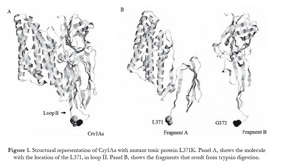
In order to test the hypothesis that virtually the whole toxin inserts as a single molecule, we have constructed a trypsin-sensitive Cry1Aa mutant, in which the Leu residue at position 371 has been replaced with Lys (L371K), producing two fragments of the toxin under denaturing conditions (Figure 1). Functional studies such as toxicity, receptor binding and insertion with wild type and the mutant protein, have led us to propose that besides domain I, domains II and III have also the ability to penetrate the membrane in support of our previous findings (Arnold et al., 2001; Alzate et al., 2006; Nair et al., 2008; Alzate et al., 2009).
Materials and methods
Mutant construction and screening. The construction of pOS4102 (Cry1Aa), by subcloning the Cry1Aa1 gene (Schnepf et al., 1985) from Bt HD-1 into pKK223-3 and over expression in Escherichia coli, has been described previously (Ge et al., 1989). To target the putative loop region in domain II, we sub-cloned the 354 bpEcoRI-SacI fragment from pOS4102 into pBluescript KS-, to obtain pBS350. Site-directed mutagenesis was performed with Muta-Gene M13 in vitro Mutagenesis kit (Bio-Rad) following manufacturers protocol. Mutagenic oligonucleotides were obtained from Sandoz Agro. Inc (Palo Alto, California). Potential mutant colonies were screened by the dideoxy sequencing method (Sanger et al., 1977), using sequenase kit (U.S Biochemical). After mutagenesis and selection, the 354 bp EcoRI-SacI fragment was sub-cloned into expression vector pOS4102. The resulting construct was named L371K.
Toxin purification and activation. The mutant gene, L371K, was overexpressed in host strain MV1190. δ-endotoxin was purified from E. coli as described (Alzate et al., 2009). The purified crystal proteins were solubilized in sodium carbonate buffer (50 mM Na>2CO3/Na2HCO2; 10 mM dithiothreitol, pH 9.5). Protoxin concentration was determined by Coomassie blue protein assay reagent (Pierce). The wild type (Cry1Aa) and the mutant (L371K) protoxins were digested with 3% (w/w) of trypsin at 37oC for 4hrs. Activated toxins were analyzed as described (Alcantara et al., 2001). Trypsin-activated toxin was purified by liquid chromatography on a HiLoad 16/60 Superdex 200 column (Pharmacia) with a Pharmacia Äkta Explorer preparative HPLC.
Structural analyses. Thermolysin digestion was carried out with the trypsin resistant core of Cry1Aa and L371K, the procedure employed was as described (Almond and Dean, 1993). Toxins in 30o g aliquots were incubated for 20 minutes at 50oC, and 58oC with 4% (w/w) of thermolysin in 50 mM Tris-HCl (pH 9.5) buffer containing 10 mM CaCl2. The reaction was terminated by adding 20 mM EDTA (final concentration). Samples were analyzed by SDS-12% PAGE and stained with Coomassie brilliant blue. The relative intensities of toxin fragments at various temperatures were determined with a UVP trans-illuminator using the Labworks™ Analysis Software (UVP, Inc. Upland, CA.) and recorded as the percentage of peptide remaining against temperature.
Toxicity bioassays. M. sexta eggs were obtained from Dr. D. L. Dahlman (Dept. of Entomology, University of Kentucky, Lexington, Kentucky). Bioassays were performed by surface contamination of artificial diet (Bio-Serve Inc., French Town, New Jersey). The diet was poured into 24-well tissue culture plates (Falcon). 50 µL of various toxin dilutions were then applied to the surface and allowed to air-dry. Six toxin concentrations were used to calculate the median lethal concentration (LC50) value with 12 M. sexta neonate larvae for each concentration. The average of three bioassays per toxin was used to calculate effective doses (LC50) and 95% fiducial limits with the Probit analysis (Finney, 1972).
Receptor binding experiments. BBMV were prepared from fifth instar M. sexta larvae by the differential magnesium precipitation method as described (Wolfersberger et al., 1987), resuspended in binding buffer (8 mM Na2HPO4, 2 mM KH2PO4, 150 mM NaCl, pH 7.4) and stored in liquid nitrogen until further use. Iodination of toxins for competition and dissociation assays was carried out as described (Lee et al., 1992). The homologous competition assay was performed by competing 1nM of 125I-labeled toxin with an increasing concentration of the same unlabeled toxin as described (Rajamohan et al., 1996). For the dissociation experiments, 50 µg of M. sextaBBMV was incubated with 2 nM of either 125I-labeled Cry1Aa or 125I-labeled L371K in 100 µl of binding buffer at room temperature. After 1hr incubation, 500-fold excess of unlabeled toxin was added to the 125I-labeled toxin-BBMV suspension. The reaction was stopped at various time intervals (0 to 60 min) by spinning down the mixture. The pellets were washed twice with 300 µl of binding buffer in order to remove any unbound toxin. The iodine content of the final pellets was determined in a gamma counter (Beckman instruments). Non-specific binding was determined by adding together labeled toxin and 500 fold excess of the corresponding unlabeled toxin to the BBMV. Non-specific binding was subtracted in the final data analysis. Binding data were analyzed with Sigma Plot (Jandel Scientific Co.).
Membrane partitioning assay. M. sexta BBMV (45 µg) was incubated with 0.120 µg of 125I-Cry1Aa or 125I- L371K. The reaction volume was adjusted to 60 µl with binding buffer supplemented with 0.1% bovine serum albumin (BSA) to avoid non-specific binding. After 150 min. of incubation at 25 oC, the suspension was centrifuged and the pellets were washed with binding buffer to remove any unbound toxin. The pellets were resuspended in 30 µl of binding buffer and 10µL of M. sexta gut juice was added. Three vials containing the same volume of mixture and no gut juice were used as control. The suspension was incubated at 25 oC for 1hr. At the end of the incubation period 5µl of 50X protease inhibitor cocktail (Roche, Molecular Biochemicals) was added to stop the reaction. The BBMV was solubilized by adding denaturing loading gel buffer (Laemmli, 1970), and boiled for 5 min. Toxin fragments were separated from BBMV on SDS-12% PAGE and analyzed by autoradiography. As a negative control mouse BBMV was incubated with either 125I-Cry1Aa or 125I-L371K and subjected to same treatment as M. sexta BBMV. Cry1Aa and L371K toxins were also treated with M. sexta gut juice (10 µl) without adding any BBMV, 60 µg of BSA were added to compensate for the BBMV peptides. Relative intensities of gut juice treated and untreated fragments were determined as described above. The data were recorded as the percentage of peptide remaining after protease digestion as compared to undigested toxin.
Results
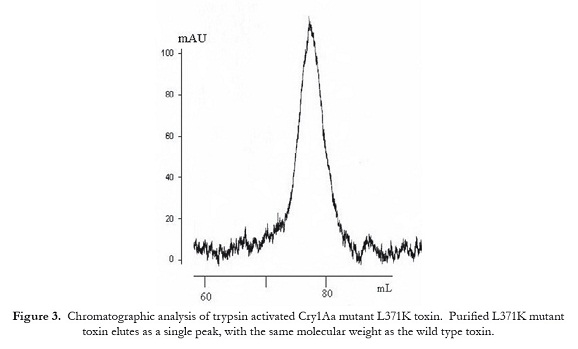
Trypsin digestion of proteins. Solubilized crystals of both, Cry1Aa and its mutant L371K, produced 130 kDa protoxin (figure 2, lanes 1 and 3, respectively). Digestion of the mutant protoxin with trypsin produces an activated toxin that separates as a single peak, eluting at the position of a 65 kDa protein molecule, by size exclusion chromatography (figure 3). Peak fractions were analyzed by SDS-PAGE (figure 2, lanes 2 and 4, respectively). The mutant protein was cleaved into two peptides with approximate molecular weights of 37 kDa (fragment A) and 28 kDa (fragment B), in contrast to a single 65 kDa for Cry1Aa toxin (figure 2). Since the trypsin-sensitive site in L371K was created in the loop2 of domain II, we consider the 37.3 kDa peptide as domain I, plus a portion of domain II (expected molecular weight, ~37.3kDa). Domain III and the remaining of domain II correspond to the 26.2 kDa fragment (calculated molecular weight, ~26.2kDa).
Structural stability of Cry1Aa and L371K. In order to determine stability alterations in the trypsin-resistant core of L371K, it was treated with thermolysin at various temperatures and compared with the wild type toxin under the same conditions. Our data shows that fragment B of L371K was more susceptible to thermolysin than fragment A (figure 4, lanes 5, 6, and 7).
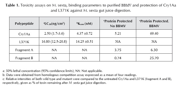
Bioassay. Trypsin-activated L371K and Cry1Aa were analyzed for toxicity against M. sexta neonate larvae. The mutant protein was active against the insect, although its toxicity was 7 times lower than the wild type (table 1).
Receptor binding studies. In order to study the binding ability of L371K toxin toward the membrane receptor ofM. sexta midgut competition assays were performed. Homologous competition experiments indicated that the mutant toxin (Kcom=14.25 ± 0.51 nM) has three-fold less initial (reversible) binding affinity than Cry1Aa toxin (Kcom =4.37 ± 0.72 nM) (Fig. 5A). In order to determine the irreversible binding parameters of the mutation to M. sexta BBMV, 125I-labeled wild type and mutant toxins were allowed to bind to the BBMV for 1h. In the presence of excess amounts of unlabeled toxin, 45% of the mutant toxin was chased off in the first 20 minutes and only 50% of the toxin remained bound to BBMV at 60 min. In contrast, the wild type toxin showed more irreversible association with only 17% being chased off after 60 min (figure 5B).
Evidence for toxin insertion into the BBMV. We investigated the regions of the Cry1Aa toxin that insert into the BBMV. It was observed that after incubation with excess M. sexta gut juice for 1h at 25 oC in the absence of BBMV, 74.2% of the Cry1Aa toxin molecules were digested (Figure 6A, lanes 1 and 2). Only 37.7% of the M. sextaBBMV-protected toxin molecules were degraded by the gut juice (figure 6A, lanes 3 and 4). It is seen that the digested fragment is slightly smaller than the undigested toxin. In agreement with a report showing that α-helix 1 is cleaved off during the insertion process (Aronson et al., 1999).
In the case of L371K toxin, the effect of M. sexta gut juice on fragments A and B was studied. In the absence ofM. sexta BBMV both fragments showed lower band intensities (figure 6B, lane 2) than the untreated toxin (figure 6B, lane 1). There was a relatively equal degradation of fragment B (49.8%) compared with intensity on lane 1, as compared to fragment A (47.4%). In contrast to these results, after incubation with M. sexta BBMV and gut juice treatment, fragment B showed less degradation (17.2%) than fragment A (66.2%; figure 6B, lanes 3 and 4).
In order to eliminate the possibility of non-specific interaction of Cry1Aa or L371K, to a lipid bilayer and thus protection from proteases, mouse BBMV was used as a control. It was observed that there was only a weak binding of either toxin to these vesicles. This may be due to the non-specific binding of toxin to the mouse BBMV. However, both Cry1Aa toxin and L371K toxin were completely degraded by gut juice proteases in the presence of mouse BBMV (figure 6, lanes 5 and 6).
Discussion
The mechanism of action of the δ-endotoxins involves binding to receptors on the microvillar membranes of the midgut, with a major portion of this interaction becoming irreversible. A direct correlation was found between toxicity and the rate of irreversible binding (Ihara et al., 1993; Liang et al, 1995). Several indirect studies suggest that irreversible binding indicates toxin insertion into the membrane (Schnepf, 1995). More specifically it has been hypothesized that domain I is the only region involved in membrane integration and channel formation (Li et al.,1991; Knowles, 1994; Gazit et al., 1998; Schwartz et al., 1997).
In this report we have attempted to test this hypothesis by performing membrane association studies with Cry1Aa and its mutant toxin L371K. We designed this trypsin-cleavable mutant based on previous biochemical studies that showed that Cry1A could be considered as two fragments linked by a protease susceptible loop that can be cleaved by chymotrypsin at amino acid residue 371 (Covents et al., 1991). Our work shows that the digestion of L371K protoxin with trypsin produces two fragments of ~37 kDa (fragment A) and ~26 kDa (fragment B) that can be resolved on SDS-PAGE. Fragment A corresponds to domain I and a portion of domain II (residues 1 to 371), whereas fragment B contains domain III and the remaining of domain II (residues 372 to 609). In solution, the two fragments remain associated as a single 65 kDa molecule.
Thermal denaturation profile of L371K shows a greater degradation of fragment B compared to fragment A in the presence of thermolysin. This may be due to the fact that fragment A mainly has domain I, which consists of amphipathic α-helices that are close-packed keeping the hydrophobic residues away from the solvent (Li et al.,1991; Grochulski et al., 1995).
In Cry1A toxins, amino acid residues 365 to 371 have been shown to affect receptor binding or membrane insertion (irreversible binding), and toxicity (Rajamohan et al., 1996; Lu et al., 1994; Rajamohan et al., 1995). The partial reduction (2 to 3 times) in the initial binding affinity of the mutant toxin L371K is likely to be caused by changes in the physicochemical properties of amino acid residue at position 371, and because of the likelihood of increased mobility of the loop due to protease cleavage. Hydrophobic amino acids at this position are required for proper binding to receptors or insertion into BBMV (Rjamohan et al., 1995). The importance of this region in the Cry1A family of δ-endotoxins is further strengthen by studies showing that substituting aliphatic, hydrophilic and small side-chain residues for Phe at position 371 reduce the irreversible association and toxicity of Cry1Ab to M. sexta (Rajamohan et al., 1995; 1996). By analogy, it can be assumed that the seven-fold reduction in toxicity forM. sexta larvae is mainly due to reduced binding or irreversible association (insertion) of L371K toxin.
Protection against exogenous proteases has been used to determine the topology of bacterial toxins in biological membranes (Aronson et al., 1999; Aronson, 2000; Arnold et al., 2001; Loseva et al., 2001; Cabiaux et al., 1994). Using a similar technique, protection of 125I-Cry1Aa and 125I-L371K toxins against M. sexta gut juice proteases was studied. It was observed that, after 1-hour incubation with M. sexta BBMV, only 37.7% of the initial concentration of Cry1Aa was digested by gut juice, while 74.2% of Cry1Aa molecules were degraded in the absence of M. sexta BBMV. However, there were no fragments smaller than the whole toxin, suggesting that the protein enters the membrane as a single molecule. This protection might be due to the insertion of the toxin into the membrane because a membrane-inserted configuration will make the toxin resistant to proteolytic cleavage. These observations in which proteinase K has been used to digest Cry1Ab and Cry1Ac (Aronson et al., 1999; Aronson, 2000; Arnold et al., 2001), under similar conditions as those presented here, suggest that the toxin enters the membrane as a single molecule.
Proteolytic digestion studies with mutant L371K showed differential resistance of fragment A and fragment B in the presence and absence of M. sexta BBMV. Both fragments were similarly degraded in solution (fragment A showed similar resistance than fragment B). The thermal stability studies, showing fragment A being more resistant to thermolysin than fragment B, are in agreement with the amphipathic α-helices of domain I being more tightly packed in the aqueous environment in the absence of the membrane. In the presence of BBMV however, fragment B showed more protection against protease digestion than fragment A, suggesting that fragment B is capable of membrane translocation by itself, without a dragging force produced by the insertion of domain I. To account for this observation we propose that at least some portions of domain II and/or domain III may become inserted into the membrane. Our hypothesis is based on studies involving the C-terminus of δ-endotoxins. It has been shown by voltage clamp and light scattering studies, that mutations of arginine residues in the conserved block 4, of domain III, causes reduction in ion-channel function of Cry1Aa toxin (Chen et al., 1993; Wolfersbergeret al., 1996; Schwartz et al., 1997; Masson et al., 2002). Furthermore mutations in domain II of Cry1Ab and Cry3Aa have been shown to affect the irreversible association with the membrane (Rajamohan et al, 1996; 1995; Wu and Dean, 1996). Our interpretation is further supported by the earlier work indicating that the whole toxin associated with BBMV is neither attacked by proteases nor bound by monoclonal antibodies (Wolfersberger et al.,1986) and it is also protected from proteinase K digestion (Aronson et al., 1999; Aronson, 2000; Arnold et al.,2001). The possibility of anti-parallel β-sheets being inserted into the lipid bilayer is demonstrated by the protease protection and analytical studies on diphtheria toxin, and aerolysin (Cabiaux et al., 1994; Parker et al.,1994). The β-sheets have been suggested to serve as scaffold, so that &alpha-helices can acquire a more favorable position to serve as the ion-channel (Hucho et al., 1994).
Our observation that fragment B (domains II and III) has a greater affinity to the BBMV than domain I does not agree with the Umbrella Model, which predicts that only a portion of domain I partitions into the membrane, while domains II and III do not. Two other possibilities exist for the differential protection of fragment B. A folding conformation on the surface of the membrane may make domains II and III inaccessible to proteases (the carpet model). However, it is difficult to defend the position that most of a protein, 65 kDa in size, or even fragment B of 26 kDa would lie on the surface and have no exposure to at least a clip by proteinase K or midgut juice. A second possibility is that the β-sheets of domain II and III may form oligomers after binding to the receptor, thus protecting the whole toxin from degradation. In this work we support a model for the membrane bound state of the δ- endotoxins, in which the whole toxin inserts into the membrane, in agreement with previous results (Aronson et al., 1999; Aronson, 2000; Arnold et al., 2001; Loseva et al., 2001; Nair et al., 2008; Alzate et al.,2007; Alzate et al., 2009) that suggests new interpretations for the mechanism of action of these important biopesticides.
Acknowledgements
This work was supported by a grant, R01 29092 to D.H.D., from the National Institutes of Health.
References
1 Alcantara, E., Alzate, O., Lee, M. K., Curtiss, A., Dean, D. H. 2001. Role of a-helix 7 of Bacillus thuringiensis Cry1Ab d-endotoxin in membrane insertion, structural stability, and ion channel activity. Biochemistry 40: 2540-2547.
2 Almond, B. D., Dean, D. H. 1993. Suppression of protein structure destabilizing mutations in Bacillus thuringiensis d-endotoxins by second site mutations, Biochemistry 32: 1040-1046.
3 Aronson, A. 2000. Incorporation of protease K into larval insect membrane vesicles does not result in disruption of function of the pore-forming Bacillus thuringiensis -endotoxins. Appl Environ Microbiol 66: 4568-4570.
4 Aronson, A. I., Geng, C. Wu, L. 1999. Aggregation of Bacillus thuringiensis Cry1A toxins upon binding to target insect larval midgut vesicles. Appl Environ Microbiol 65: 2503-2507.
5 Arnold, S., Curtiss, A., Dean, D. H. Alzate, O. 2001. The role of a proline-induced broken-helix motif in a-helix 2 of Bacillus thuringiensis d- endotoxins. FEBS Letts 490: 70-74.
6 Alzate, O., Hemann, C. F., Osorio, C., Hille, R., Dean, D. H. 2009. Ser170 of Bacillus thuringiensis Cry1Ab -endotoxin becomes anchored in a hydrophobic moiety upon insertion of this protein into M. sexta brush border membranes. BMC Biochem 10: 25.
7 Alzate, O., You, T., Claybon, M., Osorio, C., Curtiss, A., Dean, D. H. 2006. Effects of disulfide bridges in domain I of Bacillus thuringiensis Cry1Aa -endotoxin on ion-channel formation in biological membranes. Biochemistry 45: 13597-605.
8 Arnold, S., Curtiss, A., Dean, D. H. Alzate, O. 2001. The role of a proline-induced broken-helix motif in a-helix 2 of Bacillus thuringiensis d- endotoxins. FEBS Letts 490: 70-74.
9 Cabiaux, V., Quertenmont, P., Conrath, K., Brasseur, R., Capiau, C., Ruysschaert, J.-M. 1994. Topology of diphtheria toxin B fragment inserted in lipid vesicles. Mol Microbiol 11: 43-50.
10 Chen, X. J., Lee, M. K., Dean, D. H. 1993. Site-directed mutations in a highly conserved region of Bacillus thuringiensis -endotoxin affect inhibition of short circuit current across Bombyx mori midguts. Proc Natl Acad Sci USA 90: 9041-9045.
11 Convents, D., Cherlet, M., Van Damme, J., Lasters, I., Lauwereys, M. 1991. Two structural domains as a general fold of the toxic fragment of the Bacillus thuringiensis delta-endotoxins. Eur J Biochem 195: 631-635.
12 English, L., Slatin, S. L. 1992. Mini-Review: Mode of action of delta endotoxins from Bacillus thuringiensis: A comparison with other bacterial toxins. Insect Biochem Molec Biol 22: 1-7.
13 Finney, D. 1972. Statistical Methods in Biological Assay, 3rd Ed. Griffin and Co., Ltd., London.
14 Gazit, E., LaRocca, P., Sansom, M. S. P., Shai, Y. 1998. The structure and organization within the membrane of the helices composing the pore-forming domain of Bacillus thuringiensis -endotoxin are consistent with an "umbrella-like" structure of the pore. Proc Natl Acad Sci 95: 12289-12294.
15 Ge, A. Z., Shivarova, N. I., Dean, D. H. 1989. Location of the Bombyx mori specificity domain on a Bacillus thuringiensis d-endotoxin protein. Proc Natl Acad Sci 86: 4037-4041.
16 Grochulski, P., Masson, L., Borisova, S., Pusztai-Carey, M., Schwartz, J.-L., Brousseau, R., Cygler, M. 1995. Bacillus thuringiensis CryIA(a) insecticidal toxin: crystal structure and channel formation. J Mol Biol 254: 447-464.
17 Hofmann, C., Lüthy, P., Hütter, R., Pliska, V. 1988. Binding of the delta-endotoxin from Bacillus thuringiensis to brush-border membrane vesicles of the cabbage butterfly (Pieris brassicae). Eur J Biochem 173: 85-91.
18 Hucho, F., Görne-Tschelnokow, U., Strecker, A. 1994. -structure in the membrane-spanning part of the nicotinic acetylcholine receptor (or how helical are transmembrane helices?). TIBS 19: 383-387.
19 Ihara, H., Kuroda, E., Wadano, A., Himeno, M. 1993. Specific toxicity of d-endotoxins from Bacillus thuringiensis to Bombyx mori. Biosci Biotech Biochem 57: 200-204.Knowles, B. H. 1994 Mechanism of action of Bacillus thuringiensis insecticidal d-endotoxins. Adv Insect Physiol 24: 275-308.
20 Knowles, B. H., Ellar, D. J. 1987. Colloid-osmotic lysis is a general feature of the mechanism of action of Bacillus thuringiensis -endotoxins with different insect specificity. Biochim Biophys Acta 924: 509-518.
21 Laemmli, U. K. 1970. Cleavage of structural proteins during the assembly of the head of bacteriophage T4. Nature 227: 680-685.
22 Lee, M. K., Milne, R. E., Ge, A. Z., Dean, D. H. 1992. Location of a Bombyx mori receptor binding region on a Bacillus thuringiensis -endotoxin. J Biol Chem 267: 3115-3121.
23 Li, J., Carroll, J., Ellar, D. J. 1991. Crystal structure of insecticidal -endotoxin from Bacillus thuringiensis at 2.5 Å resolution. Nature 353: 815-821.
24 Liang, Y., Patel, S. S., Dean, D. H. 1995. Irreversible binding kinetics of Bacillus thuringiensis CryIA -endotoxins to gypsy moth brush border membrane vesicles is directly correlated to toxicity. J Biol Chem 270: 24719-24724.
25 Loseva, O. I., Tiktopulo, E. I., Vasiliev, V. D., Nikulin, A. D., Dobritsa, A. P., Potekhin, S. A. 2001. Structure of Cry3A -Endotoxin within Phospholipid Membranes. Biochemistry 40: 14143-14151.
26 Lu, H., Rajamohan, F., Dean, D. H. 1994. Identification of amino acid residues of Bacillus thuringiensis -endotoxin CryIAa associated with membrane binding and toxicity to Bombyx mori. J Bacteriol 176: 5554-5559.
27 Masson, L., Tabashnik, B. E., Liu, Y.-B., Brousseau, R., Schwartz, J.L. 1999. Helix 4 of the Bacillus thuringiensis Cry1Aa toxin lines the lumen of the ion channel. J Biol Chem 274: 31996-32000.
28 Masson, L., Tabashnik, B.E., Mazza, A. Préfontaine, G. Potvin, L. Brousseau, R., Schwartz, J.L. 2002 Mutagenic analysis of a conserved region of domain III in the Cry1Ac toxin of Bacillus thuringisiensis. Appl Environ Microbiol 68: 194-200.
29 Nair, M. S., Liu, X. S., Dean, D. H. 2008. Membrane insertion of the Bacillus thuringiensis Cry1Ab toxin: single mutation in domain II block partitioning of the toxin into the brush border membrane. Biochemistry 47: 5814-22
30 Parker, M. W., Buckley, J.T., Postma, J.P.M., Tucker, A.D., Leonard, K., Pattus, F. 1994 Structure of the Aeromonas toxin proaerolysin in its water- soluble and membrane-channel states. Nature 367: 292-295.
31 Rajamohan, F., Alcantara, E., Lee, M. K., Chen, X. J., Curtiss, A., Dean, D. H. 1995. Single amino acid changes in domain II of Bacillus thuringiensis CryIAb d-endotoxin affect irreversible binding to Manduca sexta midgut membrane vesicles. J Bacteriol 177: 2276-2282.
32 Rajamohan, F., Cotrill, J. A., Gould, F., Dean, D. H. 1996. Role of domain II, loop 2 residues of Bacillus thuringiensis CryIAb d-endotoxin in reversible and irreversible binding to Manduca sexta and Heliothis virescens. J Biol Chem 271: 2390-2397.
33 Sanger, R., Niklen, S., Coulson, A. R. 1977. DNA sequencing with chain-terminating inhibitors. Proc Natl Acad Sci USA 74: 5463-5467.
34 Schnepf, H. E. 1995. Bacillus thuringiensis toxins: regulation, activities and structural diversity. Current Opinion in Biotechnology 6:305-312.
35 Schnepf, H. E., Wong, H. C., Whiteley, H. R. 1985. The amino acid sequence of a crystal protein from Bacillus thuringiensis deduced from the DNA base sequence. J Biol Chem 260: 6264-6272.
36 Schwartz, J.L., Garneau, L., Savaria, D., Masson, L., Brousseau, R., Rousseau, E. 1993 Lepidopteran-specific crystal toxins from Bacillus thuringiensis form cation- and anion-selective channels in planar lipid bilayers. J Membrane Biol 132: 53-62.Schwartz, J. L., Juteau, M., Grochulski, P., Cygler, M., Préfontaine, G., Brousseau, R. Masson, L. 1997. Restriction of intramolecular movements within the CrylAa toxin molecule of Bacillus thuringiensis through disulfide bond engineering. FEBS Letts 410: 397-402.
37 Schwartz, J. L., Lu, Y. J., Sohnlein, P., Brousseau, R., Laprade, R., Masson, L., Adang, M. J. 1997. Ion channels formed in planar lipid bilayers by Bacillus thuringiensis toxins in the presence of Manduca sexta midgut receptors. FEBS Lett 412: 270-276.
38 Schwartz, J. L., Potvin, L., Chen, X. J., Brousseau, R., Laprade, R., Dean, D. H. 1997 Single site mutations in the conserved alternating arginine region affect ionic channels formed by CryIAa, a Bacillus thuringiensis toxin. Appl Environ Microbiol 63: 3978-3984.
39 Tojo, A., Aizawa, K. 1983. Dissolution and degradation of Bacillus thuringiensis d-endotoxin by gut juice protease of the silkworm Bombyx mori. Appl Environ Microbiol 45: 576-580.
40 Wolfersberger, M. G., Chen, X. J., Dean, D. H. 1996. Site-directed mutations in the third domain of Bacillus thuringiensis d-endotoxin CryIAa affects its ability to increase the permeability of Bombyx mori midgut brush border membrane vesicles. Appl Environ Microbiol 62: 279-282.
41 Wolfersberger, M. G., Hofmann, C., Lüthy, P. 1986. Interaction of Bacillus thuringiensis delta-endotoxin with membrane vesicles isolated from lepidopteran larval midgut in Bacterial protein toxins (Falmagne, P., Alouf, J. E., Fehrenbach, F. J., Jeljaszewicz, J. & Thelestam, M., eds.), pp. 237-238, Guslav Fischer Verlag, Stuttgard, Germany.
42 Wolfersberger, M., Lüthy, P., Maurer, A., Parenti, P., Sacchi, F. V., Giordana, B., Hanozet, G. M. 1987. Preparation and partial characterization of amino acid transporting brush border membrane vesicles from the larval midgut of the cabbage butterfly (Pieris brassicae). Comp Biochem Physiol 86A: 301- 308.
43 Wu, S.-J., Dean, D. H. 1996 Functional significance of loops in the receptor binding domain of Bacillus thuringiensis CryIIIA d-endotoxin. J Mol Biol 255, 628-640
Cómo citar
APA
ACM
ACS
ABNT
Chicago
Harvard
IEEE
MLA
Turabian
Vancouver
Descargar cita
Visitas a la página del resumen del artículo
Descargas
Licencia
Derechos de autor 2010 Revista Colombiana de Biotecnología

Esta obra está bajo una licencia internacional Creative Commons Atribución 4.0.
Esta es una revista de acceso abierto distribuida bajo los términos de la Licencia Creative Commons Atribución 4.0 Internacional (CC BY). Se permite el uso, distribución o reproducción en otros medios, siempre que se citen el autor(es) original y la revista, de conformidad con la práctica académica aceptada. El uso, distribución o reproducción está permitido desde que cumpla con estos términos.
Todo artículo sometido a la Revista debe estar acompañado de la carta de originalidad. DESCARGAR AQUI (español) (inglés).


