


Publicado
STRUCTURE AND HISTOCHEMISTRY OF THE FLORAL NECTARY OF Bauhinia monandra (FABACEAE)
Estructura e histoquímica del nectario floral de Bauhinia monandra (Fabaceae)
Estrutura e histoquímica do nectário floral de Bauhinia monandra (Fabaceae)
DOI:
https://doi.org/10.15446/abc.v28n1.88610Palabras clave:
anatomy, flower, Leguminosae, medicinal plant, secretory structure (en)anatomía, estructura secretora, flor, Leguminosae, planta medicinal (es)
anatomia, estrutura secretora, flor, Leguminosae, planta medicinal (pt)
Descargas
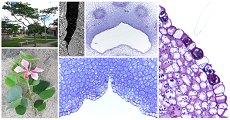
The structure and histochemistry of the floral nectary of Bauhinia monandra Kurz (Fabaceae) were investigated. Besides possessing medicinal properties, this tree is also used in the recovery of degraded areas and urban arborization. Nectaries samples were obtained from newly bloomed flowers. The nectary was located on the tubular hypanthium. This tube was partially coated by a nectary epidermis, whose cells had secretory features such as a relatively large nucleus, a dense cytoplasm, and small vacuoles. Subjacent to the nectary epidermis, there was a nectary parenchyma with eight to fifteen layers of cells which also have secretory features. Both the nectary epidermis and nectary parenchyma possessed starch grains. Subjacent to the nectary parenchyma passed collateral to amphicribral concentric vascular bundles constituted by more phloem than xylem. Although these vascular bundles did not emit terminations directed to the nectary parenchyma, the arrangement of the latter about the former suggests the supply of nectar precursors by the vascularisation. In the basal region of the hypanthium tube occurred modified stomata which were probably the main route of nectar release; and tector trichomes, possibly involved in the nectar retention.
La estructura e histoquímica del nectario floral de Bauhinia monandra Kurz (Fabaceae) fueron investigadas. Además de poseer propiedades medicinales, este árbol también se utiliza en la recuperación de áreas degradadas y en la arborización urbana. Se obtuvieron muestras de nectarios de flores recién abiertas. El nectario se ubicó en el hipanto tubular. Este tubo estaba parcialmente cubierto por una epidermis nectarífera, cuyas células tenían características secretoras, como un núcleo relativamente grande, un citoplasma denso y pequeñas vacuolas. Subyacente a la epidermis nectarífera, había un parénquima nectarífero con ocho a quince capas de células que también tenían características secretoras. Tanto la epidermis nectarífera como el parénquima nectarífero contenían granos de almidón. Subyacente al parénquima nectarífero había haces vasculares colaterales o concéntricos anficribrales constituidos por más floema que xilema. Aunque estos haces vasculares no emitieron terminaciones dirigidas al parénquima nectarífero, la disposición de este último en relación con los primeros sugiere el suministro de precursores de néctar por la vascularización. En la región basal del tubo de hipanto había estomas modificados que probablemente eran la ruta principal de liberación de néctar; y tricomas tectores, posiblemente activos en la retención de néctar.
Referencias
Antunes MN, Pereira FR, Leitão CAE. Structural characterization of the leaf of Bauhinia monandra Kurz (Fabaceae – Cercidoideae). Braz Arch Biol Technol. 2021;64:e21200618. Doi: https://doi.org/10.1590/1678-4324-2021200618.
Beaumont KP, Mackay DA, Whalen MA. Ant defence of a dioecious shrub, Adriana quadripartita (Euphorbiaceae), with extrafloral nectaries. Aust J Bot. 2016;64:539-546. Doi: https://doi.org/10.1071/BT16034.
Bentley B, Elias T. The biology of nectaries. New York: Columbia University Press; 1983. 259 p.
Bernardello, G. A systematic survey of floral nectaries. In: Nicolson SW, Nepi M, Pacini E, editor(s). Nectaries and nectar. Dordrecht: Springer; 2007. p. 19-128. DOI: https://doi.org/10.1007/978-1-4020-5937-7_2
Burnie G, Forrester S, Greig D, Guest S, Harmony M, Hobley S, Jackson G, Lavarack P, Letgett M, McDonald R, Macoboy S, Molyneux B, Moodie D, Moore J, Newman D, North T, Pienaar K, Purdy G, Silk J, Ryan S, Schien G. Botanica: the illustrated A-Z of over 10,000 garden plants and how to cultivate them. Sydney: Könemann; 2004. 1020 p.
Carmo-Oliveira R, Oliveira PE, Morretes BL. Appendicular origin and structure of the spur of Vochysiaceae flowers. Acta Bot Bras. 2017;31:433-444. Doi: https://doi.org/10.1590/0102-33062017abb0117.
Castellanos C, Forero E. El género Bauhinia L. sensu stricto (Leguminosae: Cercidoideae: Cercideae) en Colombia. In: Forero E, Castellanos C, editor(s). Estudios en leguminosas colombianas III. Bogotá: Academia Colombiana de Ciencias Exactas, Físicas y Naturales; 2019, p. 23-73.
Cocucci AA, Galetto L, Sersic A. El syndrome floral de Caesalpinia gilliesii (Fabaceae – Caesalpinioideae). Darwiniana. 1992;31:111-135.
Coutinho IAC, Meira RMSA. Structural diversity of extrafloral nectaries in Chamaecrista sect. Apoucouita. Botany. 2015;93:379-388. Doi: https://doi.org/10.1139/cjb-2014-0227.
Coutinho IAC, Valente VMM, Meira RMS. Ontogenetic, anatomical and histochemical study of the extrafloral nectaries of Sapium biglandulosum (Euphorbiaceae). Aust J Bot. 2010;58:224–232. Doi: https://doi.org/10.1071/BT09200.
Davis AR. Searching and breeding for structural features of flowers correlated with high nectar-carbohydrate production. Acta Hortic. 2001;561:107-121. Doi: https://doi.org/10.17660/ActaHortic.2001.561.16.
Davis AR, Pylatuik JD, Paradis JC, Low NH. Nectarcarbohydrate production and composition vary in relation to nectary anatomy and location within individual flowers of several species of Brassicaceae. Planta. 1998;205:305-318. Doi: https://doi.org/10.1007/s004250050325.
Etcheverry AV, Figueroa-Castro D, Figueroa-Fleming T, Alemán MM, Juárez VD, López-Spahr D, Yáñez CN, Gómez CA. Generalised pollination system of Erythrina dominguezii (Fabaceae: Papilionoideae) involving hummingbirds, passerines and bees. Aust J Bot. 2012;60:484-494. Doi: https://doi.org/10.1071/BT11325.
Fahn, A. On the structure of floral nectaries. Bot Gaz. 1952; 113:464-470. Doi: https://doi.org/10.1086/335735.
Fahn A. Secretory tissues in plants. London: Academic Press; 1979. 302 p.
Falcão PF, Melo-de-Pinna GF, Leal IR, Almeida-Cortez JS. Morphology and anatomy of extrafloral nectaries in Solanum stramonifolium (Solanaceae). Can J Bot. 2003;81:859-864. Doi: https://doi.org/10.1139/b03-083.
Gabe M. Techniques histologiques. Paris: Masson and Co.; 1968. 1113 p.
Gonzalez AM, Marazzi B. Extrafloral nectaries in Fabaceae: filling gaps in structural and anatomical diversity in the family. Bot J Linn Soc. 2018;187:26-45. Doi: https://doi.org/10.1093/botlinnean/boy004.
Heil M. Nectar: generation, regulation and ecological functions. Trends Plant Sci. 2011;16:191-200. Doi: https://doi.org/10.1016/j.tplants.2011.01.003.
Hokche O, Ramirez N. Pollination ecology of seven species of Bauhinia L. (Leguminosae: Caesalpinioideae). Ann Missouri Bot Gard. 1990;77:559-572. Doi: https://doi.org/102307/2399520.
Johansen DA. Plant microtechnique. New York: McGraw-Hill; 1940. 523 p.
Karnovsky MJ. A formaldehyde-glutaraldehyde fixative of high osmolality for use in electron microscopy. J Cell Biol. 1965;27:137-138.
Lau CPY, Saunders RMK, Ramsden L. Floral biology, breeding systems and population genetic structure of three climbing Bauhinia species (Leguminosae: Caesalpinioideae) in Hong Kong, China. J Trop Ecol. 2009;25:147-159. Doi: https://doi.org/10.1017/S0266467408005762.
Leitão CAE. Working optimally with serial sections in glycol methacrylate resin. Braz Arch Biol Technol. 2018;61:e18180103. Doi: https://doi.org/10.1590/1678-4324-2018180103.
Leitão CAE, Dolder MAH, Cortelazzo AL. Anatomy and histochemistry of the nectaries of Rodriguezia venusta (Lindl.) Rchb. f. (Orchidaceae). Flora. 2014;209:233-243. Doi: https://doi.org/10.1016/j.flora.2014.03.002.
Lewis G, Schrire, B, Mackinder, B, Lock, M.. Legumes of The World. Kew: Royal Botanic Gardens; 2005. 577p.
LPWG. A new subfamily classification of the Leguminosae based on a taxonomically comprehensive phylogeny. Taxon. 2017;66:44-77. Doi: https://doi.org/10.12705/661.3.
Maia V. Técnica histológica. São Paulo: Atheneu; 1979. 246p.
Moreira FF, Vaz AMSF, Mendonça CBF, Gonçalves-Esteves V. The systematic value of pollen morphology in trees and shrubs species of Bauhinia L. (Caesalpinioideae - subg. Bauhinia – sect. Pauletia) occurring in Brazil. Acta Bot Bras. 2013;27:400-417. Doi: https://doi.org/10.1590/S0102-33062013000200014.
Nepi M. Nectary structure and ultrastructure. In: Nicolson SW, Nepi M, Pacini E, editor(s). Nectaries and nectar. Dordrecht: Springer; 2007. p. 129-166. DOI: https://doi.org/10.1007/978-1-4020-5937-7_3
Nepi M, Ciampolini F, Pacini E. Development and ultrastructure of Cucurbita pepo nectaries of male flowers. Ann Bot. 1996;78:95-104. Doi: https://doi.org/10.1006/anbo.1996.0100.
Nilsson LA. The evolution of flowers with deep corolla tubes. Nature. 1988;334:147-149. Doi: https://doi.org/10.1038/334147a0.
Paiva EAS. Anatomy, ultrastructure, and secretory activity of the floral nectaries in Swietenia macrophylla (Meliaceae). Am J Bot. 2012;99:1910-1917. Doi: https://doi.org/10.3732/ajb.1200122.
Paiva EAS. How does the nectar of stomata-free nectaries cross the cuticle? Acta Bot Bras. 2017;31:525-530. Doi: https://doi.org/10.1590/0102-33062016abb0444.
Paiva EAS, Machado SR. Ontogenesis, structure and ultrastructure of Hymenaea stigonocarpa (Fabaceae: Caesalpinioideae) colleters. Rev Biol Trop. 2006;54:943-950. Doi: https://doi.org/10.15517/rbt.v54i3.13692.
Paiva EAS, Machado SR. The floral nectary of Hymenaea stigonocarpa (Fabaceae, Caesalpinioideae): structural aspects during floral development. Ann Bot. 2008;101:125-133. Doi: https://doi.org/10.1093/aob/mcm268.
Paiva EAS, Martins LC. Structure of the receptacular nectary and circadian metabolism of starch in the antguarded plant Ipomoea cairica (Convolvulaceae). Plant Biol. 2014;16:244-251. Doi: https://doi.org/10.1111/plb.12038.
Pearse AGE. Histochemistry: theoretical and applied, Vol I. London: Longman Group Limited; 1980. 759 p.
Pizzolato P, Lillie RD. Mayer’s tannic acid-ferric chloride stain for mucins. J Histochem Cytochem. 1973;21:56-64. Doi: https://doi.org/10.1177/21.1.56.
Quijano JGC, Muñoz DER, Alejandro, MAM. Uso y conocimiento de Bauhinia monandra Kurz en una zona urbana de Quintana Roo. Rev Etnobiología 2018;16:48-57.
Rachmilevitz T, Fahn, A. Ultrastructure of nectaries of Vinca rosea L., Vinca major L. and Citrus sinensis Osbeck cv. Valencia and its relation to the mechanism of nectar secretion. Ann Bot. 1973;37:1-9. Doi: https://doi.org/10.1093/oxfordjournals.aob.a084662.
Radice S, Galati BG. Floral nectary ultrastructure of Prunus persica (L.) Batch cv. Forastero (Newcomer), an Argentine peach. Plant Syst Evol. 2003;238:23-32. Doi: https://doi.org/10.1007/s00606-002-0279-9.
Ribeiro VC, Leitão CAE. Utilisation of Toluidine blue O pH 4.0 and histochemical inferenceds in plant sections obtained by free-hand. Protoplasma. 2020;257:993-1008. Doi: https://doi.org/10.1007/s00709-019-01473-0.
Salatino A, Blatt CTT, Santos DYAC, Vaz AMSF. Foliar flavonoids of nine species of Bauhinia. Rev Bras Bot. 1999;22:17-20. Doi: https://doi.org/10.1590/S0100-84041999000100003.
Santos OG, Pereira RCA, Sousa FJB, Paiva LGG, Bezerra MGA. Análise do crescimento de mudas de Bauhinia monandra, Kurz. Hortic Bras. 2014;31:S2611-S2618.
Silva MS, Coutinho IAC, Araújo MN, Meira RMSA. Morphoanatomy of nectaries of Chamaecrista (L.) Moench sections Chamaecrista, Caliciopsis and Xerocalyx (Leguminosae: Caesalpinioideae). Acta Bot Bras. 2017;31:445-458. Doi: https://doi.org/10.1590/0102-33062017abb0101.
Sinou C, Forest F, Lewis GP, Bruneau A. The genus Bauhinia s.l. (Leguminosae): a phylogeny based on the plastid trnLtrnF region. Botany. 2009;87:947-960. Doi: https://doi.org/10.1139/B09-065.
Stpiczyn´ska M, Davies KL, Gregg A. Nectary structure and nectar secretion in Maxillaria coccinea (Jacq.) L.O. Williams ex Hodge (Orchidaceae). Ann Bot. 2003;93:87-95. Doi:https://doi.org/10.1093/aob/mch008.
Stpiczyn´ska M, Davies KL, Kamin´ska M. Comparative anatomy of the nectary spur in selected species of Aeridinae (Orchidaceae). Ann Bot. 2011;107:327-345. Doi: https://doi.org/10.1093/aob/mcq246.
Stpiczyn´ska M, Kamin´ska M, Zych M. Nectary structure in dichogamous flowers of Polemonium caeruleum L. (Polemoniaceae). Acta Biol Crac. 2012;54:61-68. Doi: https://doi.org/10.2478/v10182-012-0019-6.
Stpiczyn´ska M, Milanesi C, Faleri C, Cresti M. Ultrastructure of the nectary spur of Platanthera chlorantha (Custer) Rchb. (Orchidaceae) during successive stages of nectar secretion. Acta Biol Crac. 2005;47:111-119.
Subramanian RB, Inamdar JA. The structure, secretion and biology of nectaries in Tecomaria capensis Thunb (Bignoniaceae). Phytomorphology. 1989;39:69-74. Doi: https://doi.org/10.2307/2445877.
Torres-Colín R, Stefano RD, Can LL. El género Bauhinia (Fabaceae, Caesalpinioideae, Cercideae) en la península de Yucatán (México, Belice y Guatemala). Rev Mex Biodivers. 2009;80:293-301. Doi: https://doi.org/10.22201/ib.20078706e.2009.002.625.
Vesprini JL, Pacini E, Nepi M. Floral nectar production in Helleborus foetidus: an ultrastructural study. Botany. 2012;90:1308-1315. Doi: https://doi.org/10.1139/b2012-101.
Vidal BC. Dichroism in collagen bundles stained with Xylidine Ponceau 2R. Ann Histochim. 1970;15:289-296.
Vidal BC. Acid glycosaminoglycans and endochondral ossification: microspectrophotometric evaluation and macromolecular orientation. Cell Mol Biol. 1977;22:45-64.
Whiteman P. The quantitative measurement of Alcian blueglycosaminoglycan complexes. Biochem J. 1973;131:343-350. Doi: https://doi.org/10.1042/bj1310343.
Yamasaki E, Sakai S. Wind and insect pollination (ambophily) of Mallotus spp. (Euphorbiaceae) in tropical and temperate forests. Aust J Bot. 2013;61:60-66. Doi: https://doi.org/10.1071/bt12202.
Zhang X, Zhao L. Morphology, structure and ultrastructure of staminal nectary in Lamprocapnos (Fumarioideae, Papaveraceae). Flora. 2018;242:128-136. Doi: https://doi.org/10.1016/j.flora.2018.03.015.
Cómo citar
APA
ACM
ACS
ABNT
Chicago
Harvard
IEEE
MLA
Turabian
Vancouver
Descargar cita
CrossRef Cited-by
1. Andrey Sinjushin. (2025). Floral Nectaries in Leguminosae: Structure, Diversity, and Possible Evolution. The Botanical Review, 91(1), p.51. https://doi.org/10.1007/s12229-024-09305-4.
2. Andrews V.S. Silva, Steven D. Johnson, Vidal F. Mansano, Louis P. Ronse De Craene, Giseli D. Pedersoli, Juliana V. Paulino. (2024). Puzzling androecium development in Bauhinia galpinii (Fabaceae) facilitates wing pollination by butterflies. Perspectives in Plant Ecology, Evolution and Systematics, 65, p.125832. https://doi.org/10.1016/j.ppees.2024.125832.
3. Lukas Gabriel Macedo Pessanha de Souza, Marcus José de Azevedo Falcão, João Paulo Basso-Alves, Vidal de Freitas Mansano. (2025). Floral developmental insights into two species of Erythrina (Fabaceae: Papilionoideae: Phaseoleae) pollinated by hummingbirds and passerines. Journal of Plant Research, 138(2), p.253. https://doi.org/10.1007/s10265-024-01610-8.
Dimensions
PlumX
Visitas a la página del resumen del artículo
Descargas
Licencia
Derechos de autor 2023 Acta Biológica Colombiana

Esta obra está bajo una licencia internacional Creative Commons Atribución-NoComercial-CompartirIgual 4.0.
1. La aceptación de manuscritos por parte de la revista implicará, además de su edición electrónica de acceso abierto bajo licencia Attribution-NonCommercial-ShareAlike 4.0 (CC BY NC SA), la inclusión y difusión del texto completo a través del repositorio institucional de la Universidad Nacional de Colombia y en todas aquellas bases de datos especializadas que el editor considere adecuadas para su indización con miras a incrementar la visibilidad de la revista.
2. Acta Biológica Colombiana permite a los autores archivar, descargar y compartir, la versión final publicada, así como las versiones pre-print y post-print incluyendo un encabezado con la referencia bibliográfica del articulo publicado.
3. Los autores/as podrán adoptar otros acuerdos de licencia no exclusiva de distribución de la versión de la obra publicada (p. ej.: depositarla en un archivo telemático institucional o publicarla en un volumen monográfico) siempre que se indique la publicación inicial en esta revista.
4. Se permite y recomienda a los autores/as difundir su obra a través de Internet (p. ej.: en archivos institucionales, en su página web o en redes sociales cientificas como Academia, Researchgate; Mendelay) lo cual puede producir intercambios interesantes y aumentar las citas de la obra publicada. (Véase El efecto del acceso abierto).


