Published
Citrus huanglongbing: validation of Real-Time PCR (qPCR) for the detection of Candidatus Liberibacter asiaticus and Candidatus Liberibacter americanus in Colombia
Citrus huanglongbing: validación de PCR en tiempo real para la detección de Candidatus Liberibacter asiaticus y Candidatus Liberibacter americanus en Colombia
DOI:
https://doi.org/10.15446/agron.colomb.v32n3.44069Keywords:
Diaphorina citri, HLB, Psyllidae, qPCR, 16S rDNA (en)Diaphoroina citri, HLB, Psyllidae, qPCR, 16S rDNA (es)
Downloads
Doi: 10.15446/agron.colomb.v32n3.44069
1 National Phytosanitary Laboratory Diagnostics, Tibaitata Research Center, Instituto Colombiano Agropecuario (ICA). Mosquera (Colombia). jorge.angel@ica.gov.co
Received for publication: 19 June, 2014. Accepted for publication: 27 November, 2014.
ABSTRACT
Citrus huanglongbing (HLB) is the most destructive citrus disease. Two of the three known HLB-associated Candidatus Liberibacter species were recently found to be present in the Americas. In this study, eggs, nymphs and adults of Diaphorina citri Kuwayama (Hemiptera: Liviidae) and suspect citrus plant materials were collected in 25 municipalities in the departments of Cundinamarca, Santander, Valle del Cauca, Meta and Quindio (Colombia). The detection sensitivity, specificity and assay performance of the 16S rDNA-based real-time PCR (qPCR) were validated for the field survey of the disease in Colombia. The validation confirmed the reliability and robustness of the real-time PCR method for the detection of HLB bacteria in host citrus plant tissues and the vector D. citri. The diagnosis was performed for Candidatus Liberibacter asiaticus (Ca. L. asiaticus) and for Candidatus Liberibacter americanus (Ca. L. americanus) on 168 citrus plant material samples and 239 insect samples. Neither Ca. L. asiaticus nor Ca. L. americanus were detected in the host plants or insects vector, confirming the absence of the disease in the citrus-producing areas of Colombia.
Key words: Diaphorina citri, HLB, Psyllidae, qPCR, 16S rDNA.
RESUMEN
La enfermedad de los cítricos conocida como huanglongbing (HLB) es considerada como la más destructiva para este cultivo. De las tres especies de Candidatus Liberibacter asociadas a HLB, dos han sido recientemente reportadas en América. En el presente trabajo, huevos, ninfas y adultos de Diaphorina citri Kuwayama (Hemiptera: Liviidae) y material de plantas de cítricos sospechosas fueron colectadas en 25 municipios de los departamentos de Cundinamarca, Santander, Valle del Cauca, Meta y Quindio (Colombia). La detección, sensibilidad, especificidad de los ensayos realizados a partir de la región 16s del ADN ribosomal, mediante la prueba de PCR en tiempo real para la detección de la bacteria causante de HLB, fue validada para el monitoreo de la enfermedad en Colombia. La validación confirmó la confiabilidad y robustez del método de PCR en tiempo real para la detección de la bacteria en tejido de plantas de cítricos y en el insecto vector D. citri y se realizó el diagnóstico para Candidatus Liberibacter asiaticus (Ca. L. asiaticus) y para Candidatus Liberibacter americanus (Ca. L. americanus) en 168 muestras de tejido vegetal y en 239 muestras de insectos. Ninguna de las dos variantes de la bacteria fue detectada en plantas e insectos, confirmando la ausencia de la enfermedad en las áreas citrícolas de Colombia.
Palabras clave: Diaphoroina citri, HLB, Psyllidae, qPCR, 16S rDNA.
Introduction
The disease known as citrus huanglongbing (HLB) is caused by the non-cultivated alpha subdivision of proteobacteria, with the Candidatus status "Candidatus Liberibacter", which lives in the phloem of citrus plants (Tsai and Liu, 2000; Tsai et al., 2002) and is disseminated through vegetative propagation and insect vectors, making it difficult to control (Hung et al., 2004; Manjunath et al., 2008). HLB was detected initially in countries on the Asian and African continents and, more recently, in countries in the Americas, such as Argentina (Senasa, 2013), Costa Rica (SFE, 2011), Belize (Manjunath et al., 2010), Cuba (Martínezet al., 2009), Mexico (NAPPO, 2009), Dominican Republic (Matos et al., 2009), the USA (Halbert, 2005; Manjunath et al., 2008) and Brazil (Teixeira et al., 2005), with large losses for citrus farmers (Bové, 2006).
Three Candidatus sp. of the pathogen have been described that affect citrus crops, of which Ca. Liberibacter asiaticus is the most widely distributed (Halbert and Manjunath, 2004, Teixeira et al., 2005). The American and Asian variants are transmitted by Diaphorina citri; additionally, the Asian variant is more tolerant to high temperatures, close to 30°C (Garnier et al., 2000). The African variant, caused by Candidatus Liberibacter africanus, develops between the temperatures of 22 and 25°C and is transmitted by Trioza erytreae (Del Guercio) (Bové, 2006; Lin et al., 2010). The American variant was detected for the first time in the state of Sao Paulo (Brazil), and the name proposed for this new HLB etiologic agent was Candidatus Liberibacter americanus (Teixeira et al. 2005).
The Instituto Colombiano Agropecuario, ICA, reported the Diaphorina citri psyllid vector for first time in 2007 in nursery plants and on citrus farms in the departments of Valle del Cauca and Tolima in Colombia (Ebratt et al., 2011a). It was later determined that the insect was present in all of the Andean regions in the departments of Risaralda, Caldas, Quindío, Antioquia, Norte de Santander, Santander, Huila, Cauca, Nariño and Cundinamarca, in the Caribbean regions in the departments of Córdoba, Cesar, Bolívar, Atlántico, and in the Orinoquia regions in the departments of Casanare, Meta and Vichada, with a potential infestation of 95% of the citrus production area in Colombia (Ebratt et al., 2011a). This fact was further worsened due to the closeness of countries where the vector insect has been detected and where the presence of the HLB disease has been confirmed, which is why Colombia is ranked as having a high phytosanitary risk of presenting this serious pathology on citrus plantations.
Although the visual symptoms favor the detection of the presence of HLB (Roistacher, 1991) on citrus plantations, it is only by using more sophisticated detection methods that are based on electronic microscopy, Enzyme-Linked Immuno-Sorbent Assays with monoclonal antibodies (ELISA), HLB specific fluorescent marking substances (Schwarz, 1968). Recently, the implementation of PCR and real-time PCR methods that have been used in many countries for the detection of the three causal agents of HLB based on the 16S ribosomal DNA region and other regions of the bacterial genome (Lin et al., 2010), that the presence of the disease can be truly diagnosed.
For all the reasons mentioned above, the present study aimed to determine the presence and geographic distribution of D. citri in the Andean and Orinoquia regions of the departments of Cundinamarca, Santander, Valle del Cauca, Meta and Quindio and determine the presence of HLB through the evaluation of citrus leaf tissue and D. citri insect vectors using conventional and real-time PCR with the use of specific primers based on some genomic sequences, such as the 16S ribosomal DNA region, as a contribution to the implementation and validation of a diagnostic method in the sampling and detection processes for this disease in Colombia.
Materials and methods
Capture and taxonomic identification of Psyllids
Nymph and adult psyllids were collected between December of 2012 and December of 2013 from plants of the Rutaceae family. In the Andean and Orinoquia regions, 262 farms were sampled. These farms were located in 25 producing municipalities in the departments of Cundinamarca, Santander, Valle del Cauca, Quindio and Meta (Colombia). Each of the collected samples were made up of psyllid nymphs or adults and were saved in vials with 95% ethanol that were labeled with the identification data and geographic location of the sites. Additionally, a form with data on the citrus planted area, the species used, age and observed natural enemies of D. citri and farm management was completed.
The taxonomic identification was done at the facilities of the National Phytosanitary Diagnostics Laboratory of the ICA. The content of each vial was transferred to Petri dishes with 75% ethanol and each of the specimens were observed using a stereoscope and microscope to detail the morphological diagnostic characteristics suggested in the keys for the immature stages proposed by Blackwell (2005) and Burckhardt (1987) (Figs. 1 and 2).


Plant sample
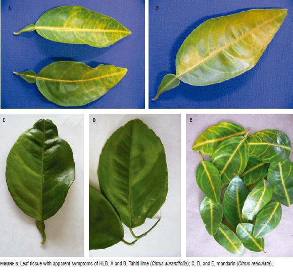
Branches from asymptomatic adult trees and from adult trees with apparent HLB symptoms that were found with different phenological stages (vegetative, flowering and harvesting) of the following species: orange (Citrus sinensis), mandarin (Citrus reticulata), Tahiti lime (Citrus aurantifolia), grapefruit (Citrus paradasi), satsuma mandarin (Citrus unshiu), Volkamer lemon (Citrus volkameriana), orange jessamine (Murraya paniculata) and Tabog (Swinglea glutinosa), were collected from the same plants where the presence of psyllids was observed. A total of 168 samples of 10 to 20 leaves were collected, which were saved in paper bags and stored at -20°C until the implementation of the DNA extraction process (Fig. 3).
Detection of Ca. L. asiaticus and Ca. L. americanus on psyllids and leaf tissue with PCR
The methods used for the DNA extraction were those previously reported for both leaf tissues and psyllids. For the detection of HLB on psyllids, the protocol reported by Manjunath et al. (2008) was used, and for the detection on leaf tissue, the method reported by Murray and Thompson (1980) was used. For the detection of the bacteria in psyllids, qPCR with the specific primers Cit 295 - Cit 298 tested for the Asian variant, Cit 297 - Cit 298 for the American variant, and a combination with the probe Cit 409-FAM in both cases was used (Li et al., 2006). For the internal control of the reaction while monitoring the quality of the DNA extraction of the psyllids, the primers Cit 418 - Cit 419 were used, which amplify a fragment of the wingless gene (wg) of D. citri, which codes for a secreted, diffusible glycoprotein, in addition to the probe Cit 420-HEX (Manjunath et al., 2008). On the plant tissue, the detection of these bacterial variants was done with the same primers and specific probe, and the internal control of the reaction was done with the primers Cit 315 - Cit 317, which amplify a fragment of the cytochrome oxidase gene, together with the probe Cit 316-Cy3 (Li et al., 2006) (Tab. 1). The reactions in the real-time PCR to determine the presence of the Asian and American variants were carried out with a reaction volume of 25 mL, according to the conditions of amplification reported by Li et al. (2006).

Additionally, an alternative verification method of conventional PCR technique was done through a preliminary test with positive controls provided by Fundecitrus (Brazil), in which region 16S of the bacterial DNA of Ca. L. americanus and Ca. L. asiaticus was been inserted in the plasmid TOPO TA cloning (Invitrogen, Carlsbad, CA). This method was carried in a reaction volume of 25 mL, in which, in the case of the controls for the American variant primers, GB1 - GB3 were used along with the conditions reported by Teixeira et al. (2005). Likewise, the procedures used for the Asian variant were done with the primers OI1 - OI2c and conditions according to Jagoueix et al. (1996) (Tab. 2).

Validation of the real-time PCR protocol
Preparation of the samples for the validation of the model
Due to an absence of insects and leaf tissue infected with HLB in Colombia and the difficulty of importing this infected material stemming from bio-security and preventing a possible HLB bacterial infection in Colombia, negative insect and leaf tissue material, previously tested by qPCR, were inoculated with DNA plasmid containing the sequence of the 16S rDNA gene of Ca. L. asiaticus or Ca. L. americanus with the objective of obtaining positive samples in order to conduct the different experiments during the validation method. For this purpose, bacterial DNA fragments of approximately 1,500 pb that corresponded to the sequence of the 16S gene of Ca. L. asiaticus and Ca. L. americanus were independently cloned using TOPO® TA Cloning Kits (Invitrogen, Carlsbad, CA) and with competent cells using One Shot® TOP10 chemically competent E. coli (Invitrogen, Carlsbad, CA). Subsequently, 10 dilutions were done for each variant from the plasmid DNA that was positive for Ca. L. asiaticus and Ca. L. americanus, for which NanoDrop ND 1000 spectrophotometer (Nano-Drop Technologies, Wilmington, DE) was used with a starting concentration of 1,500 ng mL -1 until reaching 1.5 fg mL-1. The number of copies present in the different concentrations of plasmid DNA was determined with the equation reported by Lee et al. (2006) (Tab. 3).

Ten midrib plant samples with 100 mg of Citrus aurantifolia (Thaiti lime) that were negative for HLB were used in order to obtained positive samples, with ten replicates for each one. The first replication of each sample was inoculated with 5 mL of the first dilution of the positive plasmid dilution, thereby independently obtaining a 10 x 10 matrix for each of the two variants (Table 4). Afterwards, the sample was soaked and the DNA extraction was carried out through the method described by Murray and Thompson (1980) and the respective detection of bacteria with real-time PCR using the specific indicators reported for Ca. L. asiaticus and Ca. L. americanus (Li et al. 2006; Manjunath et al. 2008).

Establishment of the 90% detection limit of CandisatusLiberibacter sp. The 90% detection limit was calculated using the formula for the final-point method of Spearman-Kärber, LD90% = m-d (S-0.5), (NordVal, 2009), in which m is the highest dilution dose with 100% presence (ng mL-1 or No. of copies/mL); d is the distance between the doses or dilution factor (0.1); S is the sum of fractions P (presence) from the highest dilution with 100% P to 0% P, divided by 100; and 0.5 is a constant. This procedure was carried out with the plant and insect samples in order to detect the bacterium.
Determination of the sensitivity, specificity, efficiency, false positives, false negatives, and selectivity of the real-time PCR method
The sensitivity, specificity, efficiency, false positives, false negatives and selectivity of the technique on insect and plant tissue for Ca. L. asiaticus and Ca. L. americanus were determined by the inoculation of the previous dilution obtained in the detection limit for each variant with the different possible sample combinations. Three different midrib samples of plant leaf and three samples of insect tissue (six psyllids in each sample) were soaked in liquid nitrogen. Four replicates of 100 mg were taken from each soaked sample, which were used to obtain the four possible sample combinations using inoculation with five mL of plasmid DNA of the previous dilution used in obtaining the detection limit of each variant (Tab. 5). Subsequently, the DNA extraction was done using the protocol described by Manjunath et al. (2008) for insects and by Murray and Thompson (1980) for foliar tissue, with the respective analysis with real-time PCR for the detection of the Asian and American variants; the results were registered in a matrix for the presence or absence in the plant tissue and insect tissue (Tab. 6).


Results and discussions
Taxonomic identification of the psyllids
The presence of the insect vector was confirmed in the Andean and Orinoquia geographical regions of the sampled departments of Cundinamarca, Tolima, Santander, Quindio and Meta. In 66% of the visited sites, D. citri was found in different stages of development on the young growth of Rutaceus plants (Tab. 7); however, it was only during the new shoots season, after the first rains, that the different stages of development were observed on the same plants (Figs. 1 and 2). During the dry season, only the adult stage was observed on the observed citrus plantations, except on Swinglea glutinosa plants (Blanco), used as live barriers, and on Orange Jessamine, Murraya paniculata (L.) Jack., used as an ornamental plant. These observations are consistent with those made by Gómez (2009) and Ebratt et al. (2011a).

Of the 239 psyllid samples, 598 adults were obtained, of which 58% were female, and 2,695 were nymphs (Tab. 5). The semi-quiesence of the nymphs and the presence of buds on the rutacea plants facilitated the capture of the nymphs. Cundinamarca, Valle del Cauca and Meta presented the higher number of captured individuals at 1,086, 673, and 580, respectively; D. citri predominated in the municipalities that had characteristics of a dry, tropical forest (Ebratt, et al., 2011b).
Detection of Ca. L. asiaticus and Ca. L. americanus on psyllids and leaf tissue using qPCR and conventional PCR
All of the 168 samples of leaf tissue with apparent symptoms (Fig. 3) and the 239 samples of psyllids collected in the departments of Cundinamarca, Santander, Valle del Cauca, Meta and Quindio were diagnosed as negative for the presence of Ca. L. asiaticus and Ca. L. americanus using real-time PCR; the negative controls did not generate an amplification curve while the positive controls did with a Ct that started amplification between 15 and 22 for the two variants in the foliar and insect tissues (Figs. 4A, 4B, 5A and 5B). Likewise, all of the DNA samples from the leaf tissue as well as from the insects amplified their respective DNA quality internal control, which demonstrated the absence of an inhibition of the reaction (Figs. 4C and 5C). The absence of the HLB disease in the analyzed citrus farms in Colombia indicated that the variants of the bacteria have not yet been found in these analyses. However, due to the large insect populations that are closely associated with the citrus plants and the fact that the disease is already present in neighboring countries, it is necessary to maintain permanent surveillance programs in order to take measures that avoid the establishment of the disease in this country or to avoid rapid propagation throughout the national territory the moment the presence of any of the bacterial variants that cause the disease is detected. The conventional PCR methodology was established as an alternative with the use of GB1 - GB3 indicators, which amplified a fragment of 1,027 bp in the positive control of the Americana variant, and OI1 - OI2c indicators, which amplified a fragment of 1,160 bp for the positive control of the Asian variant were used (Figs. 6 and 7). These results facilitate complimentary analysis when a sequence examination is required for the two variants.


Validation of the detection method for Ca. L. asiaticus and Ca. L. americanus using real-time PCR
The standard curves for Ca. L. asiaticus and Ca. L. americanus were made by using dilutions 2 to 7 of the plasmid DNA for each variant, as described in Tab. 3. The standard curve for Ca. L. asiaticus obtained a correlation coefficient of 0.994 and an efficiency of 90.6%. Likewise, the standard curve for Ca. L. americanus obtained a correlation coefficient of 0.997 and an efficiency of 97.2% (Fig. 8). These data demonstrated the effectiveness, efficiency, and linearity of the method employed for the detection of the two analyzed variants.

In terms of the absence of positive samples in Colombia, which were sought for in the present study, it resulted from the approximation of the conditions of a real sample, carrying out an inoculation of negative tissues with positive plasmid DNA, as discussed in the materials and methods section. The inoculated samples were analyzed through real-time PCR with indicators specific for each variant, with the obtained results registered in the presence-absence matrixes (Tab. 8). Using the Spearman-Kärber formula for final points, it was determined that the 90% detection limit for Ca. L. asiaticus was 14.91 ng mL-1, equivalent to 2.49·109 copies, in the case of the insect material and 14.89 ng mL-1 (2.49·109 copies) for the citrus leaf tissue. The detection limit for Ca. L. americanus was 1.36 ng mL-1 (2.49·108 copies) in the insect material and 1.25 ng mL-1 (2.49·108 copies) in the plant tissue (Tab. 9).


Three plant samples and three insect samples were inoculated in each of the possible sampling combinations starting from the previous dilution until the detection limit (150 ng mL-1) with the positive controls of Ca. L. americanus and Ca. L. asiaticus, for a total of 12 plant samples and 12 insect samples, on which DNA extraction was performed with subsequent detection using real-time PCR, wherein only true positives and true negatives were found (Tab. 10). A sensitivity of 100%, a specificity of 100%, an efficiency of 100%, an assignment of false positives of 0%, an assignment of false negatives of 0%, and a selectivity of -0.3 for the two variants of the disease, both in the plant and insect tissue, were determined, which indicated the reliability and robustness of the real-time PCR technique utilized in the present study for the detection of region 16S of Ca. L. americanus and Ca. L. asiaticus, in both in the leaf and insect tissues.

Through this validation process, it was possible to determine that the real-time PCR technique for the detection of the American and Asian variants in the citrus-producing regions of Colombia is reliable and highly sensitive and can be used for further monitoring of HLB, with all its controls, both on citrus leaf tissues and on the insect vector.
Conclusions
The HLB insect vector Diaphorina citri was found in the 25 tested municipalities of Cundinamarca, Santander, Valle del Cauca, Meta and Quindio, in the egg, nymph and/or adult stages.
During the development of this project, using real-time PCR, it was confirmed that all of the insect and leaf tissue samples were negative for the presence of Ca. L asiaticus and Ca. L americanus, which indicates the absence, to date, of this disease in the sampled citrus-producing regions.
It was determined that the detection limit for the real-time PCR technique for region 16S of Ca. L. asiaticus was 14.89 ng mL-1 for the leaf tissue and 14.91 ng mL-1 for the insect tissue; and 1.25 ng mL-1 for the leaf tissue and 1.36 ng mL-1 for the insect tissue for region 16S of Ca. L. americanus.
The results for sensitivity, specificity, efficiency, false positives, false negatives and selectivity for the real-time PCR technique used for the detection of Ca. L. asiaticus and Ca. L. americanus on plant and insect tissues presented reliability and robustness for the molecular detection of the two variants of the disease known as citrus HLB.
Acknowledgements
The authors express their appreciation to Keremane Manjunath, a researcher with the United States Department of Agriculture (USDA) and to Chandrika Ramadugu, a researcher from the Department of Botany and Plant Sciences at the University of California, Riverside (USA), for their technical support and for providing the DNA positive controls of citrus plants infected with Ca. L. americanus and Ca. L. asiaticus bacteria; to Diva do Carmo Teixeira, a researcher with the Fund for the Defense of Citrus Cultures (Fundecitrus) of Brazil, who kindly provided the DNA positive controls of citrus plants from that country, infected with the Ca. L. americanus and Ca. L. asiaticus bacteria; to the Administrative Department of Science and Technology and Research (Departamento Administrativo de Ciencia, Tecnología e Investigación-Colciencias), for funding of this research from 2012 to 2013; to the Colombian Agricultural Institute (Instituto Colombiano Agropecuario - ICA) and its departmental branches in Cundinamarca, Santander, Valle del Cauca, Meta and Quindío; and to all the producers in the visited regions for their collaboration and interest in this research.
Literature cited
Blackwell, P. 2005. Diaphorina citri PM7/52(1).Bulletin 35. Organisation Européenneet Méditerranéenne pour la Protection des Plantes; European and Mediterranean Plant Protection Organization (OEPP/EPPO), Paris. pp. 271-333.
Bové, J.M. 2006. Huanglongbing: a destructive, newly-emerging, century-old disease of citrus. J. Plant Pathol. 88, 7-37. Doi: 10.4454/jpp.v88i1.828
Burckhardt, D. 1987. Jumping plant lice (Homoptera: Psylloidea) of the temperate neotropical region. Part 2: Psyllidae (subfamilies Diaphorininae, Acizziinae, Ciriacreminae and Psyllinae). Zool. J. Linn. Soc. 90, 145-205. Doi: 10.1111/j.1096-3642.1987.tb01353.x
Ebratt R., E.E., L.T. Rubio G., V.A. Costa, E.M. Zambrano G., A.P. Castro Á., and M.Y. Santamaría G. 2011a. Primer registro de Tamarixia radiata (Waterston, 1922) (Hymenoptera: Eulophidae) en Colombia. Rev. Fac. Nal. Agr. Medellin 64, 6141-6146.
Ebratt R., E.E., L.T. Rubio-González; V.A. Costa, Á.P. Castro-Ávila, E.M. Zambrano G., and J.E. Ángel-Díaz. 2011b. Diaphorina citri (Kuwayama, 1907) and Tamarixia radiata (Waterson, 1922) in citrus crops of Cundinamarca, Colombia. Agron. Colomb. 29, 487-493.
Garnier, M., S. Jagoueix-Eveillard, P.R. Cronje, H.F. Le Roux, and J.M. Bové. 2000. Genomic characterization of a liberibacter present in an ornamental rutaceous tree, Calodendrum capense, in the Western Cape Province of South Africa. Proposal of 'Candidatus Liberibacter africanus subsp. capensis'. Int. J. Syst. Evol. Microbiol. 50, 2119-2125. Doi: 10.1099/00207713-50-6-2119
Gómez T., M.L. 2009. Estudos bioecológicos de Tamarixia radiata (Waterston, 1922) (Hymenoptera: Eulophidae) para o controle de Diaphorina citri Kuwayama, 1907 (Hemiptera: Psyllidae). PhD thesis. Escola Superior de Agricultura Luiz de Queiroz (ESALQ), Universidade de São Paulo, Piracicaba, Brazil.
Halbert, S.E. 2005. The discovery of huanglongbing in Florida. In: Gottwald, T.R., W.N. Dixon, J.H. Graham, and P. Berger (eds.). Proc. 2nd Int. Citrus Canker and Huanglongbing Res. Workshop.Florida Citrus Mutual. Orlando, FL.
Halbert, S.E. and K.L. Manjunath. 2004. Asian citrus psyllids (Sternorrhyncha: Psyllidae) and greening disease of citrus: a literature review and assessment of risk in Florida. Fla. Entomol. 87, 330-353. Doi: 10.1653/0015-4040(2004)087[0330:ACPSPA]2.0.CO;2
Hung, T.-H., S.-C. Hung, C.-N. Chen, M.-H. Hsu, and H.-J. Su. 2004. Detection by PCR of Candidatus Liberibacter asiaticus, the bacterium causing citrus huanglongbing in vector psyllids: application to the study of vector-pathogen relationships. Plant Pathol. 53, 96- 102. Doi: 10.1111/j.1365-3059.2004.00948.x
ISO, International Organization for Standardization. 2011. ISO 22119: microbiology of food and animal feeding stuffs - Real-time polymerase chain reaction (PCR) for the detection of food-borne pathogens - General requirements and definitions. Geneva, Switzerland.
Jagoueix, S., J.M. Bové, and M. Garnier. 1996. PCR detection of the two 'Candidatus' Liberibacter species associated with greening disease of citrus. Mol. Cell. Probes 10, 43-50. Doi: 10.1006/mcpr.1996.0006
Lee, C., J. Kim, S.G. Shin, and S. Hwang. 2006. Absolute and relative QPCR quantification of plasmid copy number in Escherichia coli. J. Biotechnol. 123, 273- 280. Doi: 10.1016/j.jbiotec.2005.11.014
Li, W., J.S. Hartung, and L. Levy. 2006. Quantitative real-time PCR for detection and identification of Candidatus Liberibacter species associated with citrus huanglongbing. J. Microbiol. Methods 66, 104-115. Doi: 10.1016/j.mimet.2005.10.018
Lin, H., C. Chen, H. Doddapaneni, Y. Duan, E.L. Civerolo, X. Bai, and X. Zhao. 2010. A new diagnostic system for ultra-sensitive and specific detection and quantification of Candidatus Liberibacter asiaticus, the bacterium associated with citrus Huanglongbing. J. Microbiol. Methods 81, 17-25. Doi: 10.1016/j.mimet.2010.01.014
Manjunath, K.L., S.E. Halbert, C. Ramadugu, S. Webb, and R.F. Lee. 2008. Detection of 'Candidatus Liberibacter asiaticus' in Diaphorina citri and its importance in the management of citrus huanglongbing in Florida. Phytopathology 98, 387-396. Doi: 10.1094/PHYTO-98-4-0387
Manjunath, K.L., C. Ramadugu, V.M. Majil, S. Williams, M. Irey, and R.F. Lee. 2010. First report of the citrus huanglongbing associated bacterium 'Candidatus Liberibacter asiaticus' from sweet orange, Mexican lime, and Asian citrus psyllid in Belize. Plant Dis. 94, 781. Doi: 10.1094/PDIS-94-6-0781A
Martínez, Y., R. Llauger, L. Batista, M. Luis, A. Iglesia, C. Collazo, I. Peña, J.C. Casín, J. Cueto, and L.M. Tablada. 2009. First report of 'Candidatus Liberibacter asiaticus' associated with Huanglongbing in Cuba. Plant Pathol. 58, 389. Doi: 10.1111/j.1365-3059.2008.01997.x
Matos, L., M.E. Hilf, and J. Camejo. 2009. First report of 'Candidatus Liberibacter asiaticus' associated with citrus Huanglongbing in the Dominican Republic. Plant Dis. 93, 668. Doi: 10.1094/PDIS-93-6-0668B
Murray, M.G. and W.F. Thompson. 1980. Rapid isolation of high molecular weight plant DNA. Nucl. Acids Res. 8, 4321-4326. Doi: 10.1093/nar/8.19.4321
NAPPO, North American Plant Protection Organization. 2009. Detection of Huanglongbing ('Candidatus Liberibacter asiaticus') in the municipality of Tizimin, Yucatan, Mexico. Official pest reports. In: Phytosanitary Alert System, http://www.pestalert.org/oprDetail.cfm?oprID=384; consulted: November, 2014.
NordVal. 2009. Protocol for the validation of alternative microbiological methods. Oslo.
Roistacher, C.N. 1991. Techniques for biological detection of specific citrus graft-transmissible diseases. pp. 13-158. In: Roistacher, C.N. (ed.). Graft-transmissible diseases of citrus. Handbook for detection and diagnosis. FAO, Rome.
Schwarz, R.E. 1968. Indexing of greening and exocortis through fluorescent marker substances. pp. 118-124. In: Price, W.C. (ed.). Proc. 4th Conf. Int. Org. Citrus Virol. University of Florida, Gainesville, USA.
Senasa, Servicio Nacional de Sanidad y Calidad Agroalimentaria. 2013. La amenaza del HLB (Huanlongbing de los cítricos) para la citricultura nacional. In: http://www.senasa.gov.ar/contenido.php?to=n&in=11&io=18493; consulted: November, 2014.
SFE, Servicio Fitosanitario del Estado. 2011. Comunicado de prensa: autoridades del SFE confirman presencia de "Dragón Amarillo" en árboles de la Zona Norte. In: Ministerio de Agricultura y Ganadería de Costa Rica https://www.sfe.go.cr/documentos/comunicados/2011/21_02_2011_SFE_confirma_presencia_HLB_en_zona_norte.pdf; consulted: September, 2014.
Teixeira, D.C., J.L. Danet, S. Eveillard, E.C. Martins, W.C.J. Junior, P.T. Yamamoto, S.A. Lopes, R.B. Bassanezi, A.J. Ayres, C. Saillard, and J.M. Bové, 2005. Citrus huanglongbing in São Paulo State, Brazil: PCR detection of the 'Candidatus' Liberibacter species associated with the disease. Mol. Cell. Probes 19, 173-179. Doi: 10.1016/j.mcp.2004.11.002
Tsai, J.H. and Y.H. Liu. 2000. Biology of Diaphorina citri (Homoptera: Psyllidae) on four host plants. J. Econ. Entomol. 93, 1721-1725. Doi: 10.1603/0022-0493-93.6.1721
Tsai, J.H., J.J. Wang, and Y.H. Liu. 2002. Seasonal abundance of the Asian citrus psyllid, Diaphorina citri (Homoptera: Psyllidae) in southern Florida. Fla. Entomol. 85, 446-451. Doi: 10.1653/0015-4040(2002)085[0446:SAOTAC]2.0.CO;2
References
Blackwell, P. 2005. Diaphorina citri PM7/52(1).Bulletin 35. Organisation Européenneet Méditerranéenne pour la Protection des Plantes; European and Mediterranean Plant Protection Organization (OEPP/EPPO), Paris. pp. 271-333.
Bové, J.M. 2006. Huanglongbing: a destructive, newly-emerging, century-old disease of citrus. J. Plant Pathol. 88, 7-37. Doi: 10.4454/jpp.v88i1.828
Burckhardt, D. 1987. Jumping plant lice (Homoptera: Psylloidea) of the temperate neotropical region. Part 2: Psyllidae (subfamilies Diaphorininae, Acizziinae, Ciriacreminae and Psyllinae). Zool. J. Linn. Soc. 90, 145-205. Doi: 10.1111/j.1096-3642.1987.tb01353.x
Ebratt R., E.E., L.T. Rubio G., V.A. Costa, E.M. Zambrano G., A.P. Castro Á., and M.Y. Santamaría G. 2011a. Primer registro de Tamarixia radiata (Waterston, 1922) (Hymenoptera: Eulophidae) en Colombia. Rev. Fac. Nal. Agr. Medellin 64, 6141-6146.
Ebratt R., E.E., L.T. Rubio-González; V.A. Costa, Á.P. Castro-Ávila, E.M. Zambrano G., and J.E. Ángel-Díaz. 2011b. Diaphorina citri (Kuwayama, 1907) and Tamarixia radiata (Waterson, 1922) in citrus crops of Cundinamarca, Colombia. Agron. Colomb. 29, 487-493.
Garnier, M., S. Jagoueix-Eveillard, P.R. Cronje, H.F. Le Roux, and J.M. Bové. 2000. Genomic characterization of a liberibacter present in an ornamental rutaceous tree, Calodendrum capense, in the Western Cape Province of South Africa. Proposal of 'Candidatus Liberibacter africanus subsp. capensis'. Int. J. Syst. Evol. Microbiol. 50, 2119-2125. Doi: 10.1099/00207713-50-6-2119
Gómez T., M.L. 2009. Estudos bioecológicos de Tamarixia radiata (Waterston, 1922) (Hymenoptera: Eulophidae) para o controle de Diaphorina citri Kuwayama, 1907 (Hemiptera: Psyllidae). PhD thesis. Escola Superior de Agricultura Luiz de Queiroz (ESALQ), Universidade de São Paulo, Piracicaba, Brazil.
Halbert, S.E. 2005. The discovery of huanglongbing in Florida. In: Gottwald, T.R., W.N. Dixon, J.H. Graham, and P. Berger (eds.). Proc. 2nd Int. Citrus Canker and Huanglongbing Res. Workshop.Florida Citrus Mutual. Orlando, FL.
Halbert, S.E. and K.L. Manjunath. 2004. Asian citrus psyllids (Sternorrhyncha: Psyllidae) and greening disease of citrus: a literature review and assessment of risk in Florida. Fla. Entomol. 87, 330-353. Doi: 10.1653/0015-4040(2004)087[0330:ACPSPA]2.0.CO;2
Hung, T.-H., S.-C. Hung, C.-N. Chen, M.-H. Hsu, and H.-J. Su. 2004. Detection by PCR of Candidatus Liberibacter asiaticus, the bacterium causing citrus huanglongbing in vector psyllids: application to the study of vector-pathogen relationships. Plant Pathol. 53, 96- 102. Doi: 10.1111/j.1365-3059.2004.00948.x
ISO, International Organization for Standardization. 2011. ISO 22119: microbiology of food and animal feeding stuffs - Real-time polymerase chain reaction (PCR) for the detection of food-borne pathogens - General requirements and definitions. Geneva, Switzerland.
Jagoueix, S., J.M. Bové, and M. Garnier. 1996. PCR detection of the two 'Candidatus' Liberibacter species associated with greening disease of citrus. Mol. Cell. Probes 10, 43-50. Doi: 10.1006/mcpr.1996.0006
Lee, C., J. Kim, S.G. Shin, and S. Hwang. 2006. Absolute and relative QPCR quantification of plasmid copy number in Escherichia coli. J. Biotechnol. 123, 273- 280. Doi: 10.1016/j.jbiotec.2005.11.014
Li, W., J.S. Hartung, and L. Levy. 2006. Quantitative real-time PCR for detection and identification of Candidatus Liberibacter species associated with citrus huanglongbing. J. Microbiol. Methods 66, 104-115. Doi: 10.1016/j.mimet.2005.10.018
Lin, H., C. Chen, H. Doddapaneni, Y. Duan, E.L. Civerolo, X. Bai, and X. Zhao. 2010. A new diagnostic system for ultra-sensitive and specific detection and quantification of Candidatus Liberibacter asiaticus, the bacterium associated with citrus Huanglongbing. J. Microbiol. Methods 81, 17-25. Doi: 10.1016/j.mimet.2010.01.014
Manjunath, K.L., S.E. Halbert, C. Ramadugu, S. Webb, and R.F. Lee. 2008. Detection of 'Candidatus Liberibacter asiaticus' in Diaphorina citri and its importance in the management of citrus huanglongbing in Florida. Phytopathology 98, 387-396. Doi: 10.1094/PHYTO-98-4-0387
Manjunath, K.L., C. Ramadugu, V.M. Majil, S. Williams, M. Irey, and R.F. Lee. 2010. First report of the citrus huanglongbing associated bacterium 'Candidatus Liberibacter asiaticus' from sweet orange, Mexican lime, and Asian citrus psyllid in Belize. Plant Dis. 94, 781. Doi: 10.1094/PDIS-94-6-0781A
Martínez, Y., R. Llauger, L. Batista, M. Luis, A. Iglesia, C. Collazo, I. Peña, J.C. Casín, J. Cueto, and L.M. Tablada. 2009. First report of 'Candidatus Liberibacter asiaticus' associated with Huanglongbing in Cuba. Plant Pathol. 58, 389. Doi: 10.1111/j.1365-3059.2008.01997.x
Matos, L., M.E. Hilf, and J. Camejo. 2009. First report of 'Candidatus Liberibacter asiaticus' associated with citrus Huanglongbing in the Dominican Republic. Plant Dis. 93, 668. Doi: 10.1094/PDIS-93-6-0668B
Murray, M.G. and W.F. Thompson. 1980. Rapid isolation of high molecular weight plant DNA. Nucl. Acids Res. 8, 4321-4326. Doi: 10.1093/nar/8.19.4321
NAPPO, North American Plant Protection Organization. 2009. Detection of Huanglongbing ('Candidatus Liberibacter asiaticus') in the municipality of Tizimin, Yucatan, Mexico. Official pest reports. In: Phytosanitary Alert System, http://www.pestalert.org/oprDetail.cfm?oprID=384; consulted: November, 2014.
NordVal. 2009. Protocol for the validation of alternative microbiological methods. Oslo.
Roistacher, C.N. 1991. Techniques for biological detection of specific citrus graft-transmissible diseases. pp. 13-158. In: Roistacher, C.N. (ed.). Graft-transmissible diseases of citrus. Handbook for detection and diagnosis. FAO, Rome.
Schwarz, R.E. 1968. Indexing of greening and exocortis through fluorescent marker substances. pp. 118-124. In: Price, W.C. (ed.). Proc. 4th Conf. Int. Org. Citrus Virol. University of Florida, Gainesville, USA.
Senasa, Servicio Nacional de Sanidad y Calidad Agroalimentaria. 2013. La amenaza del HLB (Huanlongbing de los cítricos) para la citricultura nacional. In: http://www.senasa.gov.ar/contenido.php?to=n&in=11&io=18493; consulted: November, 2014.
SFE, Servicio Fitosanitario del Estado. 2011. Comunicado de prensa: autoridades del SFE confirman presencia de "Dragón Amarillo" en árboles de la Zona Norte. In: Ministerio de Agricultura y Ganadería de Costa Rica https://www.sfe.go.cr/documentos/comunicados/2011/21_02_2011_SFE_confirma_presencia_HLB_en_zona_norte.pdf; consulted: September, 2014.
Teixeira, D.C., J.L. Danet, S. Eveillard, E.C. Martins, W.C.J. Junior, P.T. Yamamoto, S.A. Lopes, R.B. Bassanezi, A.J. Ayres, C. Saillard, and J.M. Bové, 2005. Citrus huanglongbing in São Paulo State, Brazil: PCR detection of the 'Candidatus' Liberibacter species associated with the disease. Mol. Cell. Probes 19, 173-179. Doi: 10.1016/j.mcp.2004.11.002
Tsai, J.H. and Y.H. Liu. 2000. Biology of Diaphorina citri (Homoptera: Psyllidae) on four host plants. J. Econ. Entomol. 93, 1721-1725. Doi: 10.1603/0022-0493-93.6.1721
Tsai, J.H., J.J. Wang, and Y.H. Liu. 2002. Seasonal abundance of the Asian citrus psyllid, Diaphorina citri (Homoptera: Psyllidae) in southern Florida. Fla. Entomol. 85, 446-451. Doi: 10.1653/0015-4040(2002)085[0446:SAOTAC]2.0.CO;2
How to Cite
APA
ACM
ACS
ABNT
Chicago
Harvard
IEEE
MLA
Turabian
Vancouver
Download Citation
CrossRef Cited-by
1. Ashis K. Das, Susheel K. Sharma, Neha Thakre. (2019). Plant Biotechnology: Progress in Genomic Era. , p.597. https://doi.org/10.1007/978-981-13-8499-8_26.
2. Daiver Cardona-Salgado, Yves Dumont, Olga Vasilieva. (2025). Natural population dynamics of Asian citrus psyllid, Diaphorina citri, and its control based on pheromone trapping. Mathematical Biosciences, 390, p.109540. https://doi.org/10.1016/j.mbs.2025.109540.
3. Edisson Chavarro-Mesa, Enrique Delahoz-Dominguez, Mary Fennix-Agudelo, Wendy Miranda-Castro, Jorge Evelio Angel-Diaz. (2020). Preliminary machine learning model for citrus greening disease (Huanglongbing-HLB) prediction in Colombia. 2020 IEEE Colombian Conference on Applications of Computational Intelligence (IEEE ColCACI 2020). , p.1. https://doi.org/10.1109/ColCACI50549.2020.9247900.
4. Yesenia García, Yaneth Patricia Ramos, Paola Andrea Sotelo, Takumasa Kondo. (2016). Biology of Diaphorina Citri (Hemiptera: Liviidae) Under Glass House Conditions In Palmira, Colombia . Revista Colombiana de Entomología, 42(1), p.36. https://doi.org/10.25100/socolen.v42i1.6667.
5. A Zafarullah, S Naz, F Osman. (2021). COMPARISON OF A CONVENTIONAL PCR AND QUANTITATIVE REAL TIME PCR (qPCR) ASSAY FOR THE DETECTION OF HUANGLONGBING DISEASE ASSOCIATED WITH ‘CANDIDATUS LIBERIBACTER ASIATICUS’ IN CITRUS VARIETIES. The Journal of Animal and Plant Sciences, 31(5), p.1453. https://doi.org/10.36899/JAPS.2021.5.0346.
6. Kaili Xia, Zengwei Feng, Xianjiao Zhang, Yang Zhou, Honghui Zhu, Qing Yao. (2023). Potential functions of the shared bacterial taxa in the citrus leaf midribs determine the symptoms of Huanglongbing. Frontiers in Plant Science, 14 https://doi.org/10.3389/fpls.2023.1270929.
7. Takumasa Kondo, Hugo Cesar Aredondo-Bernal, Yenifer Campos Patiño, Laura Isabel Colorado Herrera, Camila Yesenia García Córdoba, Guillermo González F., Mauricio Fernando Martínez, Denys Yohana Mora-Herrera, Yaneth Patricia Ramos Villafañe, Paola Andrea Sotelo Cardona, Rubilma Tarazona Velásquez. (2017). Protocolo de cría y liberación de Tamarixia radiata (Waterston) (Hymenoptera: Eulophidae). https://doi.org/10.21930/agrosavia.manual.7402414.
8. Luis Alberto Olvera-Vargas, Ángel de Jesús Quiroz Gaspar, David Israel Contreras-Medina, Noé Aguilar-Rivera. (2020). Análisis de riesgo potencial de Huanglongbing a través de tecnología geoespacial en Colombia. Ciencia & Tecnología Agropecuaria, 21(3), p.1. https://doi.org/10.21930/rcta.vol21_num3_art:1552.
9. Catalina Chaves-Sierra, María Camila Rodriguez-Cruz, Fernan Santiago Mejia-Alvarado, Carolina Ramírez-Higuera, Antonio Mejía-Eslava, Hernán Mauricio Romero. (2024). Identification of ‘Candidatus Liberibacter asiaticus’, the Huanglongbing Bacterium, in Citrus from Colombia. Plant Disease, 108(5), p.1169. https://doi.org/10.1094/PDIS-10-23-2003-SC.
10. Yanyun Hong, Yongyang Luo, Jianglan Yi, Ling He, Liangying Dai, Tuyong Yi, Zonghua Wang. (2019). Screening nested-PCR primer for ‘Candidatus Liberibacter asiaticus’ associated with citrus Huanglongbing and application in Hunan, China. PLOS ONE, 14(2), p.e0212020. https://doi.org/10.1371/journal.pone.0212020.
11. Juan Camilo Cifuentes-Arenas, George Andrew Charles Beattie, Leandro Peña, Silvio Aparecido Lopes. (2019). Murraya paniculata and Swinglea glutinosa as Short-Term Transient Hosts of ‘Candidatus Liberibacter asiaticus’ and Implications for the Spread of Huanglongbing. Phytopathology®, 109(12), p.2064. https://doi.org/10.1094/PHYTO-06-19-0216-R.
Dimensions
PlumX
Article abstract page views
Downloads
License
Copyright (c) 2014 Agronomía Colombiana

This work is licensed under a Creative Commons Attribution-NonCommercial-ShareAlike 4.0 International License.
© Centro Editorial de la Facultad de Ciencias Agrarias, Universidad Nacional de Colombia
Reproduction and quotation of material appearing in the journal is authorized provided the following are explicitly indicated: journal name, author(s) name, year, volume, issue and pages of the source. The ideas and observations recorded by the authors are their own and do not necessarily represent the views and policies of the Universidad Nacional de Colombia. Mention of products or commercial firms in the journal does not constitute a recommendation or endorsement on the part of the Universidad Nacional de Colombia; furthermore, the use of such products should comply with the product label recommendations.
The Creative Commons license used by Agronomia Colombiana journal is: Attribution - NonCommercial - ShareAlike (by-nc-sa)

Agronomia Colombiana by Centro Editorial of Facultad de Ciencias Agrarias, Universidad Nacional de Colombia is licensed under a Creative Commons Reconocimiento-NoComercial-CompartirIgual 4.0 Internacional License.
Creado a partir de la obra en http://revistas.unal.edu.co/index.php/agrocol/.