


Publicado
Current state of the art and enduring issues in anthropometric data collection
Estado actual de la técnica y cuestiones perdurables en la recogida de datos antropométricos
DOI:
https://doi.org/10.15446/dyna.v83n197.57586Palabras clave:
anthropometry, body scanners, errors, reliability (en)la antropometría, escáneres corporales, errores, confiabilidad (es)
Descargas
DOI: https://doi.org/10.15446/dyna.v83n197.57586
Current state of the art and enduring issues in anthropometric data collection
Estado actual de la técnica y cuestiones perdurables en la recogida de datos antropométricos
Sara Bragança a, Pedro Arezes a, Miguel Carvalho b & Susan P. Ashdown c
a Department of Production and
Systems, University of Minho, Guimarães, Portugal. saraabraganca@gmail.com,
parezes@dps.uminho.pt
b Department of Textile Engineering, University
of Minho, Guimarães, Portugal. migcar@det.uminho.pt
c Fiber Science & Apparel Design, Cornell
University, Ithaca, NY, USA. spa4@cornell.edu
Received: November 30th, 2015. Received in revised form: March 17th, 2016. Accepted: April 12th, 2016.
This work is licensed under a Creative Commons Attribution-NonCommercial-NoDerivatives 4.0 International License.
Abstract
The study of
human body size and shape has been a topic of research for a very
long time. In the past, anthropometry used traditional measuring techniques to record
the dimensions of the human body and reported variance in body dimensions as a function
of mean and standard deviation. Nowadays, the study of human body dimensions
can be carried out more efficiently using three-dimensional body scanners,
which can provide large amounts of anthropometric data more quickly than traditional techniques can. This paper presents a description of the broad range of issues
related to the collection of anthropometric data using three-dimensional body
scanners, including the different types of technologies available and their
implications, the standard scanning process needed for effective data
collection, and the possible sources of measurement errors that
might affect the reliability and validity of the data collected.
Keywords: anthropometry; body scanners; errors; reliability.
Resumen
El estudio del tamaño y la forma del cuerpo
humano ha sido un tema de investigación durante un tiempo muy largo. En el
pasado, la antropometría utilizó técnicas de medición tradicionales para
registrar las dimensiones del cuerpo humano y reportó la variación en las
dimensiones del cuerpo en función de la media y la desviación estándar. Hoy en
día, el estudio de las dimensiones del cuerpo humano se puede llevar a cabo
utilizando maneras más eficientes, como los escáneres tridimensionales del
cuerpo, que pueden proporcionar grandes cantidades de datos antropométricos más
rápidamente que las técnicas tradicionales. En este trabajo se presenta una
descripción de la amplia gama de temas relacionados con la recogida de datos
antropométricos utilizando escáneres tridimensionales del cuerpo, incluyendo
los diferentes tipos de tecnologías disponibles y sus implicaciones, el proceso
de digitalización estándar necesario para la captura efectiva de datos, y las
posibles fuentes de los errores de medición que podrán afectar la fiabilidad y
validez de los datos recogidos.
Palabras clave: la antropometría; escáneres corporales; errores; confiabilidad.
1. Introduction
In traditional anthropometry the determination of human body dimensions can be achieved using a range of devices. Ever since Richer first used calipers in 1890, a standard set of anthropometric instruments has been used [1]. Simple, quick, relatively non-invasive tools include scales (to determine weight), measuring tapes (to measure circumferences and linear body surface dimensions), anthropometers (to measure height and various transverse widths and depths of the body), spreading calipers (also to measure widths and depths of the body), sliding compasses (to measure short distances, e.g., on the nose, ears or hands), and head spanners (to measure the height of the head) [1]. All of these devices usually require calibration and the measurements taken are only as accurate as the techniques used by the person who takes them. Therefore, it is generally necessary to take multiple measurements and to calculate average values. Additionally, there are typically differences between measurements taken by different people, although this can be reduced with consistent training.
With these traditional methods of collecting anthropometric data, the measuring process is time-consuming, expensive and error-prone. Moreover, traditional methods require the person being measured to adopt standardized postures that are prescribed when measurements are taken (and to maintain them during the measurement process). These standard measuring postures, defined in ISO 7250 [2], are based on the studies of several authors such as Kroemer and Kroemer [3], who explain the standard method of measuring a subject in detail. The primary measuring posture is referred to as the "anatomical position", in which the participant's body is placed in a defined, straight, upright posture, with the body segments at either 180, 0, or 90 degrees to each other. Participants are required to stand erect; heels together; buttocks, shoulder blades, and the back of head touching a vertical surface; arms vertical, fingers straight. The head is positioned in the Frankfurt plane; with the pupils on the same horizontal level; the right tragion and the lowest point of the right orbit are likewise aligned horizontally. When measurements are taken of a seated subject, the surfaces of the seat and the foot support must be arranged so that the thighs and feet are horizontal and the lower legs vertical. The measurements should be taken in the morning, because the human body tends to decrease in height during the day, and this practice can help remove one source of variation [4].
With the appearance of new ways of acquiring surface data, anthropometry has gained a new way of performing a deeper investigation of human body size and shape. These new digital shape analysis tools make it possible to acquire data on complex geometrical features, such as curvatures or partial volumes. Using these tools to acquire anthropometric data has the potential to be more practical, reliable, fast and -in comparison with traditional anthropometry- less expensive.
The study of the human body as a 3D object using digital capture tools began in 1973 with a light sectioning technique proposed by Lovesey [5]. This was labor intensive, as the interpretation of data was extremely time consuming. That technology evolved into what is now known as a three-dimensional body scanner.
A whole body scanner is an optical 3D measuring system that produces a digital copy of the surface geometry of the human body [6]. In most cases, three-dimensional body scanners capture the visible surface of the body by using optical techniques, in combination with light sensitive devices, and do not require physical contact with the body. The subject being scanned usually wears form-fitting clothing during the process. Despite the fact that there is no need for the measurer to touch the participants' body, there are still some privacy issues. On the one hand there is more privacy because the body is not touched but, on the other, the recognizable image-capture of the semi-nude body results in sensitive personal images and data that may be stored in insecure circumstances and can potentially be made available across the Internet [7]. Nevertheless, with recent advances in anthropometry and in digital human-shape reconstruction it is possible to provide a different perspective on the collection of anthropometric measurements.
This paper presents an overview of how anthropometric data is collected using three-dimensional body scanners. The available types of technology are discussed and the standard processes used for scanning test participants are described. In addition, the main causes of error are identified, including some discussion of the landmarking issue.
2. Use of 3D body scanners for collecting anthropometric data
One of the earliest 3D body scanning systems was the Loughborough Anthropometric Shadow Scanner (LASS), a shadow scanning method developed by Loughborough University in the UK [8]. This system was developed and used to digitize the human body, but the data had to be manipulated before body measurements could be derived from the scan. The original shadow data collection methods are different from other conventional structured lighting approaches since they require very little hardware other than a camera, a desk-lamp, a pencil and a checkerboard. LASS was an automated, computerized 3D measurement system based on triangulation, where the subjects stood on a rotating platform that was turned 360º in measured angular increments. Brooke-Wavell et al. [9] compared anthropometric measurements taken using the LASS with measurements acquired using traditional anthropometry and concluded that the results were similar. For women, statistical differences were found between various measurements (neck and chest circumferences, waist width, depth and height), whilst for men a significant difference was only found in the case of measurements of waist depth. These variations were explained as being due to landmarking and to difficulties in making horizontal measurements with a tape measure.
Body scanner hardware and software technologies have developed greatly since the early 1990s. Today, several alternative body scanning systems are available, using a variety of technologies.
2.1. Types of imaging techniques
Currently, several types of imaging techniques are used to create full body images. These imaging technologies, include: (i) 2D silhouette images converted into 3D models, (ii) white light phase-based image capture, (iii) laser-based image capture, and (iv) radio wave linear array image capture [10-12]. More recently, systems have been developed that use infrared light sources.
Body scanning systems normally consist of one or more light sources, one or more vision or capturing devices, soft-ware, computer systems and monitor screens to visualize the data capture process [6]. The major scan technologies in use are those employing laser and non-laser light. According to Daanen & Ter Haar [13], in 2013 the various types of technology used in three-dimensional body scanners were:
- Laser line systems: A laser line is projected onto the body from all sides and is viewed by cameras that are triangulated at a fixed angle. The advantage of a single line is that it is easily detected by the sensor which can compute how the projected 2D line is deformed over the 3D surface very accurately. The sequentially captured 3D lines (generally taken at 1 or 2 mm increments) are then merged to form the complete 3D image.
- Structured light systems: A structured light system projects a structured light pattern onto the surface of the body from the front and from the back, and a full 3D image is calculated using the deformed pattern. The light pattern may consist of dots, bars, or any other pattern. The advantage of a structured light scanner is the speed with which it is capable of capturing data from the whole body. Structured light scanning is so fast that it can actually be used for 4D scanning: i.e. real time 3D scanning at 200 Hz. This offers opportunities to couple the registration of movements with 3D shape analysis.
- Multi-view camera system: A 3D image is acquired from two or more cameras. A stereo-camera records two images at the same time from a different viewpoint. From the content of the two images the depth to the body can be calculated and converted into a dense 3D image in real-time. The advantage of a stereo-camera system is that no laser line or light pattern is transmitted, meaninng that environmental light cannot interfere with the pattern. However, using a laser line or patterns enables a higher resolution, more accurate, 3D image to be produced.
- Millimeter waves: Both active and passive millimeter wave scanners are available. Active scanners use the reflection patterns of millimeter waves projected onto the body. Passive scanners process the millimeter waves that are emitted by the human skin. Millimeter waves offer the advantage that they pass through most clothing ensembles but not the skin. Thus, the shape of the body can be captured without the client being required to undress. This offers an advantage in terms of time and effort, but may introduce an ethical problem because the private parts of the subjects can be seen. Millimeter wave scanners are currently employed at airports for the detection of metal parts under garments and offer an alternative to low radiation x-ray scanners.
Many studies are available that compare the various types of existing body scanners. Daanen et al., Daanen & Ter Haar and Olds & Honey [6,13-15] discuss the use of 3D whole body scanners in anthropometry as a whole, giving a good overview of the evolution of body scanning technology and the different scanners in use at the time their articles were written. Olds & Honey affirm that scanners using white light are generally faster and cheaper than laser scanners, but can produce lower quality scans, with areas of data missing. Despite concluding that body scanners are expensive, require technical expertise, and cannot measure skin-folds or compressed bone lengths, they agree that they offer the ability to collect greater amounts of data, can extract data when the subjects are no longer present and are able to use the it directly in computer-aided design software applications. Mckinnon & Istook [16] compared two scanners available from the company TC2 at the time they were writing (2001), finding that the newer design was an improvement on the older version, as it produced data that replicated information obtained using traditional measurement methods more closely. They then anticipated that the extraction of fast and accurate anthropometric data would be possible in the future, as is in fact the case, 15 years later.
2.2. Scanning process
3D scanning offers a technique for capturing body dimensions in a fast and reproducible way. However, the position of the subject in the scanning volume is important if reliable data that can be used in an anthropometric database are to be obtained. The postures used for measurements in traditional anthropometry are not suitable for body scanning because they occlude large areas at the axillae or the crotch Instead, they require a scan posture with abducted arms and legs that affords the image capture devices a view of the inside surfaces of the limbs and torso. Kouchi et al. [17] state that this change to the basic posture may alter some measurements when compared to those acquired using standard anthropometric tools. Although occluded areas are smaller when arms and legs are abducted, these postures can also result in changes to the shape of the shoulders and to body dimensions around the shoulders and hips. In this study the authors concluded that the acromial height remains stable as long as the abduction angle is smaller than 20°, and the biacromial breadth smaller when the abduction angle is greater, approximately, than 5°.
As scanning systems are different from one another in the number and placement of image capture devices, the optimal scanning position may vary from system to system. When the optimal position is determined, it should be described precisely and used for all subjects. ISO Standard 20685 [18] suggests the four postures identified in Fig. 1. For all postures, quiet respiration (normal breathing) should be adopted. The shoulders should be straight without being stiff, and muscles should not be tense.
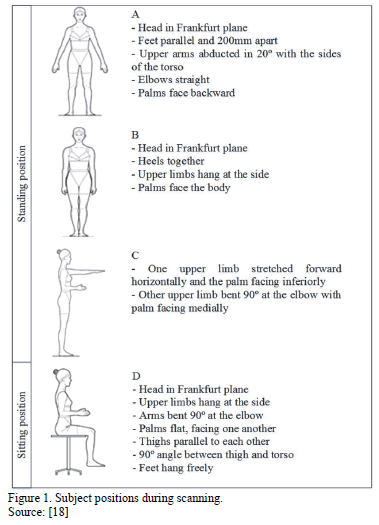
The positions adopted by subjects during the scanning process should be adapted to the study being conducted. As such, in the literature it is possible to find positions that differ from those indicated in ISO 20685. This is the case presented by Ashdown et al. [19] who argued (in a case in which a laser scanner with 8 paired cameras was stationed at four points equally spaced around the body) that the subjects' feet should be positioned about 30cm apart with the arms abducted from the body. This is a fundamental aspect of a good scan because other positions often result in holes or missing data for some portion of the body or obscure another area (such as under the arms) or for areas where the cameras cannot record data (such as surfaces parallel to the floor). Additionally, surfaces such as hair and dark-textured clothing decrease the quality of the scan by scattering the light and preventing the cameras from capturing a complete set of data points. A study by Tomkinson & Shaw [20] showed that most direct 3D scan measurements of standing posture had good repeatability, except the head and neck postures, whose repeatability was poor, as a result of significant postural errors. In this case they recommend that researchers aim at reducing postural and technical errors by strictly adhering to measurement protocols, undergoing extensive tester training, choosing appropriate test-retest intervals, minimizing diurnal variability, and taking multiple measurements.
2.3. The different body scanners
There are two types of body scanners: high-end scanning systems that produce high-quality scans for sizing surveys and in-shop or in-house inexpensive scanning systems that produce lower quality scans for retail use [17]. Based on a publication from 2013 [13], several 3D whole body scanning systems are currently available on the market, including those presented in Table 1.

There are also new ways of creating three-dimensional images using systems that were not initially designed for the purpose. This is the case of Microsoft Kinect, which can be used for numerous other applications besides games. The Microsoft Kinect sensor is one of a class of devices known as depth cameras in the category of structured light systems [21]. Kinect may be considered a 3D markerless motion capture system because it provides a simplified skeleton in real time, without the need for special clothes or other equipment. Despite the fact that it cannot be used for extremely accurate studies [22-24], it may be deployed when there is no need for high levels of accuracy, for example in clothing or shoe sizing, indirect fat measurement, or clinical rehabilitation [25, 26].
2.4. Applications
Body scanners are used for a wide variety of applications. Jones & Rioux [14] divided their applications into:
- Medical: body deformity; glaucoma; orthodontics; orthopedics; surgery; lung function studies; custom prostheses; breast topography; pediatrics; medical management;
- Human systems engineering: work environment; population anthropology; helmets and face masks; gloves; clothing; human morphology; human motion analysis; forensic imaging; hearing studies;
- Virtual reality and communications: three-dimensional portraits; computer animation of human models.
More recently, according to Ashdown et al. [19], 3D scans have been used to create virtual models of customers for the apparel industry, which consumers can use to try on clothing virtually.
Another important use of body scanners is the creation of anthropometric databases. The first large-scale 3D anthropometry survey project carried out was the Civilian American and European Surface Anthropometry Resource (CAESAR). The CAESAR database contains anthropometric variability of men and women, with ages ranging from 18 to 65 years old. Representatives were asked to ensure the database contained samples for various weights, ethnic groups, gender, geographic regions, and socio-economic status. The study was conducted between April 1998 and early 2000 and included three scans per person for (i) standing posture, (ii) full-coverage posture and (iii) relaxed seating posture. The data collection methods were standardized and documented so that the database may be continually expanded and updated. High-resolution body scans were made using three-dimensional body scanners from Cyberware and Vitronics.
In addition to the CAESAR project, many other studies, such as Size UK, Size USA, Size Spain and Size China, have been conducted to classify entire populations using data collected with 3D body scanners.
3. Reliability and validity of anthropometric data
When using anthropometric data it is important to test its reliability and validity, as these factors may influence both the measurements and the interpretation of the results obtained. Appannah et al. [27] argue that the validity of data is defined by the ability to achieve the "true value" of a measurement, while Johnson et al. [28] defined reliability as the ability to repeat, reproduce, or consistently obtain the same measurement under identical conditions. According to Mueller & Martorell [29], the reliability of a measurement relies on precision and dependability, the former being the most important determinant. Also important is intra-observer reliability, described as the ability of the same observer to obtain consistent measurement, and inter-observer reliability - ability of different measurers to obtain similar measurement. Kouchi et al. [17] stated that for anthropometric data users there are three essential quality parameters:
- Validity of the data, meaning that the target population is well defined by the subject population of an anthropometric survey;
- Comparability of measurement items, implying that the exact same method is used when taking the same one dimensional measurement;
- Accuracy and precision of measurements, which are affected by factors such as instruments, measurer skills or, even, the participants themselves.
3.1. Sources of errors
Despite the importance of all these issues (because of their impact on measurement error) they are sometimes neglected when conducting an investigation. In most studies the error limits are set prior to data collection, whilst the performance of the measurer is evaluated during the collection process (comparing it with previously defined standards).
Both in traditional anthropometry methods and in 3D anthropometry, there are some factors that may affect the incidence of errors. Kouchi et al. [30] presented a list of some of these factors (Table 2), where they state that the two principal sources of error are related to the devices used and to the persons involved in the data collection process.

The traditional instruments used when collecting anthropometric data are usually simple to calibrate and as such, they are only unreliable if they were poorly designed (e.g., a tape measure that is made from a material that will stretch). On the other hand the calibration of a 3D scanning system can be compromised either by the hardware or software. Most scanning systems have a calibration process that will verify and correct the calibration of the scanner by measuring a simple geometric shape of known dimensions.
The skill of measurement collection processes resides not only in the ability to produce several consistent measurements, but also in the ability to accurately identify the locations of the various landmarks. However, these two factors are very difficult to separate and assess individually as no "true values" are present in the human body [29]. Despite the fact that the repeatability of posture is marked as being a factor caused by the participants, a proper measuring posture and its repeatability are factors that are also related to the measurer, as they can be controlled if the measurer provides proper instructions [30]. As such, it may be said that observer error is the cause of most errors in traditional anthropometry since it includes imprecision in landmark location, subject positioning, and instrument usage [31]. The same authors also discuss the fact that when multiple observers are involved, this error can be accentuated, as happens in most large-scale anthropometric surveys, where the landmarking process is conducted by a single person but the body measurements are done by several [22].
Although when traditional methods are used the steps to achieving actual usable anthropometric data are not very complex, they are much more complex when a 3D body scanner is employed, as shown in Fig. 2. This is the reason why 3D anthropometry presents so many more factors that may influence the existence of errors in measurements.

Ever since 3D body scanners first appeared there have been authors who have tried to assess the accuracy and precision of the measurements derived from them. Attempts have been made to evaluate them in terms of: (i) comparability with measurements from traditional methods [32-34]; (ii) repeatability of scan-derived measurements [35,36]; (iii) and repeatability of scan-derived landmark locations obtained from the same image [35]. However, Kouchi et al. [30] state that the quality parameters of these studies are not usually consistent, because there is no explicit required accuracy standard and no widely accepted quality evaluation protocol.
Both Table 2 and Fig. 2 show that an important part of the three-dimensional anthropometry is based on landmarking. Most studies evaluate errors in body measurements rather than errors in landmark locations. However, Kouchi et al. [30] showed that the assessment of accuracy using measurement errors underestimates errors in landmark locations. This happens because when the same image is used, the scan-derived landmark locations are not always identical, but may vary according to the methodology - marker stickers indicating landmarks or landmark locations calculated from surface data - and to the way that markers are identified - by an operator, or calculated using software.
The following section goes into more detail about landmarking.
3.2. Landmarking
According to the manual of the International Society for the Advancement of Kinanthropometry [37], a landmark is an identifiable skeletal point which generally lies close to the body's surface and is the marker that identifies the exact location of a measurement site. They are found by palpation or measurement and can be used to define anatomical correspondence between individuals. Most commonly used landmarks are located on specific bones or are easily identifiable by soft tissue features such as nipples or the navel [17].
Wrongly identifying a body landmark is the main cause of observer error in the collection of anthropometric data [30]. As such, in any anthropometry-based study it is extremely important to agree on the body measurements to be recorded and the common points on the body to be identified.
In anthropometry, the same landmark is frequently used to measure several body dimensions. The first step in traditional landmarking is to mark the locations on the body sites that will be measured on the participants' skin using a non-smearing, skin pencil or skin-safe, washable, ink that can be easily removed using makeup remover [38, 39]. Anthropometrists usually use a small cross or dot as a marking symbol (Fig. 3).

Locating the required body landmarks can be a very difficult and time-consuming task. This may be especially problematic in people with more body fat over the bony landmarks [15], or people in wheelchairs with whom it can be hard to gain access to the required landmarks [32]. According to Paquette [41], in 1988, four hours were required to physically landmark, measure, and record the data of one individual in an anthropometric survey of US Army personnel.
The traditional anthropometric measuring procedure has not changed much since this study was carried out in the late 1980s.
For the marking of landmark locations it is fundamental that participants adopt the correct posture for the measurement or the landmark location might be compromised and the results biased. Kouchi et al. [17] give the example of landmarking the tip of the spinous process of the seventh cervical vertebra, which can be easily palpated at the back of the base of the neck when the participant has the neck bent forward. They state that if the location were marked in this posture, the mark on the skin would slide away from it when participants lift their heads for orientation in the Frankfurt plane, compromising the reliability of the results.
As landmarking is the basis for obtaining valid results, it is important to quantify the measurement errors caused by landmarking. Despite the fact that the repeatability of landmarking has been considered an important factor that, if handled incorrectly, contributes to errors in anthropometry measurement, there are few studies that quantify the phenomenon. Kouchi et al. [30] argued that landmarks with large intra-observer errors also had large inter-observer errors. Additionally, they found that the errors in body dimensions were smaller than landmarking errors in 23 of the 35 measurements analyzed, suggesting that the magnitude of landmarking errors would likely be underestimated by examining errors in body dimensions.
When using 3D body scanners, the landmarking process is also crucial for the correct correspondence of anatomical locations between subjects and across scans. Moreover, measurements derived from reliable landmarks can be used for statistical analysis, for reconstructing variation in human body shape or even for creating homologous models [42, 43]. As such, the poor identification of landmark locations characteristic of 3D anthropometry has a significant effect on the derived data that is used to define participant body dimensions and to effect shape analysis. Landmarks can be placed manually (with traditional markers, for scanners that can sense color differentiation, or small hemispherical objects stuck to the skin for scanners that only capture surface geometry - Fig. 4). Or they may be identified by the scanner's software system, in which case they are automatically identified from body surface geometries. Fig. 5 shows a set of landmarks automatically derived from a scan based on surface geometries.


Before starting the scan measurement process it is necessary to identify some body parts, in order to achieve an appropriate reconstruction from the captured datapoints a. The human model is usually divided at armpits and crotch (a process known as segmentation). Five body parts, including head and torso, both arms, and both legs can be identified. There are many ways to perform this segmentation, for example, Nurre et al. [45] proposed a cusp algorithm for segmenting the 3D scanning data of a human body while Wang et al. [46] applied fuzzy logic concepts to locate the armpits and crotch, and then to separate the arms and legs from the trunk.
The landmarking process is the most problematic aspect of 3D anthropometry since the landmarks placed on bony structures that are found below the skin surface, and palpated by the anthropometrist (in traditional methods) cannot be accurately identified from the surface shape of the scan. As this is a difficult process, and integral to reliable data collection, some attention has been given to developing methods to enable correct landmark identification. Some studies present algorithms for automatically calculating landmark locations [47,48] while others propose alternatives for automatically detecting and calculating 3D coordinates of markers positioned by experienced anthropometrists [46]. However, as creating a good criterion for evaluating the performance of an algorithm is very difficult, the performance of very experienced anthropometrists is used as a criterion for evaluating the performance of algorithms used for calculating scan-derived body dimensions [30].
According to Wang et al. [46], landmark identification methods may in general terms be classified as: (i) premarking, (ii) human body mapping, (iii) geometry analysis and (iv) approximate determination of height location. Often the positions of the landmarks can be easily identified on the scanning image with the human eye, a process that is difficult to program into software. By using color information obtained from the cameras in the scanning heads, manually placed landmarks can also be identified by analyzing the RGB information in the scanned image [6,46,49].
However, the procedure of placing markers on the body surface is time consuming, relatively invasive, and may involve human error. Kouchi et al. [30] discuss the fact that the amount of error in identifying landmarks is not well known. They examined the landmarking of 40 individuals carried out by experienced and novice markers and compared the differences in measurements taken with reference to the landmarks identified. Differences in measurements obtained that were due to intra- and inter- observer error were sufficiently large that it was suggested that the explicit definition of landmarks in more detail might reduce landmarking errors.
Therefore, the possibility of developing marker-free techniques for landmarking becomes an important issue for analyzing 3D whole body scanning data. For the method of automated landmarking, analyzing the geometry of the human body using techniques such as silhouette analysis is a logical approach [50]. Douros et al. [51] used the method of reconstructing curves and surfaces to locate the landmarks. Allen et al. [42] proposed a method for identifying landmarks efficiently that employed a template mapping approach, which makes use of information from the existing database of 3D human models. Lu and Wang [52] used four algorithms (silhouette analysis, minimum circumference determination, gray-scale detection, and human-body contour plots) to locate 12 landmarks and 3 characteristic lines automatically, making it possible to obtain 104 body dimensions.
According to Kouchi et al. [30], the identification of the 3D coordinates of landmark locations can be done entirely manually, entirely automatically, or using a mix of both techniques:
- Deciding landmark locations on the body: a measurer decides landmark locations manually or a system calculates them automatically;
- Obtaining 3D coordinates of markers: an operator manually picks the centers of marker stickers or a system recognizes stickers automatically and calculates 3D coordinates;
- Naming landmarks (or labeling): either an operator or a system automatically names each marker.
Although selecting a manual method to decide landmark locations on the human body and placing a marker is time consuming, so too is doing it semi-automatically by validating marker centers and naming markers. If on the one hand, automatic calculations of landmark locations save time, on the other, they may not always match the landmark locations identified by experienced anthropometrists.
4. Conclusions
Creating anthropometric databases typically requires considerable resources (time, knowledge, funds, equipment and people). To overcome these limitations, technological developments in recent years, using three-dimensional digital forms, has made it possible to advance the study of human size and shape using fewer resources. 3D body scanners make anthropometric data acquisition more practical, faster, and less expensive. It also has the potential to produce valid and reliable measurements. With these advances it is now possible to explore the possibilities of anthropometric measurement processes and create new perspectives on its use.
3D scanning systems have evolved over the last few years. From LASS to Kinect, the technology is always advancing. The type of technology used differs from scanner to scanner. There are four main types of technology: - laser line systems, structured light systems, multi-view camera systems and millimeter wave systems. Currently, there is a significant range of three-dimensional body scanners available in the market (nine major products developed in different parts of the globe).
Depending on the technology and product used, the results and applications are different. Different products may have different reliability issues, potentially compromising the applicability of the data. Depending on the desired application -i.e., highly precise anthropometric data or less accurate data for apparel applications- a variety of body scanners may be used. For example, the Microsoft Kinect, which is less accurate, may be used for apparel applications, while the Vitrus Smart LC, because it is more precise, can be used for compiling an anthropometric database.
The same logic can be applied when selecting traditional methods versus 3D anthropometry, as which method should be used will depend on the type of data required (one-dimensional measurements or three-dimensional surface shapes) and on how it will be applied.
Acknowledgments
This work is financed by FEDER funds through the Competitive Factors Operational Program (COMPETE) POCI-01-0145-FEDER-007043 and POCI-01-0145-FEDER-007136 and by national funds through FCT - the Portuguese Foundation for Science and Technology, under the projects UID/CEC/00319/2013 and UID/CTM/000264 respectively.
References
[1] Simmons, K.P. and Istook, C L., Body measurement techniques: Comparing 3D body-scanning and anthropometric methods for apparel applications. Journal of Fashion Marketing and Management: An International Journal, 7(3), pp. 306-332, 2003. DOI: 10.1108/13612020310484852
[2] ISO-7250., Basic human body measurements for technological design, 1996.
[3] Kroemer, K. and Kroemer, H.J., Engineering physiology: Bases of human factors/ergonomics. New York: John Wiley & Sons, 1997.
[4] Montagu, M.F. and Broek, J.C., A handbook of anthropometry. Springfield: Charles C Thomas Publisher, 1960. DOI: 10.1037/12018-000
[5] Lovesey, E.J., A method for determining facial contours by shadow projection. Royal Aircraft Establishment Technical Report TR66192, 1966.
[6] Daanen, H.A.M. and van de Water, G.J., Whole body scanners. Displays, 19(3), pp. 111-120, 1998. DOI: 10.1016/S0141-9382(98)00034-1
[7] Bindahman, S., Zakaria, N. and Zakaria, N., 3D body scanning technology: Privacy and ethical issues, Proceeding of the International Conference on Cyber Security, Cyber Warfare and Digital Forensic (IEEE CyberSec), pp. 150-154, 2012.
[8] Jones, P.R.M., West, G.M., Harris, D.H. and Read, J.B., The loughborough anthropometric shadow scanner (LASS). Endeavour, 13(4), pp. 162-168, 1989. DOI: 10.1016/S0160-9327(89)80014-6, 10.1016/S0160-9327(89)80004-3
[9] Brooke-Wavell, K., Jones, P.R.M. and West, G.M., Reliability and repeatability of 3-D body scanner (LASS) measurements compared to anthropometry. Annals of Human Biology, 21(6), pp. 571-577, 1994. DOI: 10.1080/03014469400003572
[10] Bragança, S., Arezes, P. and Carvalho, M., An overview of the current three-dimensional body scanners for anthropometric data collection, Occupational Safety and Hygiene III, pp. 149-153, 2015. DOI: 10.1201/b18042-32
[11] Treleaven, P. and Wells, J., 3D body scanning and healthcare applications. Computer, 40(7), pp. 28-34, 2007. DOI: 10.1109/MC.2007.225
[12] Istook, C.L. and Hwang, S., 3D body scanning systems with application to the apparel industry. Journal of Fashion Marketing and Management, 5(2), pp. 120-132, 2001. DOI: 10.1108/EUM0000000007283
[13] Daanen, H.A.M. and Haar, F.B., 3D whole body scanners revisited. Displays, 34(4), pp. 270-275, 2013. DOI: 10.1016/j.displa.2013.08.011
[14] Jones, P.R.M. and Rioux, M., Three-dimensional surface anthropometry: Applications to the human body. Optics and Lasers in Engineering, 28(1), pp. 89-117, 1997. DOI: 10.1016/S0143-8166(97)00006-7
[15] Olds, T. and Honey, F., The use of 3D whole-body scanners in anthropometry. Proceedings of the 9th International Conference of the International Society for the Advancement of Kinanthropometry, pp. 1-12, 2006.
[16] Mckinnon, L. and Istook, C., Comparative analysis of the image twin system and the 3T6 body scanner. Journal of Textile and Apparel, Technology and Management, 1(2), pp. 1-7, 2001.
[17] Kouchi, M., Gupta, D. and Zakaria, N., Anthropometric methods for apparel design: Body measurement devices and techniques. Anthropometry, Apparel Sizing and Design, pp. 67-94, 2014. DOI: 10.1533/9780857096890.1.67
[18] ISO-20685., 3-D scanning methodologies for internationally comptible anthropometric databases, 2010.
[19] Ashdown, S.P., Loker, S., Schoenfelder, K. and Lyman-Clarke, L., Using 3D scans for fit analysis. Journal of Textile and Apparel, Technology and Management, 4(1), pp. 1-12, 2004.
[20] Tomkinson, G.R. and Shaw, L.G., Quantification of the postural and technical errors in asymptomatic adults using direct 3D whole body scan measurements of standing posture. Gait and Posture, 37(2), pp. 172-177, 2013. DOI: 10.1016/j.gaitpost.2012.06.031
[21] Shotton, J., Fitzgibbon, A., Cook, M., Sharp, T., Finocchio, M., Moore, R. and Blake, A., Real-time human pose recognition in parts from single depth images. Studies in Computational Intelligence, 411(1), pp. 119-135, 2013. DOI: 10.1007/978-3-642-28661-2_5
[22] Ye, M., Wang, X., Yang, R., Ren, L. and Pollefeys, M., Accurate 3D pose estimation from a single depth image. 2011 International Conference on Computer Vision, pp. 731-738, 2011. DOI: 10.1109/ICCV.2011.6126310
[23] Clark, R.A., Pua, Y.H., Fortin, K., Ritchie, C., Webster, K.E., Denehy, L. and Bryant, A.L., Validity of the Microsoft Kinect for assessment of postural control. Gait and Posture, 36(3), pp. 372-377, 2012. DOI: 10.1016/j.gaitpost.2012.03.033
[24] Braganca, S., Carvalho, M., Xu, B., Arezes, P. and Ashdown, S., A validation study of a kinect based body imaging (KBI) device system based on ISO 20685:2010. Proceedings of the 5th International Conference on 3D Body Scanning Technologies, pp. 372-377, 2014.
[25] Fernández-Baena, A., Susín, A. and Lligadas, X., Biomechanical validation of upper-body and lower-body joint movements of kinect motion capture data for rehabilitation treatments. Proceedings of the 2012 4th International Conference on Intelligent Networking and Collaborative Systems, pp. 656-661, 2012. DOI: 10.1109/iNCoS.2012.66
[26] Weiss, A., Hirshberg, D. and Black, M.J., Home 3D body scans from noisy image and range data. Proceedings of the IEEE International Conference on Computer Vision, pp. 1951-1958, 2011 DOI: 10.1109/iccv.2011.6126465
[27] Appannah, G., Haniff, J., Mohammad Nor, N.S., Wong, N.F., Kee, C. C., Zainuddin, A.A. and Yusoff, A.F., Reliability, technical error of measurements and validity of instruments for nutritional status assessment of adults in Malaysia. Malaysian Journal of Nutrition. Nutrition Society of Malaysia, 2008.
[28] Johnson, T.S., Engstrom, J.L. and Gelhar, D.K., Intra-and interexaminer reliability of anthropometric measurements of term infants. Journal of Pediatric Gastroenterology and Nutrition, 24(5), pp. 497-505, 1997. DOI: 10.1097/00005176-199705000-00001
[29] Mueller, W.H. and Martorell, R., Reliability and accuracy of measurement. Anthropometric Standardization Reference Manual, pp. 83-86, 1988.
[30] Kouchi, M. and Mochimaru, M., Errors in landmarking and the evaluation of the accuracy of traditional and 3D anthropometry. Applied Ergonomics, 42(3), pp. 518-527, 2011. DOI: 10.1016/j.apergo.2010.09.011
[31] Bennett, K.A. and Osborne, R.H., Interobserver measurement reliability in anthropometry. Human Biology, 58(5), pp. 751-759, 1986.
[32] Sims, R.E., Marshall, R., Gyi, D.E., Summerskill, S.J. and Case, K., Collection of anthropometry from older and physically impaired persons: Traditional methods versus TC 2 3-D body scanner. International Journal of Industrial Ergonomics, 42(1), pp. 65-72, 2012. DOI: 10.1016/j.ergon.2011.10.002
[33] Han, H., Nam, Y. and Choi, K., Comparative analysis of 3D body scan measurements and manual measurements of size Korea adult females. International Journal of Industrial Ergonomics, 40(5), pp. 530-540, 2010. DOI: 10.1016/j.ergon.2010.06.002
[34] Lu, J.M. and Wang, M.J.J., The evaluation of scan-derived anthropometric measurements. IEEE Transactions on Instrumentation and Measurement, 59(8), pp. 2048-2054, 2010. DOI: 10.1109/TIM.2009.2031847
[35] Kouchi, M. and Mochimaru, M., Evaluation of Accuracy in Traditional and 3D Anthropometry, 2008.
[36] Robinette, K.M. and Daanen, H.A.M., Precision of the CAESAR scan-extracted measurements. Applied Ergonomics, 2006. DOI: 10.1016/j.apergo.2005.07.009
[37] International Society for the Advancement of Kinanthropometry, available at www.isakonlinve.com
[38] Roebuck, J.A., Kroemer, K.H.E. and Thomson, W.G., Engineering Anthropometry Methods, 1975.
[39] O'Brien, R. and Shelton, W.C., Women's Measurements for Garment and Pattern Construction, 1941.
[40] Marfell-Jones, T.O.A.S., L.C.M., Stewart, A. and Marfell-Jones, M., International Standards for Anthropometric Assessment, 2006.
[41] Paquette, S., 3D scanning in apparel design and human engineering. IEEE Computer Graphics and Applications, 16(5), pp. 11-15, 1996. DOI: 10.1109/38.536269
[42] Allen, B., Curless, B. and Popović, Z., The space of human body shapes. ACM Transactions on Graphics. ACM, 2003.
[43] Mochimaru, M. and Kouchi, M., Statistics for 3D human body forms. Proceedings of the Human Factors and Ergonomics Society Annual Meeting. SAGE Publications, 2000. DOI: 10.4271/2000-01-2149, 10.1177/154193120004403846
[44] Human Solutions, AnthoScan - Body Dimensions Acquired Easily, [Online]. Available at: http://www.human-solutions.com/download/pdf/ANTHROSCAN_en.pdf
[45] Nurre, J.H., Connor, J., Lewark, E.A. and Collier, J.S., On segmenting the three-dimensional scan data of a human body. IEEE Transactions on Medical Imaging, 19(8), pp. 787-797, 2000. DOI: 10.1109/42.876304
[46] Wang, M.-J.J., Wu, W.-Y., Lin, K.-C., Yang, S.-N. and Lu, J.-M., Automated anthropometric data collection from three-dimensional digital human models. The International Journal of Advanced Manufacturing Technology, 2007. DOI: 10.1007/s00170-005-0307-3
[47] Azouz, B.Z., Shu, C. and Mantel, A., Automatic locating of anthropometric landmarks on 3D human models. Proceedings of the Third International Symposium on 3D Data Processing, Visualization and Transmission, 2006.
[48] Leong, I.F., Fang, J.J. and Tsai, M.J., Automatic body feature extraction from a marker-less scanned human body. CAD Computer Aided Design, 39(7), pp. 568-582, 2007. DOI: 10.1016/j.cad.2007.03.003
[49] Burnsides, D., Boehmer, M. and Robinette, K., 3-D landmark detection and identification in the CAESAR project. Proceedings Third International Conference on 3-D Digital Imaging and Modeling, pp. 393-398, 2001.
[50] Buxton, B., Dekker, L., Douros, I. and Vassilev, T., Reconstruction and interpretation of 3D whole body surface images. Scanning, 2000.
[51] Douros, I., Dekker, L. and Buxton, B., Reconstruction of the surface of the human body from 3D scanner data using B-splines. Proceedings of the Electronic Imaging '99, pp. 234-245, 1999. DOI: 10.1117/12.341065
[52] Lu J. and Wang, M., Automated anthropometric data collection using 3D whole body scanners. Expert Systems with Applications, 2008. DOI: 10.1016/j.eswa.2007.07.008
S. Bragança, is a PhD. student in the Department of Production and Systems, School of Engineering, University of Minho, Portugal. She completed her MSc. in Industrial Engineering and Management in 2012, also at the University of Minho, Portugal. Her MSc Thesis was in the Implementation of Standard Work and other Lean Production tools. Her current research focuses on ergonomics and anthropometry. ORCID: orcid.org/0000-0002-4765-3856
P. Arezes, has a PhD. in Industrial and Systems Engineering from the University of Minho, Portugal, where he is currently a full professor in Ergonomics and Human Factors. He is also a visiting fellow at MIT's AgeLab in the USA. He leads the Human Engineering research group and coordinates the Engineering Design and Advanced Manufacturing (EDAM) area of MIT's Portugal Program at the University of Minho, where he is also chair of the Steering Board of the PhD program "Leaders for Technical Industries (LTI)". ORCID: orcid.org/0000-0001-9421-9123
M. Carvalho, graduated in Textile Engineering; he has an MSc. in Design and Marketing and a PhD. in Textile Engineering from the University of Minho, Portugal. Since 1993 he has been a researcher at 2C2T the University of Minho's Center of Science and Textile Technology. He is involved in the supervision of research projects in the areas of clothing and textile design; comfort; pattern design; anthropometrics; ergonomics and development of functional/multi-functional materials and textile products; and interactive textiles with applications in the health, automobile, sport and other sectors. ORCID: orcid.org/0000-0001-8010-6478
S. Ashdown, is Helen G. Canoyer Professor in the Department of Fiber Science & Apparel Design at Cornell University. Her research group investigates the impact of new technologies on apparel design, with a focus on sizing and fit, functional apparel design, and the use of the 3-D full-body scanners as a research tool for apparel designers. ORCID: orcid.org/0000-0002-0276-4122
Cómo citar
IEEE
ACM
ACS
APA
ABNT
Chicago
Harvard
MLA
Turabian
Vancouver
Descargar cita
CrossRef Cited-by
1. Andreja Rudolf, Sonja Šterman, Andrej Cupar. (2024). Development of a textile sheet mask design for facial care based on a 3D face model of an average woman. Journal of Engineered Fibers and Fabrics, 19 https://doi.org/10.1177/15589250241254443.
2. Xiaoyu Cai, Bingfei Gu, Huazhou He. (2021). Classification analysis of young female students’ waist–abdomen–hip based on body photos. Textile Research Journal, 91(11-12), p.1409. https://doi.org/10.1177/0040517520979742.
3. Yupeng Hu, Yuanping Xia, Bingfei Gu. (2023). An image-based shape analysis approach and its application to young women’s waist-hip-leg position. Ergonomics, 66(12), p.2074. https://doi.org/10.1080/00140139.2023.2184366.
4. Sara Bragança, Ignacio Castellucci, Eric Costa, Pedro Arezes, Miguel Carvalho. (2020). Anthropometric data for wheelchair users: a systematic literature review. International Journal of Occupational Safety and Ergonomics, 26(1), p.149. https://doi.org/10.1080/10803548.2019.1567974.
5. Kayna Hobbs-Murphy, Isabel Olmedo-Nockideneh, William J. Brazile, Kristen Morris, John Rosecrance. (2024). Intra-rater and inter-rater reliability of 3D facial measurements. Applied Ergonomics, 116, p.104218. https://doi.org/10.1016/j.apergo.2023.104218.
6. Michael Thelwell, Neil Masters, Robert Appleyard, Alice May Bullas. (2024). Advanced Body Measurement Techniques Can Complement Current Methods of Cytotoxic Chemotherapy Dose Prescription. Applied Sciences, 14(2), p.834. https://doi.org/10.3390/app14020834.
7. Joe D. Derouchey, Grant R. Tomkinson, Jesse L. Rhoades, John S. Fitzgerald. (2020). Reliability of the Styku 3D Whole-Body Scanner for the Assessment of Body Size in Athletes. Measurement in Physical Education and Exercise Science, 24(3), p.228. https://doi.org/10.1080/1091367X.2020.1791124.
8. Andreja Rudolf, Zoran Stjepanovič, Andrej Cupar. (2021). Study Regarding the Kinematic 3D Human-Body Model Intended for Simulation of Personalized Clothes for a Sitting Posture. Materials, 14(18), p.5124. https://doi.org/10.3390/ma14185124.
9. Elvia Luz González-Muñoz, Rosalio Avila Chaurand, John A. Rey Galindo, Gabriel Ibarra Mejia. (2022). Proceedings of the 21st Congress of the International Ergonomics Association (IEA 2021). Lecture Notes in Networks and Systems. 223, p.96. https://doi.org/10.1007/978-3-030-74614-8_11.
10. Nurashikin Saaludin, Amna Saad, Cordelia Mason. (2022). Digital Manufacturing Technology for Sustainable Anthropometric Apparel. , p.71. https://doi.org/10.1016/B978-0-12-823969-8.00011-3.
11. C. Viviani, P.M. Arezes, S. Bragança, J. Molenbroek, I. Dianat, H.I. Castellucci. (2018). Accuracy, precision and reliability in anthropometric surveys for ergonomics purposes in adult working populations: A literature review. International Journal of Industrial Ergonomics, 65, p.1. https://doi.org/10.1016/j.ergon.2018.01.012.
12. Yuanping Xia, Jiayu Shao, Beibei Zhang, Shouning Jin, Yuqing Li, Bingfei Gu. (2022). Individualized generation of young women's crotch curve based on body images. International Journal of Industrial Ergonomics, 90, p.103296. https://doi.org/10.1016/j.ergon.2022.103296.
13. G. Bravo, S. Bragança, P.M. Arezes, J.F.M. Molenbroek, H.I. Castellucci. (2018). A literature review of anthropometric studies of school students for ergonomics purposes: Are accuracy, precision and reliability being considered?. WORK: A Journal of Prevention, Assessment & Rehabilitation, 60(1), p.3. https://doi.org/10.3233/WOR-182719.
Dimensions
PlumX
Visitas a la página del resumen del artículo
Descargas
Licencia
Derechos de autor 2016 DYNA

Esta obra está bajo una licencia internacional Creative Commons Atribución-NoComercial-SinDerivadas 4.0.
El autor o autores de un artículo aceptado para publicación en cualquiera de las revistas editadas por la facultad de Minas cederán la totalidad de los derechos patrimoniales a la Universidad Nacional de Colombia de manera gratuita, dentro de los cuáles se incluyen: el derecho a editar, publicar, reproducir y distribuir tanto en medios impresos como digitales, además de incluir en artículo en índices internacionales y/o bases de datos, de igual manera, se faculta a la editorial para utilizar las imágenes, tablas y/o cualquier material gráfico presentado en el artículo para el diseño de carátulas o posters de la misma revista.


