Using a non-reducing sugar in the green synthesis of gold and silver nanoparticles by the chemical reduction method
Uso de una azúcar no reductora en la síntesis verde de nanopartículas de oro y plata por medio de reducción química
DOI:
https://doi.org/10.15446/dyna.v85n206.72136Palabras clave:
metal nanoparticles, sucrose, hydrolysis, reduction oxide reaction, salt of carboxylic acid (en)nanopartículas metálicas, sacarosa, hidrólisis, óxido reducción, sales de ácido carboxílico (es)
Descargas
Recibido: 18 de mayo de 2018; Revisión recibida: 21 de junio de 2018; Aceptado: 24 de julio de 2018
Abstract
The use of more biocompatible and renewable chemical compounds to obtain metal nanoparticles with desired properties and characteristics becomes an alternative route for the reduction of environmental risks and to minimize the incompatibility of these structures when interacting with biological models for their possible application in the health area. The purpose of this research was focused on the use of sucrose, as a reducing agent for gold and silver nanoparticles using different volumes of sodium hydroxide. The nanoparticles obtained were characterized by UV-visible spectrometry, transmission electron microscopy TEM and Fourier transform infrared spectroscopy FTIR, which allowed to determine the surface plasmon resonance, experimental and theoretical particle sizes, morphology and structural changes in the reducing agent, as well as the influence of sodium hydroxide in the synthesis process. The results obtained confirm the generation of gold and silver nanoparticles by the previous formation of reducing sugars and oxidation of the functional group from glucose to salts of carboxylic acid.
Keywords:
metal nanoparticles, sucrose, hydrolysis, reduction oxide reaction, salt of carboxylic acid.Resumen
El uso de compuestos químicos más biocompatibles y renovables para la obtención de nanopartículas metálicas con propiedades y características deseadas, se convierte en una ruta alternativa para la reducción de riesgos ambientales y del grado de incompatibilidad de estas estructuras al interactuar con modelos biológicos para su posible aplicación en el área de la salud. El propósito de este trabajo se centró en el uso de sacarosa, como agente reductor de nanopartículas de oro y plata al emplear diferentes volúmenes de hidróxido de sodio. Las nanopartículas obtenidas fueron caracterizadas mediante espectrometría UV-visible, microscopía electrónica de transmisión TEM y espectroscopia infrarroja por transformada de Fourier FTIR, la cual permitió determinar los plasmones de resonancia superficial, tamaños de partícula experimentales y teóricos, morfología y cambios estructurales en el agente reductor, así como la influencia del hidróxido de sodio en el proceso de síntesis. Los resultados obtenidos confirman la formación de nanopartículas de oro y plata mediante la previa formación de azúcares reductores. Así mismo, la oxidación del grupo funcional de la glucosa a sales de ácido carboxílico.
Palabras clave:
nanopartículas metálicas, sacarosa, hidrólisis, óxido reducción, sales de ácido carboxílico.1. Introduction
Nanoparticles have been potentiated for the research and development of biosensors, catalytic processes, and biomedical applications, among others. The interest in these materials is due to the fact that is possible to obtain new properties and characteristics by modifying their size and structure depending on the production process employed [1-3]. Currently, different physical and chemical processes are used to synthesize metal nanoparticles (MNPs) to produce particles with characteristics and properties related to the application area. Two kinds of methodologies are used to obtain nanostructures, i) top-down strategies, in which dimensionally bulk materials are collapsed little-by-little by manual or automated processes until reaching nanoscale [4], and ii) bottom-up where the molecular modification of a precursor agent in order to obtain atoms, which by nucleation processes result in monodisperse nanostructures [5]. However, most of these methods are often expensive, labor-intensive, potentially hazardous to the environment and highly cytotoxic [6]. Therefore, there is a need to carry out new processes to obtain nanoparticles with controlled size and shape, with high performance, high purity, and environmentally friendly conditions and with low levels of cytotoxicity [7,8].
On the other hand, the partial or total substitution of hazardous substances with sustainable processes is known as green chemistry, which arises from the growing demand for more sustainable processes, and is intended to minimize or prevent waste from the reactions involved in the processes of obtaining nanostructured materials while preserving their efficacy [9-11]. This is because, with the obtained characteristics, it is possible to use more biocompatible and renewable chemicals for the development of MNPs with physical, chemical, optical, catalytic and/or electrical properties for their possible application in different areas of science such as medicine [12], biology [13], chemistry [14], and physics [15], among others.
In the search of a nanomaterial for biomedical applications, chitosan (CS), a natural, non-toxic, and biodegradable compound derived from the deacetylation of chitin extracted from the cell walls of some fungi, crustacean exoskeletons and insects, has been studied. [16,17]. Presently, CS is used in the synthesis of gold and silver nanoparticles as a reducing and stabilizing agent at different concentrations of precursor agents, in order to obtain nanomaterials with optical and structural controllable properties, by evaluating with characterization techniques such as UV-visible, infrared spectroscopy by Fourier transform - FTIR, scanning electron microscopy, among others [2].
In addition, the use of gallic acid, present in a variety of plants such as spinach, grapes, green tea, oak bark, red wine and white wine, has been studied as well as chitosan as a reducing agent and stabilizer in the synthesis of gold and silver nanoparticles. In order to compare the catalytic capacity of this material in the process of reducing 4-nitrophenol in the presence of sodium borohydride [18].
Furthermore, the use of ascorbic acid known as vitamin C, present in several fruits and vegetables and used as a dietary supplement, is used as a reducing agent and stabilizer of gold and silver nanoparticles, to evaluate its influence under different conditions of pH for obtaining stable nanostructures and with defined optical properties [19].
On the other hand, the use of sodium alginate, a natural hydrophilic anionic polysaccharide, extracted from brown marine algae [20], which is used in the synthesis of silver nanoparticles as a reducing agent and in turn as a stabilizer of the same, due to this coating provided through the Van der Waals forces. Moreover, its antibacterial effect against Gram-positive (Staphylococcus aureus) and Gram-negative (Pseudomonas aeruginosa) microorganisms have been evaluated for possible use in medical devices or wound dressings [21].
Based on the foregoing, this investigation used a non-reducing sugar such as sucrose, a product of the extraction and refining of sugarcane, as a reducing agent of gold and silver nanoparticles, with the aim to evaluate its influence under different volumes of sodium hydroxide to obtain colloidal nanostructures for its possible application in a cellular microenvironment.
2. Methodology
For this investigation, gold trichloride hydrochloride (HAuCl4∙3H2O 99.995 % trace metals basis) was obtained from Aldrich (Sigma-Aldrich), silver nitrate (AgNO3 pure, pharma grade) was obtained from PanReac (PanReac Applichem). Sodium hydroxide (NaOH pellets) was acquired from Merck (Merck Millipore). Sucrose was obtained at a local market.
2.1. Synthesis of silver nanoparticles (AgNPs)
A 1 mM silver nitrate (AgNO3) solution was prepared and mixed with 0.2 % sucrose solution. Then, sodium hydroxide (NaOH) 0.1 M was added at volumes of 0 ml, 0.25 ml, 0.5 ml, 0.75 ml and 1.0 ml, the solutions were mixed at 2000 rpm and left to rest at room temperature until a change in color was observed. Finally, the pH reading was performed on each of the samples obtained, using an Ohaus Starter 2100 pH bench meter at standard conditions for temperature and pressure. In addition, blank solutions from the mixture of reducing agent and the different volumes of sodium hydroxide employed in the reaction were prepared. All assays were performed in triplicate.
2.2. Synthesis of gold nanoparticles (AuNPs)
A 0.5mM chloroauric acid (HAuCl4) was prepared and mixed with 0.1M sucrose solution. Then, 0.1M NaOH was added at volumes of 0 ml, 0.1 ml, 0.15 ml, 0.20 ml and 0.25 ml. Next, the solutions were carried in a water bath at 60 °C by 30 minutes. Once the samples reached standard conditions for temperature and pressure, the pH was read using an Ohaus Starter 2100 pH bench meter. In addition, blank solutions from the mixture of reducing agent and the different volumes of sodium hydroxide employed in the reaction were prepared. All assays were performed in triplicate.
2.3. UV-visible spectrophotometry analysis
Gold and silver nanoparticles were analyzed in a SHIMADZU UV-1800 UV-visible spectrophotometer in a range of 300 - 800 nm; as a control, reducing agent solutions without precursor agent were used. The UV-visible absorbance data obtained for each of the working solutions were subtracted from their respective blank. The result of this subtraction resulted in the resonance associated with the type of metallic particle.
2.4. Fourier transform infrared spectroscopy (FTIR) analysis
The samples were analyzed in an IRAffinity-1S FTIR Spectrophotometer in the range of 500-4000 cm-1 to determine the presence of a reducing agent, using attenuated total reflectance (ATR) modulus, with a resolution of 4 cm-1, 128 scans and using Happ-Genzel as apodization function.
2.5. Transmission electron microscopy (TEM) analysis
Analysis by transmission electron microscopy (TEM) was carried out using a Tecnai F20 Super Twin TMP transmission electron microscope operated at 200 kV with the propose to determinate the morphology and particle size.
2.6. Statistical analysis
The synthesis of metal nanoparticles and UV-visible measurements were performed in triplicate in three independent times. In addition, a multivariate statistical analysis was carried out using the Statgraphics Centurion XVI software, which allowed obtaining the population mean, the standard deviation and 95% confidence intervals. For the statistical analysis of the particle size from the TEM images, the ImageJ® software was used, obtaining the population mean, the standard deviation and 95% confidence intervals.
3. Results and discussion
3.1. Non-reducing sugar as a reducing agent in the formation of metal nanoparticles
A reducing sugar is one that through its carbonyl group can donate electrons to molecules that act as oxidants, which accept these electrons. Due to its glycosidic bond, which prevents the opening of the ring of any of its two monosaccharaides, glucose, and fructose, it is not possible to act as reducing sugar of metal nanoparticles in the absence of hemiacetal groups without a process through which the ring opening of either of the two monosaccharaides [22].
This is achieved by a process of hydrolysis of the sucrose molecule, as shown in Fig. 1, in which the dissolution of this structure into glucose and fructose is generated [23]. At the same time, the glucose and fructose molecules may be present in a cyclic or open form.
Figure 1: Sucrose hydrolysis into glucose and fructose, in its open and closed form.
The presence of carbonyl groups, especially aldehydes in the case of glucose and ketones for fructose, facilitate the synthesis of metal nanoparticles, where glucose is easier to oxidize than fructose, due to the presence of the hydrogen atom in its main group, becoming a strong reducing agent and responsible for the reduction of gold and silver ions to their metallic states [24]. See eq. (1) and (2).


For a reduction of gold and silver salts to occur, it is necessary that the reducing agent donates electrons, which will be captured by the oxidizing agents. This is known as a reduction-oxidation reaction, in which the structure acting as a reducer, in this case, glucose, passed from an aldehyde group to form a gluconic acid or salt of gluconic acid, related with pH values in the synthesis process (see Fig. 2), an oxidation reaction. While those structures that act as oxidants, gold salts, and silver are reduced and will result in nanometric structures.
Figure 2: Oxidation of glucose in gluconic acid and gluconic acid salt.
In this research, synthesis of nanoparticles of gold and silver by a non-reducing sugar as sucrose was in an alkaline medium, by the addition of different volumes of sodium hydroxide. Upon which, the expected products in the synthesis are salts of gluconic acid because of the oxidation of glucose and the formation of gold and silver nanoparticles. Also, the use of sodium hydroxide allows for the presence of reducing sugars in a short period of time, due to a low dissolution rate of sucrose, requiring the use of acids, alkaline or enzymatic agents, which increase the reaction rate [25,26].
3.2. UV-visible spectroscopy
To demonstrate the formation of gold and silver nanoparticles in the different solutions obtained, these were analyzed by UV-visible spectrophotometry, to identify their respective surface plasmon resonance, which indicates the wavelength in where the greatest electronic movement is presented, translated into an increase in absorbance value [27]. Generally, absorbance peaks have a presence at wavelengths between 400 - 500 nm for silver nanoparticles [27,28] and 500 - 600 nm for gold nanoparticles [29,30]. The absorbance spectra obtained in this investigation are shown below (see Figs. 3 and 4).
Figure 3: UV-visible spectra of silver nanoparticles at different volumes of sodium hydroxide. As control samples, solutions without reducing agent was used.
Figure 4: UV-visible spectra of gold nanoparticles at different volumes of sodium hydroxide. As control samples, solutions without reducing agent was used. *The results presented correspond to the population mean of the replicas made. With n = 3
From Fig. 3 and 4, it is possible to demonstrate that sucrose in presence of different volumes of sodium hydroxide promotes the formation of gold and silver nanoparticles, from hydrolysis and reduction-oxidation reactions, combined with the presence of the absorbance peaks for each kind of metal between the characteristic wavelength ranges.
In addition, it was possible to show that as of a certain volume of sodium hydroxide is possible to obtain gold and silver nanoparticles. This chemical behavior can be expressed in eq. (3.1) - (3.3) for silver and eq. (4.1) - (4.3) for gold, where is necessary the oxidation of number of glucose moles for the reduction of a particular number of salts precursors moles.


From the above equations and as of the addition of 0.25 ml and 0.15 ml of sodium hydroxide for silver and gold respectively [8], it is possible to obtain nanoparticles (see Figs. 5 and 6). Therefore, the presence of sodium hydroxide besides favoring the formation of silver and gold nanoparticles, at the same time increases the pH values, which significantly decreases the efficiency and formation rate of the nanostructures [31]. This behavior can be observed in the absorption spectra of Fig. 3 and 4, where with volumes of sodium hydroxide of 0.5 ml for silver and 0.15 ml for gold, there is no significant variation in the absorbance values (see Tables 1 and 2).
Figure 5: Gold nanoparticles at different volumes of sodium hydroxide.
Figure 6: Silver nanoparticles at different volumes of sodium hydroxide.
Source: The authors
Table 1: Wavelengths and absorbance of silver nanoparticles at different volumes of sodium hydroxide.

Source: The authors
Table 2: Wavelengths and absorbance of gold nanoparticles at different volumes of sodium hydroxide.

On the other hand, sodium hydroxide plays an important role in the morphology of nanoparticles; this attribute can be evidenced in TEM images (section 3.4). This was possible to evince it from the behavior of the absorption spectrum of gold, where the presence of a second band (see Fig. 4) at volumes of sodium hydroxide of 0.20 and 0.25 ml, can present a different morphology to that obtained by using 0.15 ml of sodium hydroxide [32,33]. To confirm the foregoing, the samples were analyzed after four weeks, with the purpose of evidencing changes in the absorption spectra of the gold and silver nanoparticles (see Figs. 7 and 8).
Figure 7: UV-visible spectra of silver nanoparticles at different volumes of sodium hydroxide after four weeks.
Figure 8: UV-visible spectra of gold nanoparticles at different volumes of sodium hydroxide after four weeks.
The first findings after four weeks of synthesis was found in the gold and silver absorption spectra, it is the displacement of the superficial plasmon resonance to those initially recorded (see Tables 3 and 4), which can be associated with changes in the colloidal stability of the nanoparticle solution [35]. Second aspect to highlight, is the decrease of the second gold absorption band. Which indicates that high values of sodium hydroxide at the beginning of the reaction, leads to the formation of specific types of morphologies to evidenced two bands in the absorption spectrum, but with the passage of weeks, the reactions become stabilized in only absorption band could lead to a single morphological structure, this behavior is similar to that reported by Attia et al [32].
Source: The authors
Table 3: Comparison between wavelengths and absorbance weeks 1 and 4 for silver nanoparticles at different volumes of sodium hydroxide.

Source: The authors
Table 4: Comparison between wavelengths and absorbance weeks 1 and 4 for gold nanoparticles at different volumes of sodium hydroxide.

3.3. Particle size
From the results obtained in the analysis by UV-visible spectrophotometry, it is possible to determine a particle size approximation from the absorbance and wavelength values obtained for each type of nanoparticle. Using the equations reported by Haiss [35] (5) and Paramelle [34] (6), which are shown below. The results obtained are shown in tables 5 and 6, which are presented as the population mean.


Where A spr and 𝐴 450 correspond to the absorbance obtained at the wavelength of the superficial plasmon resonance and at a wavelength of 450 nm respectively. While 𝜆 𝑚𝑎𝑥 is the wavelength of the superficial plasmon resonance. The above equations are valid for a range between [5-80nm] and [20-100nm], respectively.
Additionally, the width at medium height or FWHM was determined, from the UV-vis absorption spectra, to determine experimentally the degree of dispersion of the different synthesized nanoparticles. For this, low FWHM values indicate monodispersity, while high values reflect the polydispersity of particles [36]. The results obtained in this investigation are shown in tables 5 and 6.
Source: The authors
Table 5: The experimental particle size and FWHM of silver nanoparticles at different volumes of sodium hydroxide.

Source: The authors
Table 6: The experimental particle size and FWHM of gold nanoparticles at different volumes of sodium hydroxide.

3.4. Transmission electron microscopy analysis
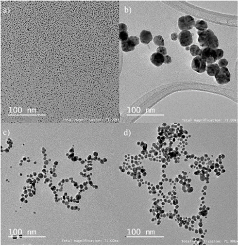
Fig. 9 shows the TEM images obtained for the gold and silver nanoparticles synthesized using sucrose as a reducing agent, in which the morphology, dispersion and average particle size can be evidenced for each type of synthesis performed.
Figure 9: TEM image of metal nanoparticles, a) Silver 0.25 ml NaOH, b) Silver 0.50 ml NaOH, c) gold 0.15 mL of NaOH and d) gold 0.20 ml NaOH.
Fig. 9 shows the different morphologies obtained from the gold and silver nanoparticles. In the case of silver, when using a volume of sodium hydroxide of 0.25 ml, the initial phase of nanoparticle formation is observed (Figure 9a), while at a volume of 0.50 ml led to formation of structures with a larger diameter and defined morphologies compared at a volume of 0.25 ml (Figure 9b). This behavior is correlated with the results presented in table 7, where average particle diameters of 3 nm are obtained from a volume of sodium hydroxide of 0.25 ml compared to the 32 nm reached at a volume of 0.5 ml.
Source: The authors
Table 7: Particle size analysis obtained by TEM for gold and silver nanoparticles.

In relation to gold nanoparticles (Fig. 9c and 9d), greater nanoparticle formation is contemplated as the volume of sodium hydroxide increases, which could be associated with a greater availability of reducing sugars present in the reaction. The increase in the volume of sodium hydroxide leads to increased polydispersity of particle as shown in Table 6, which relates to the value of width at medium height, where the value changes from 126 nm to a volume of 0.15 ml of sodium hydroxide at 142 nm at a volume of 0.20 ml.
3.5. Fourier-transform infrared spectroscopy analysis
As a complementary technique to UV-vis spectrophotometry transmission electron microscopy, Fourier transform infrared spectroscopy is used to evidence changes in the reducing agent used for the synthesis of metal nanoparticles (see Figs. 10 and 11).
Figure 10: FTIR spectra of silver nanoparticles at different volumes of sodium hydroxide. *Indicates the change in carbonyl bond intensity (-C=O) as the volume of sodium hydroxide increases in the reaction.
Figure 11: FTIR spectra of gold nanoparticles at different volumes of sodium hydroxide. *Indicates the change in carbonyl bond intensity (-C=O) as the volume of sodium hydroxide increases in the reaction.
The FTIR spectra of silver nanoparticles exhibit characteristic bands at 3283 and 1637 cm-1, which may be associated with hydroxyl groups either from sucrose, glucose, fructose or gluconic acid [31,37], while the presence of a band at 1738 cm-1 is linked to the stretching of the carbonyl group of the glucose or the gluconic acid. Increasing the volume of sodium hydroxide leads to the appearance and increase of the intensity of the band related to the stretching of the carbonyl group, generated by the breakdown of the glycosidic bond or the oxidation of glucose to gluconic acid.
Moreover, and related with the FTIR spectra of gold nanoparticles (see Fig. 10), it was found that the relative vibration of the hydroxyl group bands are presented at 3371 and 1582 cm-1, while the stretching of the carbonyl group appears at 1736 cm-1. Similarly, to that found in the silver nanoparticles FTIR spectra, the gold nanoparticle FTIR spectra exhibit a higher intensity band associated with stretching of the carbonyl group product of the reduction-oxidation reaction of metal salts and glucose molecule respectively.
4. Conclusion
In this investigation, gold and silver nanoparticles were synthesized by chemical reduction methods using a non-reducing sugar like sucrose, which, through a hydrolysis process leads to structures able of reducing gold and silver salts to its metal states by oxidizing it to salts of carboxylic acid, where these behaviors were evaluated using UV-vis spectrophotometry, TEM and FTIR spectroscopy. Moreover, the effect of sodium hydroxide in the synthesis of MNPs was evaluated, finding that sodium hydroxides, allows the oxidation of reducing sugars in periods of time less or equal to 30 minutes, but also produces different behaviors related to metal nanoparticles formation rate in relation to the volume of sodium hydroxide used, and particle size obtained, which were determined by using the equations reported by Haiss and Paramelle and the analysis of the images obtained by transmission electron microscopy.
The use of environmentally friendly materials such as sugar offers numerous benefits, ranging from the reduction of environmental risks to the integration of these nanomaterials into biologically relevant systems.
5. Ethical responsibility
Authors declare that no human experiments have been conducted for this investigation. For the investigations related in this document, the approval of the Health Research Ethics Committee of the Universidad Pontificia Bolivariana was obtained by Act dated June 8, 2016.
Author contributions
W. Agudelo worked on the experimental development, design and processing of the spectra and chemical structures of the reducing agent. W. Agudelo, Y. Montoya and J. Bustamante worked on the idea, development and analysis of results, as well as contributing to the writing, review and approval of the content of the manuscript.
Conflict of interest
The authors declare that they have no relation, condition or circumstance that constitutes a potential conflict of interest.
Acknowledgment
Thanks to the Universidad Pontificia Bolivariana for the academic support in the formation of a student resource committed in the present research work. Also, to the Department of Science, Technology and Innovation - COLCIENCIAS, Colombia, for the academic support in the formation of a student resource in the announcement of national doctorate 647 of 2014 and project 121074455524 Colciencias .
References
Referencias
Kaliaraj, G.S., Subramaniyan, B., Manivasagan, P. and Kim, S.-K., Chapter 7 - Green Synthesis of metal nanoparticles using seaweed polysaccharides. In Venkatesan, J., Anil, S. and Kim S.-K., (Edits.), Seaweed Polysaccharides. pp. 101-109, 2017. Elsevier. DOI: 10.1016/b978-0-12-809816-5.00007-4
Wei, D. and Qian, W., Facile synthesis of Ag and Au nanoparticles utilizing chitosan as a mediator agent. Colloids and Surfaces B: Biointerfaces, 62, pp. 136-142, 2008. DOI: 10.1016/j.colsurfb.2007.09.030
Tavakoli, A., Sohrabi, M. and Kargari, A., A review of methods for synthesis of nanostructured metals with emphasis on iron compounds. Chemical Papers, 61, 2007. DOI: 10.2478/s11696-007-0014-7
Razavi, M., Salahinejad, E., Fahmy, M., Yazdimamaghani, M., Vashaee, D. and Tayebi, L., Green chemical and biological synthesis of nanoparticles and their biomedical applications [online], Cham: Springer International Publishing, [online]. 2015 [consulted: may 24th of 2018]. Available at: https://link.springer.com/chapter/10.1007/978-3-319-15461-9_7
Wang, Y. and Xia, Y., Bottom-up and top-down approaches to the synthesis of monodispersed spherical colloids of low melting-point metals. Nano Letters, 4, pp. 2047-2050, 2004. DOI: 10.1021/nl048689j
Makarov, V.V., Love, A.J., Sinitsyna, O.V., Makarova, S.S., Yaminsky, I.V., Taliansky, M.E. and Kalinina, N.O., “Green” nanotechnologies: synthesis of metal nanoparticles using plants. Acta Naturae, [online]. 6, pp. 35-44. 2014. Available at: http://www.ncbi.nlm.nih.gov/pmc/ articles/PMC3999464/
Iravani, S., Green synthesis of metal nanoparticles using plants. Green Chemistry, 13, pp. 2638-2650, 2011. DOI: 10.1039/c1gc15386b
Engelbrekt, C., Sørensen, K.H., Zhang, J., Welinder, A.C., Jensen, P.S. and Ulstrup, J., Green synthesis of gold nanoparticles with starch–glucose and application in bioelectrochemistry. Journal of Materials Chemistry, 19, pp. 7839-7847, 2009. DOI: 10.1039/B911111E
Ayala, G., Oliveira-Vercik, L.C., Menezes, T.A. and Vercik, A., A simple and green method for synthesis of Ag and Au nanoparticles using biopolymers and sugars as reducing agent. 1386. Cambridge University Press (CUP), 2012. DOI: 10.1557/opl.2012.645
Silva, L.P., Reis, I.G. and Bonatto, C.C., Green synthesis of metal nanoparticles by plants: current trends and challenges. In Basiuk, V.A. and Basiuk E.V., (Edits.), Green processes for nanotechnology: from
inorganic to bioinspired nanomaterials, pp. 259-275, 2015. Cham: Springer International Publishing. DOI: 10.1007/978-3-319-15461-9_9
Duan, H., Wang, D. and Li, Y., Green chemistry for nanoparticle synthesis. Chemical Society Reviews, 44, pp. 5778-5792, 2015. DOI: 10.1039/c4cs00363b
Murthy, S.K., Nanoparticles in modern medicine: state of the art and future challenges. International Journal of Nanomedicine, [online]. 2, pp. 129-141, 2007. Available at: http://www.ncbi.nlm.nih.gov/pmc/ /PMC2673971/
De, M., Ghosh, P.S. and Rotello, V.M., Applications of nanoparticles in biology. Advanced Materials, 20, pp. 4225-4241, 2008. DOI: 10.1002/adma.200703183
Chandra, P., Singh, J., Singh, A., Srivastava, A., Goyal, R.N. and Shim, Y.B., Gold nanoparticles and nanocomposites in clinical diagnostics using electrochemical methods. Journal of Nanoparticles, 2013, pp. 1-12, 2013. DOI: 10.1155/2013/535901
Shipway, A.N., Katz, E. and Willner, I., Nanoparticle arrays on surfaces for electronic, optical and sensor applications. ChemPhysChem, 1, pp.18-52, 2000. DOI: 10.1002/1439-7641(20000804)1:1<18::AID-CPHC18>3.0.CO;2-L
Kheiri, A., Jorf, S.A., Malihipour, A., Saremi, H. and Nikkhah, M., Synthesis and characterization of chitosan nanoparticles and their effect on Fusarium head blight and oxidative activity in wheat. International Journal of Biological Macromolecules, 102, pp. 526-538, 2017. DOI: 10.1016/j.ijbiomac.2017.04.034
Zając, A., Hanuza, J., Wandas, M.,and Dymińska, L., Determination of N-acetylation degree in chitosan using Raman spectroscopy. Spectrochimica Acta Part A: Molecular and Biomolecular Spectroscopy, 134, pp. 114-120, 2015. DOI: 10.1016/j.saa.2014.06.071
Park, J., Cha, S.-H., Cho, S. and Park, Y., Green synthesis of gold and silver nanoparticles using gallic acid: catalytic activity and conversion yield toward the 4-nitrophenol reduction reaction. Journal of Nanoparticle Research, 18, 2016. DOI: 10.1007/s11051-016-3466-2
Malassis, L., Dreyfus, R., Murphy, R.J., Hough, L.A., Donnio, B. and Murray, C.B., One-step green synthesis of gold and silver nanoparticles with ascorbic acid and their versatile surface post-functionalization. RSC Advances, 6, pp. 33092-33100, 2016. DOI: 10.1039/c6ra00194g
Shao, Y., Wu, C., Wu, T., Yuan, C., Chen, S., Ding, T. and Hu, Y., Green synthesis of sodium alginate-silver nanoparticles and their antibacterial activity. International Journal of Biological Macromolecules, 111, pp. 1281-1292, 2018. DOI: 10.1016/j.ijbiomac.2018.01.012
Faried, M., Shameli, K., Ubaidillah, Miyake, M., Hara, H. and Khairudin, N.B., Green synthesis of silver nanoparticles in biopolymer stabilizer and their application as antibacterial efficacy. Author(s), 2017. DOI: 10.1063/1.4968377
Filippo, E., Serra, A., Buccolieri, A. and Manno, D., Green synthesis of silver nanoparticles with sucrose and maltose: morphological and structural characterization. Journal of Non-Crystalline Solids, 356, pp. 344-350, 2010. DOI: 10.1016/j.jnoncrysol.2009.11.021
Hurtado, R.B., Cortez-Valadez, M., Ramírez-Rodríguez, L.P., Larios-Rodriguez, E., Alvarez, R.A., Rocha-Rocha, O. and Flores-Acosta, M., Instant synthesis of gold nanoparticles at room temperature and SERS applications. Physics Letters A, 380, pp. 2658-2663, 2016. DOI: 10.1016/j.physleta.2016.05.052
Darroudi, M., Ahmad, M.M., Abdullah, A.H., Ibrahim, N.A. and Shameli, K., Green synthesis and characterization of gelatin-based and sugar-reduced silver nanoparticles. International Journal of Nanomedicine, 2011(6), pp. 569-574, 2011. DOI: 10.2147/ijn.s16867
Maryan, A.S. and Gorji, M., Synthesize of nano silver using cellulose or glucose as a reduction agent: the study of their antibacterial activity on polyurethan fibers. Bulgarian Chemical Communications, 47, pp. 151-155, 2016.
Suvarna, S., Das, U., Sunil, K.C., Mishra, S., Sudarshan, M., Saha, K.D., Narayana, Y., et al, Synthesis of a novel glucose capped gold nanoparticle as a better theranostic candidate. (Y.K. Mishra, Ed.) PLOS ONE, 12, pp. 1-15, 2017. DOI: 10.1371/journal.pone.0178202
Pettegrew, C., Dong, Z., Muhi, M.Z., Pease, S., Mottaleb, M.A. and Islam, M.R., Silver nanoparticle synthesis using monosaccharides and their growth inhibitory activity against gram-negative and positive bacteria. ISRN Nanotechnology, 2014, pp. 1-8, 2014. DOI: 10.1155/2014/480284
Ashraf, J.M., Ansari, M.A., Khan, H.M., Alzohairy, M.A. and Choi, I., Green synthesis of silver nanoparticles and characterization of their inhibitory effects on AGEs formation using biophysical techniques. Scientific Reports, 6, 2016. DOI: 10.1038/srep20414
Kikuchi, F., Kato, Y., Furihata, K., Kogure, T., Imura, Y., Yoshimura, E. and Suzuki, M., Formation of gold nanoparticles by glycolipids of Lactobacillus casei. Scientific Reports, 6, 2016. DOI: 10.1038/srep34626
Qi, Z.-m., Zhou, H.-s., Matsuda, N., Honma, I., Shimada, K., Takatsu A. and Kato, K., Characterization of gold nanoparticles synthesized using sucrose by seeding formation in the solid phase and seeding growth in aqueous solution, The Journal of Physical Chemistry B, 108, pp. 7006-7011, 2004.
Meshram, S.M., Gade, A.K., Bonde, S.R., Gupta, I.R. and Rai, M.K., Green synthesis of silver nanoparticles using white sugar. IET Nanobiotechnology, 7, pp. 28-32, 2013. DOI: 10.1049/iet-nbt.2012.0002
Attia, Y.A., Buceta, D., Requejo, F.G., Giovanetti, L.J. and López-Quintela, M.A., Photostability of gold nanoparticles with different shapes: the role of Ag clusters. Nanoscale, 7, pp. 11273-11279, 2015. DOI: 10.1039/c5nr01887k
Cobley, C.M., Skrabalak, S.E., Campbell, D.J. and Xia, Y., Shape-controlled synthesis of silver nanoparticles for plasmonic and sensing applications. Plasmonics, 4, pp. 171-179, 2009, DOI: 10.1007/s11468-009-9088-0
Pimpang, P. and Choopun, S., Monodispersity and stability of gold nanoparticles stabilized by using polyvinyl alcohol. Chiang Mai Journal of Science, 38, pp. 31-38, 2011.
Haiss, W., Thanh, N.T., Aveyard, J. and Fernig, D.G., Determination of size and concentration of gold nanoparticles from UV-Vis spectra. Analytical Chemistry, 79, pp. 4215-4221, 2007. DOI: 10.1021/ac0702084
Paramelle, D., Sadovoy, A., Gorelik, S., Free, P., Hobley, J. and Fernig, D.G., A rapid method to estimate the concentration of citrate capped silver nanoparticles, 2014.
Oliveira J.P., Prado A.R., Keijok W.J., Ribeiro M.R.N., Pontes M.J., Nogueira B.V. and Guimarães M.C.C., A helpful method for controlled synthesis of monodisperse gold nanoparticles through response surface modeling. Arabian Journal of Chemistry, 2017. DOI: 10.1016/j.arabjc.2017.04.003
Janardhanan, R., Karuppaiah, M., Hebalkar, N. and Rao, T.N., Synthesis and surface chemistry of nano silver particles. Polyhedron, 28, pp. 2522-2530, 2009. DOI: 10.1016/j.poly.2009.05.038
Cómo citar
IEEE
ACM
ACS
APA
ABNT
Chicago
Harvard
MLA
Turabian
Vancouver
Descargar cita
CrossRef Cited-by
1. Akanksha Gautam, Himanki Dabral, Awantika Singh, Sourabh Tyagi, Nipanshi Tyagi, Diksha Srivastava, Hemant R. Kushwaha, Anu Singh. (2024). Graphene-based metal/metal oxide nanocomposites as potential antibacterial agents: a mini-review. Biomaterials Science, 12(18), p.4630. https://doi.org/10.1039/D4BM00796D.
2. Alexandru Scafa Udriște, Alexandra Burdușel, Adelina-Gabriela Niculescu, Marius Rădulescu, Alexandru Grumezescu. (2024). Metal-Based Nanoparticles for Cardiovascular Diseases. International Journal of Molecular Sciences, 25(2), p.1001. https://doi.org/10.3390/ijms25021001.
3. Paulina Szczyglewska, Agnieszka Feliczak-Guzik, Izabela Nowak. (2023). Nanotechnology–General Aspects: A Chemical Reduction Approach to the Synthesis of Nanoparticles. Molecules, 28(13), p.4932. https://doi.org/10.3390/molecules28134932.
4. G. R. Vázquez-Martínez, M. A. Meraz-Rios, J. A. Balderas-López. (2022). Synthesis of glyco-gold nanoparticles stabilized with non-thioled disaccharides. MRS Advances, 7(30), p.678. https://doi.org/10.1557/s43580-022-00333-z.
5. Yuliet Montoya, Wilson Agudelo, Alejandra Garcia-Garcia, John Bustamante. (2024). Cellular viability in an in vitro model of human ventricular cardiomyocytes (RL-14) exposed to gold nanoparticles biosynthesized using silk fibroin from silk fibrous waste. OpenNano, 20, p.100218. https://doi.org/10.1016/j.onano.2024.100218.
6. Sang-Hyeon Lee, Jaeil Kim, Minho Seong, Somi Kim, Hyejin Jang, Hyung Wook Park, Hoon Eui Jeong. (2022). Magneto-responsive photothermal composite cilia for active anti-icing and de-icing. Composites Science and Technology, 217, p.109086. https://doi.org/10.1016/j.compscitech.2021.109086.
7. Zi-Han Hsu, Li Hsu, Chia-Chi Huang. (2022). Polyacrylamide Gel Scaffold Immobilized with Silver Nanoparticles as SERS Substrates and Catalysts for H2 Generation via Methanolysis of NaBH4. ACS Applied Nano Materials, 5(2), p.2258. https://doi.org/10.1021/acsanm.1c03964.
8. Sri Juari Santosa, Muhammad Hadi, Adhi Dwi Hatmanto, Salwaa Mumtaazah Darmanastri, Eny Kusrini, Khoirina Dwi Nugrahaningtyas, Anwar Usman. (2025). A novel eco-friendly method for synthesizing silver nanoparticles (AgNPs)-decorated chitosan film having high antibacterial efficacy. JCIS Open, 20, p.100155. https://doi.org/10.1016/j.jciso.2025.100155.
9. Muniyandi Maruthupandi, Nagamalai Vasimalai. (2021). Nanomolar detection of L-cysteine and Cu2+ ions based on Trehalose capped silver nanoparticles. Microchemical Journal, 161, p.105782. https://doi.org/10.1016/j.microc.2020.105782.
10. Nehil Shreyash, Sushant Bajpai, Mohd. Ashhar Khan, Yashi Vijay, Saurabh Kr Tiwary, Muskan Sonker. (2021). Green Synthesis of Nanoparticles and Their Biomedical Applications: A Review. ACS Applied Nano Materials, 4(11), p.11428. https://doi.org/10.1021/acsanm.1c02946.
11. Christian Nanga Chick, Tomoyo Misawa-Suzuki, Yumiko Suzuki, Toyonobu Usuki. (2020). Preparation and antioxidant study of silver nanoparticles of Microsorum pteropus methanol extract. Bioorganic & Medicinal Chemistry Letters, 30(22), p.127526. https://doi.org/10.1016/j.bmcl.2020.127526.
12. S. Mohammad Sajadi, Mahmoud Nasrollahzadeh, Reza Akbari. (2019). Cyanation of Aryl and Heteroaryl Aldehydes Using In‐Situ‐Synthesized Ag Nanoparticles in Crocus sativus L. Extract. ChemistrySelect, 4(4), p.1127. https://doi.org/10.1002/slct.201802853.
13. Mahdia Hamidinasab, Naeimeh Salehi, Mahsa Alsadat Mostafavi, Mohammad Ali Bodaghifard, Alireza Foroumadi, Tayebeh Akbari, Mehdi Khoobi. (2025). Carbohydrates-based (nano)catalysts in multicomponent reactions: Advances, Limitations and Prospects. Journal of Molecular Structure, 1341, p.142531. https://doi.org/10.1016/j.molstruc.2025.142531.
14. Ajinkya Nene, Massimiliano Galluzzi, Luo Hongrong, Prakash Somani, Seeram Ramakrishna, Xue-Feng Yu. (2021). Synthetic preparations and atomic scale engineering of silver nanoparticles for biomedical applications. Nanoscale, 13(33), p.13923. https://doi.org/10.1039/D1NR01851E.
15. Norfarina Bahari, Norhashila Hashim, Khalina Abdan, Abdah Mohd Akim, Bernard Maringgal, Laith Al-Shdifat. (2025). Green-synthesised silver and zinc oxide nanoparticles from stingless bee honey: Morphological characterisation, antimicrobial action, and cytotoxic assessment. Chemosphere, 370, p.143961. https://doi.org/10.1016/j.chemosphere.2024.143961.
16. Palaniselvam Kuppusamy, Sujung Kim, Sung-Jo Kim, Ki-Duk Song. (2022). Antimicrobial and cytotoxicity properties of biosynthesized gold and silver nanoparticles using D. brittonii aqueous extract. Arabian Journal of Chemistry, 15(11), p.104217. https://doi.org/10.1016/j.arabjc.2022.104217.
17. A. G. Oraibi, H. N. Yahia, K. H. Alobaidi, Shamsul Hayat. (2022). Green Biosynthesis of Silver Nanoparticles Using Malva parviflora Extract for Improving a New Nutrition Formula of a Hydroponic System. Scientifica, 2022, p.1. https://doi.org/10.1155/2022/4894642.
18. Zaenal Abidin, Wahyu Riski Rahmawati, Irma Isnafia Arief, Eti Rohaeti. (2024). Synthesis of Cu2O, Cu2O/Charcoal, and Cu2O/Activated Charcoal Composites as Antibacterial Agents . Jurnal Ilmu Pertanian Indonesia, 29(4), p.564. https://doi.org/10.18343/jipi.29.4.564.
19. C.S. NKUTHA, N.D. SHOOTO, E.B. NAIDOO. (2021). Coral Limestone Modified by Magnetite and Maghemite Nanocomposites for Sequestration of Lead(II) and Chromium(VI) Ions from Aqueous Solution. Asian Journal of Chemistry, 33(4), p.712. https://doi.org/10.14233/ajchem.2021.23027.
20. D. Saldivar-Ayala, A. Ashok, O.E. Cigarroa-Mayorga, Y.M. Hernández-Rodríguez. (2023). Tuning the plasmon resonance of Au-Ag core-shell nanoparticles: The influence on the visible light emission for inorganic fluorophores application. Colloids and Surfaces A: Physicochemical and Engineering Aspects, 677, p.132359. https://doi.org/10.1016/j.colsurfa.2023.132359.
21. M.E. Martínez-Barbosa, M.D. Figueroa-Pizano. (2024). Advances in Bionanocomposites. , p.17. https://doi.org/10.1016/B978-0-323-91764-3.00012-7.
Dimensions
PlumX
Visitas a la página del resumen del artículo
Descargas
Licencia
Derechos de autor 2018 DYNA

Esta obra está bajo una licencia internacional Creative Commons Atribución-NoComercial-SinDerivadas 4.0.
El autor o autores de un artículo aceptado para publicación en cualquiera de las revistas editadas por la facultad de Minas cederán la totalidad de los derechos patrimoniales a la Universidad Nacional de Colombia de manera gratuita, dentro de los cuáles se incluyen: el derecho a editar, publicar, reproducir y distribuir tanto en medios impresos como digitales, además de incluir en artículo en índices internacionales y/o bases de datos, de igual manera, se faculta a la editorial para utilizar las imágenes, tablas y/o cualquier material gráfico presentado en el artículo para el diseño de carátulas o posters de la misma revista.


