


Publicado
Testicular Morphohistology of Hypsiboas pulchellus (Amphibia, Hylidae) During the Breeding Season
DOI:
https://doi.org/10.15446/abc.v20n1.42978Palabras clave:
biología reproductiva, anfibio neotropical, histología testicular (es)Descargas
In this study, data concerning to the morphological testicular features of Hypsiboas pulchel-lus during the breeding season is provided, in order to know aspects of their reproductive biology. The testis, ovoid and yellowish with 4.64 ± 0.48 mm length and 2.05 ± 0.23 mm wide, are surrounded by the tunica albuginea which measures 5.60 ± 0.82 µm. The seminif-erous locules, placed internally, measure 257.47 ± 58.25 µm in diameter, and cysts with spermatogenic cells associated with Sertoli cells are distinguished in them. The interstitial tissue is scarce. The biggest cells of the germinal series are the spermatogonia I (14.34 ± 1.74 µm), from which spermatogonia II (10.14 ± 1.33 µm) originate. Spermatocytes I measure 9.34 ± 0.32 µm and have slightly condensed chromatin. The spermatocytes II are 8.12 ± 1.07 µm long. The spermatids I are spherical, grouped in rounded cysts, and measure 7.61 ± 1.45 µm. On the other hand, spermatids II are elongated (4.09 ± 0.51 µm) and not within the cysts. Towards the center of the loculus, the free, lengthened and flagellated spermatozoids are located. The morphohistology of the analyzed testicles show similarities with those observed in other neotropical amphibians, being all the cells of the spermatogenic lineage in the same locule
MORFOHISTOLOGÍA TESTICULAR DE Hypsiboas pulchellus (AMPHIBIA, HYLIDAE).
En este estudio se aportan datos sobre la morfohistología testicular de Hypsiboas pulchellus para conocer aspectos de su biología reproductiva. Las gónadas se procesaron empleando técnicas histológicas básicas, cortándose a 8 µm y tiñéndose con hematoxilina-eosina y tricrómico de Masson. Los testículos, ovoides y amarillentos de 4,64 ± 0,48 mm de largo por 2,05 ± 0,23 mm de ancho, están rodeados por la túnica albugínea que mide 5,60 ± 0,82 µm. Internamente presentan lóculos seminíferos de 257,47 ± 58,25 µm de diámetro, distinguiéndose en ellos cistos con células espermatogénicas asociadas a células de Sertoli. Existe escaso tejido intersticial. Las espermatogonias I son las células más grandes de la serie germinal (14,34 ± 1,74 µm), estas originan a las espermatogonias II (10,14 ± 1,33 µm). Los espermatocitos I presentan la cromatina levemente condensada midiendo 9,34 ± 0,32 µm. Los espermatocitos II miden 8,12 ± 1,07 µm. Las espermátidas I son esféricas, miden 7,61 ± 1,45 µm, agrupándose en cistos redondeados. Las espermátidas II, en cambio, son alargadas, no hallándoselas dentro de cistos (4,09 ± 0,51 µm). Los espermatozoides, alargados y flagelados, se hallan libres en el centro del lóculo. La morfohistología de los testículos analizados muestra características similares a las observadas en otros anfibios neotropicales, presentando todas las células del linaje espermatogénico en un mismo lóculo. Se propone profundizar y extender los análisis para determinar el tipo de ciclo gametogénico de estas poblaciones, los que serían, en principio, potencialmente continuos o discontinuos.
Nota breve
MORFOHISTOLOGÍA TESTICULAR DE Hypsiboas pulchellus (AMPHIBIA, HYLIDAE)DURANTE LA ESTACIÓN REPRODUCTIVA
Testicular Morphohistology of Hypsiboas pulchellus (Amphibia, Hylidae) During the Breeding Season
Fernando José CAREZZANO1,2, Karina DORFLINGER2, Matías ALONSO1, Sergio Pablo URQUIZA BARDONE1.
1 Cátedra de Morfología Animal. Facultad de Ciencias Exactas, Físicas y Naturales, Universidad Nacional de Córdoba. Avda. Vélez Sarsfield 299, 5000. Córdoba, Argentina.
2 Cátedra de Anatomía Comparada. Facultad de Ciencias Exactas, Físicas y Naturales, Universidad Nacional de Córdoba. Avda. Vélez Sarsfield 299, 5000. Córdoba, Argentina.
For correspondence. fercarezza@yahoo.com.
Received 8th April 2014, Returned for revision 10th June 2014, accepted 19th June 2014.
Citation / Citar este artículo como: Carezzano FJ, Dorflinger K, Alonso M, Urquiza Bardone SP. Morfohistología testicular de Hypsiboas pulchellus (Amphibia, Hylidae) durante la estación reproductiva. Acta biol. Colomb. 2015;20(1):225-228. doi: https://doi.org/10.15446/abc.v20n1.42978.
RESUMEN
En este estudio se aportan datos sobre la morfohistología testicular de Hypsiboas pulchellus durante el período reproductivo con el fin de conocer aspectos de su biología reproductiva. Los testículos, ovoides y amarillentos de 4,64 ± 0,48 mm de largo por 2,05 ± 0,23 mm de ancho, están rodeados por la túnica albugínea que mide 5,60 ± 0,82 µm. Internamente presentan lóculos seminíferos de 257,47 ± 58,25 µm de diámetro, distinguiéndose en ellos cistos con células espermatogénicas asociadas a células de Sertoli. Existe escaso tejido intersticial. Las espermatogonias I son las células más grandes de la serie germinal (14,34 ± 1,74 µm), estas originan a las espermatogonias II (10,14 ± 1,33 µm). Los espermatocitos I presentan la cromatina levemente condensada midiendo 9,34 ± 0,32 µm. Los espermatocitos II miden 8,12 ± 1,07 µm. Las espermátidas I son esféricas, miden 7,61 ± 1,45 µm, y se agrupan en cistos redondeados. Las espermátidas II, en cambio, son alargadas, no hallándoselas dentro de cistos (4,09 ± 0,51 µm). Los espermatozoides, alargados y flagelados, se hallan libres en el centro del lóculo. La morfohistología de los testículos analizados muestra características similares a las observadas en otros anfibios neotropicales, y presentan todas las células del linaje espermatogénico en un mismo lóculo.
Palabras clave: anfibio neotropical, biología reproductiva, histología testicular.
ABSTRACT
In this study, data concerning to the morphological testicular features of Hypsiboas pulchellus during the breeding season is provided, in order to know aspects of their reproductive biology. The testis, ovoid and yellowish with 4.64 ± 0.48 mm length and 2.05 ± 0.23 mm wide, are surrounded by the tunica albuginea which measures 5.60 ± 0.82 µm. The seminiferous locules, placed internally, measure 257.47 ± 58.25 µm in diameter, and cysts with spermatogenic cells associated with Sertoli cells are distinguished in them. The interstitial tissue is scarce. The biggest cells of the germinal series are the spermatogonia I (14.34 ± 1.74 µm), from which spermatogonia II (10.14 ± 1.33 µm) originate. Spermatocytes I measure 9.34 ± 0.32 µm and have slightly condensed chromatin. The spermatocytes II are 8.12 ± 1.07 µm long. The spermatids I are spherical, grouped in rounded cysts, and measure 7.61 ± 1.45 µm. On the other hand, spermatids II are elongated (4.09 ± 0.51 µm) and not within the cysts. Towards the center of the loculus, the free, lengthened and flagellated spermatozoids are located. The morphohistology of the analyzed testicles show similarities with those observed in other neotropical amphibians, being all the cells of the spermatogenic lineage in the same locule.
Keywords: neotropical amphibian, reproductive biology, testicular histology.
Desde hace años los anfibios se hallan en una pronunciada declinación numérica a nivel mundial (Blaustein et al., 1994). Por esto, conocer aspectos sobre su biología reproductiva permitirá diseñar e implementar estrategias para su protección y conservación. La biología reproductiva se define como el conjunto de caracteres morfofisiológicos y etológicos que permite la producción de una cantidad apropiada de descendientes adaptados al ambiente (Duellman y Trueb, 1994). Forman parte de dicho conjunto de características la morfohistología gonadal. El objetivo de este trabajo fue aportar datos sobre la morfohistología del testículo de Hypsiboas pulchellus durante la época reproductiva. El presente estudio se realizó en humedales próximos a la localidad de Ucacha (33#º 01´S; 63#º 30´W), provincia de Córdoba, República Argentina.
Se capturaron a mano, en horario nocturno, cuatro machos adultos de H. pulchellus el 3 de enero de 2010, en plena época reproductiva. Luego de colectados los animales fueron acondicionados en recipientes de plástico conteniendo agua, y transportados al laboratorio. Al día siguiente se anestesiaron y sacrificaron utilizando benzocaína disuelta en agua. Posteriormente se les practicó una incisión en la pared abdominal y se les extrajeron los órganos reproductores. Los testículos se fijaron en solución de Bouin durante ocho horas y se conservaron en alcohol etílico al 70 %. Los especímenes se hallan depositados en la Colección Herpetológica de la Cátedra de Anatomía Comparada de la Facultad de Ciencias Exactas, Físicas y Naturales de la Universidad Nacional de Córdoba bajo la numeración AC 811-814.
Este estudio se llevó a cabo siguiendo el protocolo establecido por la Comisión Europea (Close et al., 1995). Es importante señalar que se hizo todo lo posible para minimizar el sufrimiento durante el sacrificio de los individuos.
Macroscópicamente se midieron las gónadas empleando un calibre digital Mahr 16 ES de precisión 0,01mm de origen alemán. Luego, se deshidrataron en alcoholes de graduación creciente, se aclararon en xilol y se las incluyó en parafina (Prophet et al., 1995). Se realizaron cortes seriados a 8 µm de espesor, los que se tiñeron con hematoxilina de Harris-eosina y tricrómica de Masson.
Para el estudio morfohistológico se analizaron veinte cortes de cada espécimen a intervalos regulares y con la ayuda de una cámara digital (Zeiss AxioCam ERc 5s, de origen alemán), adosada a un microscopio triocular Zeiss Primo Star, de origen alemán, se midieron las diferentes células utilizando el programa AxioVision 4.0 V 4.8.2.0. Para la identificación de los distintos tipos de células espermatogénicas se consultaron los estudios realizados por Jamieson (2003), Ferreira et al., (2008), Carezzano y Cabrera (2010), Carezzano et al., (2013a) y Carezzano et al., (2013b).
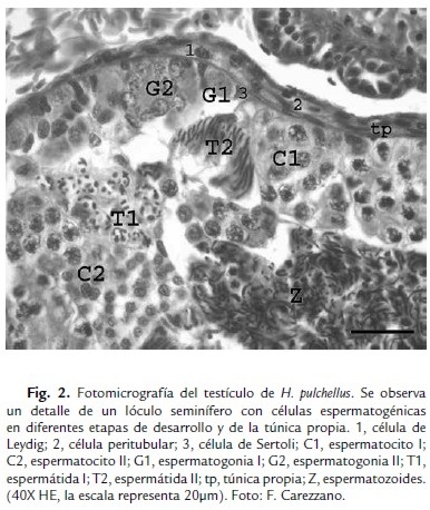
Los testículos de H. pulchellus son órganos pares, ovoides, de color amarillo claro de 4,64 ± 0,48 mm de largo por 2,05 ± 0,23 mm de ancho. Están revestidos por la túnica albugínea (5,60 ± 0,82 µm) formada por tejido conectivo denso, en el que predominan fibras de colágeno. En su interior se hallan numerosos lóculos seminíferos que miden 257,47 ± 58,25 µm de diámetro. Cada lóculo presenta varios cistos espermatogénicos. A su vez, cada cisto está formado por células germinales en una misma etapa de diferenciación, las que se asocian morfofuncionalmente a células de Sertoli (Fig. 1). Rodeando los lóculos se observa la túnica propia, formada por tejido conectivo laxo y por células peritubulares planas (Fig. 2). El tejido intersticial es escaso y en él se destacan las células de Leydig, poliédricas y con un núcleo central esférico (Fig. 2). Los cistos con espermatogonias están próximos a la pared del lóculo, pudiéndose diferenciar entre espermatogonias I y espermatogonias II. Las células más grandes corresponden a las espermatogonias I (14,34 ± 1,74 µm), se halla una por cisto y presentan escasa cromatina granular y su núcleo es multilobular; las espermatogonias II, son más pequeñas (10,14 ± 1,33 µm), y con la cromatina menos dispersa (Figs. 1 y 2). Los espermatocitos I son más chicos que sus precedentes (9,34 ± 0,32 µm) y en ellos se observan variadas formaciones y aspectos de la cromatina. Los espermatocitos II son aún más pequeños (8,12 ± 1,07 µm) y se observan más teñidos (Figs. 1 y 2). Las espermátidas son células multiformes, cuya forma varía de esférica a alargada. Se identificaron espermátidas I y II. Las primeras son células esféricas y están agrupadas en cistos redondeados, su diámetro fue de 4,09 ± 0,51 µm; mientras que las espermátidas II, son células alargadas que no se hallan dentro de cistos, pero que se organizan en paquetes asociadas a células de Sertoli (Figs. 1 y 2). En la luz de los lóculos se observan espermatozoides flagelados, células alargadas, que presentan un núcleo fuertemente basófilo debido a la intensa compactación nuclear que refleja la abundante presencia de heterocromatina (Figs. 1 y 2).

El análisis de los testículos indica que los cuatro ejemplares estaban en la misma etapa gametogénica y presentan una arquitectura similar a la descrita en otras especies de anuros neotropicales como Allobates femoralis (Asenjo et al., 2011), C. caudiverbera (Hermosilla et al, 1983), Ceratophrys ornata (Carezzano et al., 2013b), Dendropsophus minutus (Santos y Oliveira, 2007), D. nanus (Ferreira et al., 2009), Leptodactylus chaquensis (Ferreira et al., 2009), L. podicipinus (Ferreira et al., 2009), Odontophrynus americanus (Carezzano et al., 2013a), Physalaemus cuvieri (Oliveira et al., 2002), P. nattereri (Oliveira y Zieri, 2005), P. biligonigerus (Carezzano y Cabrera, 2010), Pseudis limellum (Ferreira et al., 2008, 2009), P. paradoxa (Ferreira et al., 2009) y Scinax acuminatus (Ferreira et al., 2009).
AGRADECIMIENTOS
A la Secretaría de Ciencia y Técnica de la Universidad Nacional de Córdoba por financiar el trabajo. A la Secretaría de Ambiente y Energía del gobierno de la provincia de Córdoba, República Argentina, por los permisos correspondientes para realizar los muestreos.
REFERENCIAS
Asenjo A, Ting KS, Pino, J. Morfometría de testículos y células germinales de Allobates femoralis (Boulenger 1883) (Dendrobatidae: Anura: Amphibia). The Biologist. 2011;9(2):167-175.
Blaustein A, Wake D, Sousa W. Amphibian declines: judging stability, persistence, and susceptibility of populations to local and global extinctions. Conser Biol. 1994;8(1)60-71. Doi: https://doi.org/10.1046/j.1523-1739.1994.08010060.x.
Carezzano FJ, Cabrera MR. Morfohistología del testículo de Physalaemus biligonigerus (Amphibia, Leiuperidae) en un ecosistema agrícola. Bol Asoc Herpet Esp. 2010;21:39-42.
Carezzano F, Urquiza S, Dorflinger K, Alonso M. Morfohistología testicular de Odontophrynus americanus: (Dumeril y Bibrón, 1841) (Anura, Odontophrynidae) de Argentina. The Biologist 2013a;11(1):119-129.
Carezzano F, Urquiza S, Dorflinger K, Alonso M. Morfohistología testicular de Ceratophrys ornata (Bell 1843) (Ceratophryidae: Anura: Amphibia). Iheringia, Sér Zool. 2013b;103(2):190-194. Doi: https://doi.org/10.1590/S0073-47212013000200014.
Close B, Banister K, Baumans V, Bernoth EM, Bromage N, Bunyan J, Erhardt W, Flecknell P, Gregory N, Hackbarth H, Morton D y Warwick C. Recomendaciones para la Eutanasia de los Animales de Experimentación: Parte 2; 1995. p 5-8.
Duellman WE, Trueb L. Biology of Amphibians. McGraw Hill, New York; 1994. p. 13-50.
Ferreira A, dos Santos Rosa A, Mehanna M. Organização celular dos testículos em Hylidae e Leptodactylidae, no Pantanal (Estado do Mato Grosso do Sul, Brasil). Acta Sci Biol Sci. 2009;31(4):447-452. Doi: https://doi.org/10.4025/actascibiolsci.v31i4.3332.
Ferreira A, Mehanna M, Prados CPA. Morphologic and morphometric analysis of testis of Pseudis limellum (Cope, 1862) (Anura, Hylidae) during the reproductive cycle in the Pantanal, Brazil. Biocell. 2008;32:185-194.
Hermosilla IB, Urbina AP, Cabrera JP. Spermatogenesis in the chilean frog Caudiverbera caudiverbera (Linne, 1758) (Anura, Leptodactylidae). Bol Soc Biol Concepción. 1983;54:103-115.
Jamieson BGM. Reproductive Biology and Phylogeny of Anura. Science Publishers, Inc. Enfield (NH), USA Plymouth, UK. 2003. p.119-142.
Oliveira C, Zieri R. Pigmentação testicular em Physalaemus nattereri (Steindachner) (Amphibia, Anura) com observações anatômicas sobre o sistema pigmentar extracutáneo. Rev Brás de Zool. 2005;22:454-460. Doi: https://doi.org/10.1590/S0101-81752005000200023.
Oliveira C, Zanetoni C, Zieri R. Morphological observations on the testes of Physalaemus cuvieri (Amphibia, Anura). Rev Chil Anat. 2002;20:263-268. Doi: https://doi.org/10.4067/S0716-98682002000300005.
Prophet EB, Mills B, Arrington JB y Sobin LH. Métodos histotecnológicos. Instituto de Patología de las Fuerzas Armadas de los Estados Unidos de América, Washington DC, 1995. p 31-33.
Santos LR y Oliveira C. Morfometria testicular durante o ciclo reprodutivo de Dendropsophus minutus (Peters) (Anura, Hylidae). Rev Bras Zool. 2007;24: 64-70. Doi: https://doi.org/10.1590/S0101-81752007000100008.
Referencias
Asenjo A, Ting KS, Pino, J. Morfometría de testículos y células germinales de Allobates femoralis (Boulenger 1883) (Dendrobatidae: Anura: Amphibia). The Biologist 2011;9(2):167-175. Doi: http://revistas.concytec.gob.pe/scielo.php?script=sci_arttext&pid=S1994-90732011000200003&lng=es&nrm=iso
Blaustein A, Wake D, Sousa W. Amphibian declines: judging stability, persistence, and susceptibility of populations to local and global extinctions. Conser Biol. 1994;8(1)60-71. Doi: http://dx.doi.org/10.1046/j.1523-1739.1994.08010060.x
Carezzano FJ, Cabrera MR. Morfohistología del testículo de Physalaemus biligonigerus (Amphibia, Leiuperidae) en un ecosistema agrícola. Bol Asoc Herpet Esp. 2010;21:39-42.
Carezzano F, Urquiza S, Dorflinger K, Alonso M. Morfohistología testicular de Odon-tophrynus americanus: (Dumeril y Bibrón, 1841) (Anura, Odontophrynidae) de Argentina. The Biologist 2013a;11(1):119-129. Doi: http://sisbib.unmsm.edu.pe/bvrevistas/biologist/v11_n1/pdf/a10v11n1.pdf
Carezzano F, Urquiza S, Dorflinger K, Alonso M. Morfohistología testicular de Cera-tophrys ornata (Bell 1843) (Ceratophryidae: Anura: Amphibia). Iheringia, Sér Zool. 2013b;103(2):190-194. Doi: http://dx.doi.org/10.1590/S0073-47212013000200014
Close B, Banister K, Baumans V, Bernoth EM, Bromage N, Bunyan J, Erhardt W, Flecknell P, Gregory N, Hackbarth H, Morton D y Warwick C. Recomendaciones para la Eutanasia de los Animales de Experimentación: Parte 2; 1995. p 5-8. Doi: http://sea.umh.es/files/2011/07/eutanasia2.pdf
Duellman WE, Trueb L. Biology of Amphibians. McGraw Hill, New York; 1994. p. 13-50.
Ferreira A, dos Santos Rosa A, Mehanna M. Organização celular dos testículos em Hylidae e Leptodactylidae, no Pantanal (Estado do Mato Grosso do Sul, Brasil). Acta sci., Biol. sci. 2009;31(4):447-452.
Ferreira A, Mehanna M, Prados CPA. Morphologic and morphometric analysis of testis of Pseudis limellum (Cope, 1862) (Anura, Hylidae) during the reproductive cy-cle in the Pantanal, Brazil. Biocell. 2008;32:185-194. Doi: http://www.mendoza-conicet.gov.ar/biocell/vol/pdf/32_2/04.pdf
Hermosilla IB, Urbina AP, Cabrera JP. Spermatogenesis in the chilean frog Caudiver-bera caudiverbera (Linne, 1758) (Anura, Leptodactylidae). Bol Soc Biol Con-cepción. 1983;54:103-115.
Jamieson BGM. Reproductive Biology and Phylogeny of Anura. Science Publishers, Inc. Enfield (NH), USA Plymouth, UK. 2003. p.119-142.
Oliveira C, Zieri R. Pigmentação testicular em Physalaemus nattereri (Steindachner) (Amphibia, Anura) com observações anatômicas sobre o sistema pigmentar ex-tracutáneo. Rev Brás de Zool. 2005;22:454-460. Doi: http://dx.doi.org/10.1590/S0101-81752005000200023
Oliveira C, Zanetoni C, Zieri R. Morphological observations on the testes of Physalae-mus cuvieri (Amphibia, Anura). Rev Chil Anat. 2002;20:263-268. Doi: http://dx.doi.org/10.4067/S0716-98682002000300005
Prophet EB, Mills B, Arrington JB y Sobin LH. Métodos histotecnológicos. Instituto de Patología de las Fuerzas Armadas de los Estados Unidos de América, Washing-ton DC, 1995. p 31-33.
Santos LR y Oliveira C. Morfometria testicular durante o ciclo reprodutivo de Den-dropsophus minutus (Peters) (Anura, Hylidae). Rev Bras Zool. 2007;24: 64-70. Doi: http://dx.doi.org/10.1590/S0101-81752007000100008
Cómo citar
APA
ACM
ACS
ABNT
Chicago
Harvard
IEEE
MLA
Turabian
Vancouver
Descargar cita
CrossRef Cited-by
1. Lucrecia Iruzubieta Villagra, Inés Ramos, Susana Cisint, Claudia A. Crespo, Silvia N. Fernández. (2018). Electron microscopy observations on testis and spermatozoa of Leptodactylus chaquensis (Anura, Leptodactylidae). Micron, 105, p.35. https://doi.org/10.1016/j.micron.2017.11.007.
2. MARIANA BARAQUET, FAVIO E. POLLO, MANUEL A. OTERO, PABLO R. GRENAT, NANCY E. SALAS, ADOLFO L. MARTINO. (2021). Body size, age and growth in males populations of Boana pulchella (Anura, Hylidae). Anais da Academia Brasileira de Ciências, 93(suppl 4) https://doi.org/10.1590/0001-3765202120200991.
Dimensions
PlumX
Visitas a la página del resumen del artículo
Descargas
Licencia
Derechos de autor 2015 Acta Biológica Colombiana

Esta obra está bajo una licencia internacional Creative Commons Atribución 4.0.
1. La aceptación de manuscritos por parte de la revista implicará, además de su edición electrónica de acceso abierto bajo licencia Attribution-NonCommercial-ShareAlike 4.0 (CC BY NC SA), la inclusión y difusión del texto completo a través del repositorio institucional de la Universidad Nacional de Colombia y en todas aquellas bases de datos especializadas que el editor considere adecuadas para su indización con miras a incrementar la visibilidad de la revista.
2. Acta Biológica Colombiana permite a los autores archivar, descargar y compartir, la versión final publicada, así como las versiones pre-print y post-print incluyendo un encabezado con la referencia bibliográfica del articulo publicado.
3. Los autores/as podrán adoptar otros acuerdos de licencia no exclusiva de distribución de la versión de la obra publicada (p. ej.: depositarla en un archivo telemático institucional o publicarla en un volumen monográfico) siempre que se indique la publicación inicial en esta revista.
4. Se permite y recomienda a los autores/as difundir su obra a través de Internet (p. ej.: en archivos institucionales, en su página web o en redes sociales cientificas como Academia, Researchgate; Mendelay) lo cual puede producir intercambios interesantes y aumentar las citas de la obra publicada. (Véase El efecto del acceso abierto).


