


Publicado
Atrapamiento del tendón tibial posterior en luxofractura irreductible abierta de tobillo: reporte de un caso.
Posterior tibial tendon entrapment in open irreducible ankle fracture-dislocation: a case report.
DOI:
https://doi.org/10.15446/cr.v9n1.94056Palabras clave:
Fracturas Óseas, Tibia, Peroné (es)Fractures, Bone, Tibia, Fibula (en)
Descargas
Resumen
Introducción. La presencia de luxofractura de tobillo es una patología común en urgencias; sin embargo, en casos raros de luxación de tobillo, la interposición de las estructuras musculares y tendinosas adyacentes puede dificultar la adecuada reducción de la articulación. Es poco común que el tendón tibial posterior (TP) se interponga en este tipo de fracturas, tal como sucedió en el siguiente reporte de caso, en el cual se describe a una paciente con una luxación anterior del TP.
Descripción del caso. Mujer de 32 años que sufrió una luxo fractura del tobillo izquierdo en un accidente de motocicleta. La luxación del tobillo se reparó el día del accidente; sin embargo, al ingreso a un segundo tiempo quirúrgico, 48 horas después del accidente, cuando se procedió a retirar el material de fijación externo se identificó el TP en el sitio de la fractura. Se reprodujo el mecanismo de la fractura, que a su vez demostró una luxación anterior del TP. Durante el procedimiento se logró la reducción anatómica de la luxación de la fractura y del TP y se pudo realizar la fijación interna y el cierre de la herida.
Conclusiones. No lograr la reducción anatómica debido a la luxación del TP sobre el peroné y el espacio tibial es extremadamente raro y puede provocar dolor crónico y pérdida de funcionalidad del tobillo, por lo que se recomienda determinar la ubicación de todas las estructuras adyacentes (tendones, ligamentos, músculos) y su comportamiento respecto a la luxo fractura y, en caso de ser necesario, se debe realizar la reparación anatómica temprana del TP incarcerado, ya que esto impacta directamente sobre la recuperación de la funcionalidad del órgano afectado y, por lo tanto, en el bienestar del paciente.
Abstract
Introduction: Ankle fracture-dislocation is a common condition found in emergency departments; however, in rare cases of ankle dislocation, the interposition of adjacent muscle and tendon structures may hinder proper reduction of the joint. It is uncommon to see the posterior tibial tendon (PTT) interpose in this type of fracture, as occurred in the following case report, which describes a patient with an anterior dislocation of the PTT.
Case presentation: A 32-year-old woman sustained a fracture-dislocation of the left ankle in a motorcycle accident. Ankle dislocation was repaired on the day of the accident, but on admission for a second surgery 48 hours after the accident, during which the external fixation material was removed, PTT was identified at the site of the fracture. The fracture mechanism was reproduced, demonstrating an anterior dislocation of the PTT. During the procedure, anatomical reduction of the fracture and PTT dislocation was achieved, and internal fixation and wound closure could be performed.
Conclusions: Failure to achieve anatomical reduction due to PTT dislocation on the fibula and tibial space is extremely rare and can lead to chronic pain and loss of ankle function. It is therefore recommended to establish the location of all adjacent structures (tendons, ligaments, muscles) and their behavior with respect to the fracture dislocation and, if necessary, to perform early anatomical repair of the incarcerated PTT, as this directly impacts the recovery of function in the affected organ and, consequently, the patient's well-being.
https://doi.org/10.15446/cr.v9n1.94056
Posterior tibial tendon entrapment in open irreducible ankle fracture-dislocation: a case report
Keywords: Fractures, Bone; Tibia; Fibula.
Palabras clave: Fracturas Óseas; Tibia; Peroné.
Julián David Molano-Castro
Clínica de Marly -
Orthopedics and Trauma Department
- Bogotá D.C. - Colombia.
Gabriel Eduardo Vargas-Montenegro
Universidad Nacional de Colombia -
Faculty of Medicine - Department of Surgery - Bogotá D.C. - Colombia.María
Camila Rosario Langer-Barrera
Clínica de Marly - Department of Surgery
- Bogotá D.C. - Colombia.
Corresponding author
Julián David Molano-Castro.
Departamento de Ortopedia y Traumatología,
Clínica de Marly. Bogotá D.C. Colombia.
Email: judamolca@gmail.com
Received: 02/03/2021 Accepted: 30/04/2021
Resumen
Introducción. La presencia de luxofractura de tobillo es una patología común en urgencias; sin embargo, en casos raros de luxación de tobillo, la interposición de las estructuras musculares y tendinosas adyacentes puede dificultar la adecuada reducción de la articulación. Es poco común que el tendón tibial posterior (TP) se interponga en este tipo de fracturas, tal como sucedió en el siguiente reporte de caso, en el cual se describe a una paciente con una luxación anterior del TP.
Descripción del caso. Mujer de 32 años que sufrió una luxo fractura del tobillo izquierdo en un accidente de motocicleta. La luxación del tobillo se reparó el día del accidente; sin embargo, al ingreso a un segundo tiempo quirúrgico, 48 horas después del accidente, cuando se procedió a retirar el material de fijación externo se identificó el TP en el sitio de la fractura. Se reprodujo el mecanismo de la fractura, que a su vez demostró una luxación anterior del TP. Durante el procedimiento se logró la reducción anatómica de la luxación de la fractura y del TP y se pudo realizar la fijación interna y el cierre de la herida.
Conclusiones. No lograr la reducción anatómica debido a la luxación del TP sobre el peroné y el espacio tibial es extremadamente raro y puede provocar dolor crónico y pérdida de funcionalidad del tobillo, por lo que se recomienda determinar la ubicación de todas las estructuras adyacentes (tendones, ligamentos, músculos) y su comportamiento respecto a la luxo fractura y, en caso de ser necesario, se debe realizar la reparación anatómica temprana del TP incarcerado, ya que esto impacta directamente sobre la recuperación de la funcionalidad del órgano afectado y, por lo tanto, en el bienestar del paciente.
Abstract
Introduction: Ankle fracture-dislocation is a common condition found in emergency departments; however, in rare cases of ankle dislocation, the interposition of adjacent muscle and tendon structures may hinder proper reduction of the joint. It is uncommon to see the posterior tibial tendon (PTT) interpose in this type of fracture, as occurred in the following case report, which describes a patient with an anterior dislocation of the PTT.
Case presentation: A 32-year-old woman sustained a fracture-dislocation of the left ankle in a motorcycle accident. Ankle dislocation was repaired on the day of the accident, but on admission for a second surgery 48 hours after the accident, during which the external fixation material was removed, PTT was identified at the site of the fracture. The fracture mechanism was reproduced, demonstrating an anterior dislocation of the PTT. During the procedure, anatomical reduction of the fracture and PTT dislocation was achieved, and internal fixation and wound closure could be performed.
Conclusions: Failure to achieve anatomical reduction due to PTT dislocation on the fibula and tibial space is extremely rare and can lead to chronic pain and loss of ankle function. It is therefore recommended to establish the location of all adjacent structures (tendons, ligaments, muscles) and their behavior with respect to the fracture dislocation and, if necessary, to perform early anatomical repair of the incarcerated PTT, as this directly impacts the recovery of function in the affected organ and, consequently, the patient’s well-being.
Introduction
The association between fracture and ankle dislocation is a common condition in hospital emergency rooms, posing a challenge to both general practitioners and orthopedic surgeons (1). This condition requires the reduction of articular surfaces in order to minimize damage to the adjacent soft tissues and favor early rehabilitation of the patient (1,2). However, there may be difficulties in achieving anatomical reduction during surgery (2), and failure to achieve such reduction is related to the occurrence of anterior or posterior dislocation of the PTT on the space between the fibula and the tibia (3,4). This association is extremely rare and is related to cases of chronic pain and loss of function at the talar level (5). A literature review shows that only 9 case reports have been published worldwide (6). The following is a case of PTT dislocation, its therapeutic approach, and the outcomes achieved.
Case presentation
A 32-year-old woman from Bogotá, Colombia, was involved in a traffic accident as a pedestrian and sustained a crush injury associated with rotation of the left ankle and foot, after which she was transferred to the emergency department of a tertiary care hospital in the city of Bogotá D.C., Colombia. Physical examination revealed a 3x4cm wound on the medial malleolus, classified as Gustilo type II, significant foot valgus deformity, edema, and ecchymosis of the limb. The orthopedic and trauma service requested x-ray studies of the ankle, which showed the presence of a fracture and subluxation of the tibiotalar joint, without neurovascular damage. Antibiotic therapy was started with cefazolin 1g IV every 8 hours for 7 days and gentamicin 240mg/day IV for 7 days, and the wound was washed, debrided and dressed in the emergency department. She was immediately taken to a first surgery, where she underwent lavage and debridement of the fracture site, as well as external fixation with an external fixator, achieving a partial reduction of the fracture site at the tibiotalar level.

Figure 1. Left ankle fracture-dislocation.
Source. Image obtained while conducting the study.
A second surgery was performed 48 hours after her admission, during which the external fixation material was removed, and an attempt was made to perform internal fixation of the fracture. However, an anterior tendon distally positioned over the trajectory of the posterior tibial muscle tendon was observed (Figure 1), which did not allow for a proper reduction of the syndesmosis. It was decided to remove the osteosynthesis material and the fracture mechanism was reproduced, demonstrating an anterior dislocation of the PTT. Again, reduction of the fibula and tibial malleolus was performed, achieving reduction of the syndesmosis to be subsequently fixed with the Tight Rope® system (Figure 2).

Figure 2. Ankle fracture dislocation with PTT entrapment.
Source. Image obtained while conducting the study.
Finally, the posterior tibial retinaculum was reinserted and repaired with a 2.8mm suture anchor (Figure 3).

Figure 3. Posterior tibialis tendon back to its anatomical course.
Source. Image obtained while conducting the study.
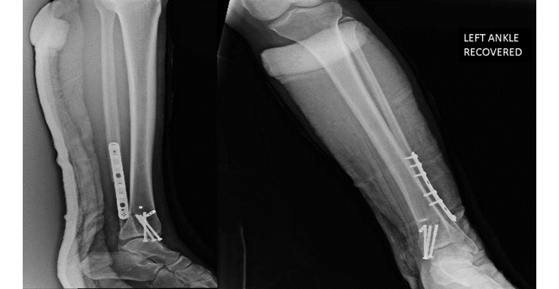
Figure 4. Internal fixation of fibula and tibia, and tibiofibular fixation (Tight Rope®).
Source. Image obtained while conducting the study.
On the other hand, since it was an open fracture, during the procedure a local flap was advanced, and the patient was immobilized for three weeks with a posterior splint until the first follow-up, when the stitches were removed. Six weeks of immobilization with an ankle brace were completed and outpatient check-ups were performed every two weeks with x-rays to monitor the consolidation process. Afterwards, comprehensive rehabilitation was undertaken with physical therapy, resulting in the recovery of gait with no limitations and without pain or reduction in the arches of mobility with respect to the contralateral limb at 12 months.
Discussion
Ankle fracture-dislocation is one of the most common conditions encountered by orthopedic surgeons in the emergency department. It can be reduced by means of closed or open techniques, both of which are effective in minimizing the occurrence of possible associated complications (7,8). However, the reduction of an ankle fracture-dislocation can be limited by the interposition of adjacent myotendinous structures, especially peroneus longus tendons (9), the flexor digitorum longus tendons and, to a lesser extent, the dislocation of the PTT towards the fracture site (3,6).
The tibialis posterior muscle originates on the posterior aspect of the tibia along the oblique line, immediately inferior to the soleus muscle, on the interosseous membrane, and on the medial aspect of the fibula; its distal tendon passes through a fibrous tunnel posterior to the medial malleolus under the cover of the flexor retinaculum. Its main functions are medial stabilization of the tibiotalar joint and reduction of inversion and pronation forces during gait (7,10).
Reports of PTT dislocation towards the fracture site are scarce, with the first report in the literature being made by Böhler in 1936 (11). Subsequent cases were reported by Parrish in 1959 (12), Walker and Farris in 1981 (13), Anderson and Hansen in 1996 (14), Ermis et al. in 2010 (3), Lacasse et al. in 2015 (15), Connors et al. in 2016 (2), Sato et al. in 2019 (6), and Tschudi et al. in 2020 (16). Consequently, the present report is one of the first 10 cases published in the scientific literature, thereby contributing to the study and treatment of this kind of condition. However, interventions are not standardized due to their low prevalence, and it remains mostly up to the treating surgeon according to their expertise.
Associations between the fracture mechanism (pronation-abduction), the concomitant valgus foot deformity and ligament and retinaculum injuries have been observed in the cases found, which would lead to favoring PTT dislocation in the joint space (7). There are cases reported in the literature in which PTT entrapment was evidenced at the level of the fracture site, preventing adequate joint reduction. High-energy trauma involving external rotation and pronation mechanisms has also been reported (7).
It is noteworthy that such dislocation usually occurs at different levels, starting at the fracture site (from the posterior malleolus or internal malleolus) until the space between the syndesmosis of the tibia and the fibula. The level of tendon entrapment is directly related to the level of energy involved in the trauma mechanism (1,16).
Delayed diagnosis of PTT entrapment at the level of the syndesmosis or fracture site could lead to early onset of osteoarthritis, limitation in patient mobility, and a possible cause of regional pain syndrome. Likewise, it has been established in the literature that this condition increases the number of surgical interventions and, therefore, the probability of infections, the probability of poor outcomes, and the inability to obtain anatomical reductions, which is considered to be the leading cause of disability due to ankle injury (15,17).
In the present case, during the second surgery, the impossibility of achieving an adequate reduction at the articular level led to a thorough exploration of the fracture site, which allowed recognizing the dislocation of the PTT, making it necessary to reconstruct the flexor retinaculum and to reposition the tendon in order to perform the definitive osteosynthesis. This allowed obtaining satisfactory outcomes in the follow-up of the patient one year after the intervention, during which time an improvement in pain was reported, as well as the recovery of the arches of motion, and an adequate consolidation without signs of osteoarthritis in radiographic studies.
Some case reports have shown that expeditious surgical management decreases the likelihood of early osteoarthritis and complex regional pain syndrome (15). Similarly, in cases in which the tendon is trapped at the articular level, a rupture of the tendon may occur and, as mentioned by Anderson et al. (14), the delay in diagnosis and incorrect anatomical reduction may lead to irreversible damage to the affected limb.
Conclusions
In cases of ankle fracture associated with irreducible dislocation of the talus, it is necessary to perform a thorough review of the vascular-nerve and posteromedial muscle-tendon elements in order to detect possible dislocations in the tibioperoneal space. The interposition of elements at this level and, therefore, the inadequate reduction of the fracture site, considerably increases the need for a new surgical intervention and the risk of complications such as infections, painful syndromes, and the possible functional loss of the limb.
Ethical considerations
Informed consent was obtained from the patient for the publication of this case report.
Conflict of interest
None stated by the authors.
Funding
None stated by the authors.
Acknowledgments
To the Orthopedics and Trauma Department of Clínica de Marly for their support in difficult cases.
References
1.Thoreau L, Kaminski L, Putineanu DC. Irreducible ankle fracture dislocation due to posterior tibialis tendon interposition: Diagnostic and clues for early management - A case report. Trauma Case Rep. 2019;20:100175. https://doi.org/gqh66r.
2.Connors JC, Coyer MA, Hardy MA. Irreducible Ankle Fracture Dislocation Due to Tibialis Posterior Tendon Interposition: A Case Report. J Foot Ankle Surg. 2016;55(6):1276-1281. https://doi.org/f9bpv7.
3.Ermis MN, Yagmurlu MF, Kilinc AS, Karakas ES. Irreducible Fracture Dislocation of the Ankle Caused by Tibialis Posterior Tendon Interposition. J Foot Ankle Surg. 2010;49(2):166-171. https://doi.org/brrckn.
4.Vosoughi AR, Glazebrook M. Interposition of Tibialis Posterior and Flexor Digitorum Longus Tendons Resulted in Irreducible Ankle Fracture-Dislocation. J Foot Ankle Surg. 2017;56(3):697-701. https://doi.org/f98nwn.
5.Al Khudairy A, Zafar MM, Padinjarathala BA. The Unexpected With Ankle Fracture: Traumatic Tibialis Posterior Tendon Dislocation: A Case report and Literature Review. Foot Ankle Spec. 2013;6(6),482-489. https://doi.org/kb6n.
6.Sato R, Tsuchida Y, Murakami H, Shirakawa T, Futamura K, Hasegawa M, et al. Fracture dislocation of the ankle with posterior tibial tendon entrapment within the tibiofibular interosseous space: A case report. Trauma Case Rep. 2019;23:100235. https://doi.org/kb6p.
7.Stevens NM, Wasterlain AS, Konda SR. Case Report: Irreducible Ankle Fracture With Posterior Tibialis Tendon and Retinaculum, Deltoid Ligament, and Anteromedial Joint Capsule Entrapment. J Foot Ankle Surg. 2017;56(4):889–893. https://doi.org/gbnq6d.
8.Schatzker J, Johnson RG. Fracture-dislocation of the ankle with anterior dislocation of the fibula. J. Trauma. 1983;23(5):420–423. https://doi.org/d57zzv.
9.Ballard DH, Campbell KJ, Blanton L, Williams JT, Sangster G, Hollister AM, et al. Tendon entrapments and dislocations in ankle and hindfoot fractures: evaluation with multidetector computed tomography. Emerg Radiol. 2016;23(4):357-363. https://doi.org/kdts.
10.Oakley J, Yewlett A, Makwana N. Avulsion fracture of the medial malleolus following posterior tibialis tendon dislocation: A case report. Foot Ankle Surg. 2011;17(2):94-97. https://doi.org/fhcrgw.
11.Böhler L. The Treatment of Fractures. BJS. 1957;44(187):544. https://doi.org/cxvzst.
12.Parrish TF. Fracture-dislocation of the ankle; an unusual cause of failure of reduction; a case report. J Bone Joint Surg Am. 1959;41-A(4):749-751.
13.Walker RH, Farris C. Irreducible fracture-dislocations of the ankle associated with interposition of the tibialis posterior tendon: case report and review of the literature of a specific ankle fracture syndrome. Clin Orthop Relat Res. 1981;(160):212-216.
14.Anderson JG, Hansen ST. Fracture-Dislocation of the Ankle With Posterior Tibial Tendon Entrapment Within the Tibiofibular Interosseous Space: A Case Report of a Late Diagnosis. Foot Ankle Int. 1996;17(2):114–118. https://doi.org/kcbz.
15.Lacasse JS, Laflamme M, Penner MJ. Irreducible Fracture-Dislocation of the Ankle Associated With Interposition of the Tibialis Posterior Tendon in the Syndesmosis: A Case Report. J Foot Ankle Surg. 2015;54(5):962-966. https://doi.org/f7pt54.
16.Tschudi S, Wittauer M, Hirschmann A, Eckardt H. (2020). Avoiding pitfalls in ankle fracture-dislocations: A case report of a dislocated tibialis posterior tendon causing persistent ankle subluxation. Foot Ankle Surg. 2021;27(6)700-709. https://doi.org/kcbv.
17.Xing W, Wang Y, Sun L, Wang L, Kong Z, Zhang C, Zhang Z. Ankle joint dislocation treating dislocated trimalleolar fractures accompanied with the complex posterior malleolus fracture without separation of the tibiofibular syndesmosis. Medicine. 2018;97(37):e12079. https://doi.org/kcbw.
Referencias
References
Thoreau L, Kaminski L, Putineanu DC. Irreducible ankle fracture dislocation due to posterior tibialis tendon interposition: Diagnostic and clues for early management - A case report. Trauma Case Rep. 2019;20:100175. https://doi.org/gqh66r.
Connors JC, Coyer MA, Hardy MA. Irreducible Ankle Fracture Dislocation Due to Tibialis Posterior Tendon Interposition: A Case Report. J Foot Ankle Surg. 2016;55(6):1276-1281. https://doi.org/f9bpv7.
Ermis MN, Yagmurlu MF, Kilinc AS, Karakas ES. Irreducible Fracture Dislocation of the Ankle Caused by Tibialis Posterior Tendon Interposition. J Foot Ankle Surg. 2010;49(2):166-171. https://doi.org/brrckn.
Vosoughi AR, Glazebrook M. Interposition of Tibialis Posterior and Flexor Digitorum Longus Tendons Resulted in Irreducible Ankle Fracture-Dislocation. J Foot Ankle Surg. 2017;56(3):697-701. https://doi.org/f98nwn.
Al Khudairy A, Zafar MM, Padinjarathala BA. The Unexpected With Ankle Fracture: Traumatic Tibialis Posterior Tendon Dislocation: A Case report and Literature Review. Foot Ankle Spec. 2013;6(6),482-489. https://doi.org/kb6n.
Sato R, Tsuchida Y, Murakami H, Shirakawa T, Futamura K, Hasegawa M, et al. Fracture dislocation of the ankle with posterior tibial tendon entrapment within the tibiofibular interosseous space: A case report. Trauma Case Rep. 2019;23:100235. https://doi.org/kb6p.
Stevens NM, Wasterlain AS, Konda SR. Case Report: Irreducible Ankle Fracture With Posterior Tibialis Tendon and Retinaculum, Deltoid Ligament, and Anteromedial Joint Capsule Entrapment. J Foot Ankle Surg. 2017;56(4):889–893. https://doi.org/gbnq6d.
Schatzker J, Johnson RG. Fracture-dislocation of the ankle with anterior dislocation of the fibula. J. Trauma. 1983;23(5):420–423. https://doi.org/d57zzv.
Ballard DH, Campbell KJ, Blanton L, Williams JT, Sangster G, Hollister AM, et al. Tendon entrapments and dislocations in ankle and hindfoot fractures: evaluation with multidetector computed tomography. Emerg Radiol. 2016;23(4):357-363. https://doi.org/kdts.
Oakley J, Yewlett A, Makwana N. Avulsion fracture of the medial malleolus following posterior tibialis tendon dislocation: A case report. Foot Ankle Surg. 2011;17(2):94-97. https://doi.org/fhcrgw.
Böhler L. The Treatment of Fractures. BJS. 1957;44(187):544. https://doi.org/cxvzst.
Parrish TF. Fracture-dislocation of the ankle; an unusual cause of failure of reduction; a case report. J Bone Joint Surg Am. 1959;41-A(4):749-751. DOI: https://doi.org/10.2106/00004623-195941040-00018
Walker RH, Farris C. Irreducible fracture-dislocations of the ankle associated with interposition of the tibialis posterior tendon: case report and review of the literature of a specific ankle fracture syndrome. Clin Orthop Relat Res. 1981;(160):212-216. DOI: https://doi.org/10.1097/00003086-198110000-00031
Anderson JG, Hansen ST. Fracture-Dislocation of the Ankle With Posterior Tibial Tendon Entrapment Within the Tibiofibular Interosseous Space: A Case Report of a Late Diagnosis. Foot Ankle Int. 1996;17(2):114–118. https://doi.org/kcbz.
Lacasse JS, Laflamme M, Penner MJ. Irreducible Fracture-Dislocation of the Ankle Associated With Interposition of the Tibialis Posterior Tendon in the Syndesmosis: A Case Report. J Foot Ankle Surg. 2015;54(5):962-966. https://doi.org/f7pt54.
Tschudi S, Wittauer M, Hirschmann A, Eckardt H. (2020). Avoiding pitfalls in ankle fracture-dislocations: A case report of a dislocated tibialis posterior tendon causing persistent ankle subluxation. Foot Ankle Surg. 2021;27(6)700-709. https://doi.org/kcbv.
Xing W, Wang Y, Sun L, Wang L, Kong Z, Zhang C, Zhang Z. Ankle joint dislocation treating dislocated trimalleolar fractures accompanied with the complex posterior malleolus fracture without separation of the tibiofibular syndesmosis. Medicine. 2018;97(37):e12079. https://doi.org/kcbw.
Cómo citar
APA
ACM
ACS
ABNT
Chicago
Harvard
IEEE
MLA
Turabian
Vancouver
Descargar cita
Licencia
Derechos de autor 2023 Case reports

Esta obra está bajo una licencia internacional Creative Commons Atribución 4.0.
Los autores al someter sus manuscritos conservarán sus derechos de autor. La revista tiene el derecho del uso, reproducción, transmisión, distribución y publicación en cualquier forma o medio.
El Formulario de Divulgación Uniforme para posibles Conflictos de Interés y los oficios de cesión de derechos y de responsabilidad deben ser entregados junto con el original.
Aquellos autores/as que tengan publicaciones con esta revista, aceptan los términos siguientes:
Los autores/as conservarán sus derechos de autor y garantizarán a la revista el derecho de primera publicación de su obra, el cual estará simultáneamente sujeto a la Licencia de reconocimiento de Creative Commons 4.0 que permite a terceros compartir la obra siempre que se indique su autor y su primera publicación en esta revista.
Los autores/as podrán adoptar otros acuerdos de licencia no exclusiva de distribución de la versión de la obra publicada (p. ej.: depositarla en un archivo telemático institucional o publicarla en un volumen monográfico) siempre que se indique la publicación inicial en esta revista.
Se permite y recomienda a los autores/as difundir su obra a través de Internet (p. ej.: en archivos telemáticos institucionales o en su página web) antes y durante el proceso de envío, lo cual puede producir intercambios interesantes y aumentar las citas de la obra publicada. (Véase El efecto del acceso abierto).


