


Publicado
DESARROLLO Y CRECIMIENTO FEMORAL DE CONEJAS DE LA LÍNEA NEW ZEALAND
Development and femoral growth of rabbits of the New Zealand line
DOI:
https://doi.org/10.15446/abc.v26n3.87221Palabras clave:
biología, biometría, bioquímica, célula, desarrollo fisiológico (es)biochemistry, biology, biometrics, cells, physiological development (en)
Descargas
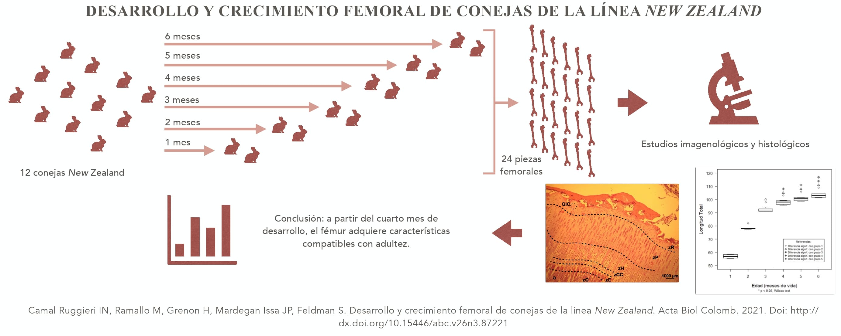
En este proyecto se investigan los cambios que acontecen en el desarrollo y crecimiento de conejos hembras de la línea New Zealand (CoNZ) en sus huesos femorales. Los animales fueron mantenidas en jaulas individuales desde las 2 semanas de edad, con comida y agua ad libitum y se sacrificaron en tiempos mensualmente consecutivos: 1, 2, 3, 4, 5, y 6 meses. Tras la obtención de las piezas femorales, y a partir de estudios imagenológicos se determinaron los ángulos del cuello femoral (Af), la longitud total (L), la densidad mineral ósea total, del centro óseo y de la metáfisis femoral (DMOt, DMOco y DMOmf respectivamente), analizándose las variaciones intergrupales por el test Wilcoxon, y corrección de Bonferroni. Se realizaron estudios histológicos de los cortes descalcificados de las piezas femorales. Los análisis sobre los Af mostraron un incremento significativo durante el primer mes mientras que L se estabilizó a partir del 4to mes. Los valores de DMOt mostraron un plateau a partir del cuarto mes, si bien las DMOco y DMOmf ya a partir del tercer mes no mostraron incrementos significativos. Histológicamente se observó para el cuarto mes ausencia de las diferentes zonas características del cartílago de crecimiento metafisiario, con presencia únicamente de un pequeño remanente de células condrales. Desde el quinto mes se observa ausencia total de cartílago, con presencia únicamente de tejido osteoide (TO). La interpretación integrada de los resultados nos permite afirmar, que a partir del cuarto mes de desarrollo, el fémur de CoNZ adquiere características compatibles con un periodo de adultez.
In this project we investigated the changes of femoral development and growth of female New Zealand rabbits (NZr). Animals were maintained in individual cages since they were two weeks old with food and water ad libitum, and were sacrificed monthly consecutively: 1, 2, 3, 4, 5 and 6. Radiological studies were made with femoral pieces to determine femoral neck angle (fnA), total length (L), total bone mineral density (tBMD), bone center mineral density (bcBMD) and femoral metaphysis bone mineral density (fmBMD). We analyzed intergroup variations with Wilcoxon test and Bonferroni correction. We also performed histologic studies with femoral pieces. The fnA analyzes showed a significant increase in the first month while L stabilized since the fourth month. tBMD showed a plateu since the fourth month, even though bcBMD and fmBMD did not show any significant changes since the third month. In histology it was observed the absence of all typical growth cartilage zones since the fourth month, with the only presence of small remaining cartilage cells. In the fifth month we observed complete absence of cartilage, and presence of osteoid tissue only. The integrated interpretation of the results allows us to affirm that since the fourth month of development the femur of NZr acquires characteristics compatible with the adulthood.
Referencias
Aragón Hernández J, Suárez Sánchez J, Pérez-Martínez M. Morphometric characteristics of female reproductive organs of New Zealand rabbits with different body weight in peripuberal period of transition. Vet Méx. 2010;41(3).
Beebe K. Alcohol/Xylene: The Unlikely Fixative/Dehydrant/Clearant. J Histotechnol. 2000;23:45–50. Doi: https://doi.org/10.1179/his.2000.23.1.45
Berendsen AD, Olsen BR. Bone development. Bone. 2015;80:14-18. Doi: https://doi.org/10.1016/j.bone.2015.04.035
Burdan F, Szumiło J, Korobowicz A, Farooquee R, Patel S, Patel A, et al. Morphology and physiology of the epiphyseal growth plate. Folia Histochem Cytobiol. 2009;47:5–16. Doi: https://doi.org/10.2478/v10042-009-0007-1
Cícero AM, Issa JPM, Feldman S. Matrices de tercera generación en la ingeniería de tejidos óseos. Actual Osteol. 2017;13(2):157-176.
Cointry, G., Capozza, R., Feldman, S. & Reina, P. (2009). ¿Los huesos son estructuras genéticas, metabólicas, biomecánicas, o todo a la vez? Actualizaciones en Osteología, 5(3), 185-195. http://www.osteologia.org.ar/?s=ver_articulo&id=132
Coletta D, Ibañez-Fonseca A, Missana L, Jammal MV, Vitelli EJ, Aimone M, et al. Special Focus: Strategic Directions In Osteoinductionand Biomimetics: Bone regeneration mediated by a bioactive and biodegradable ECM like hidrogel based on elastin-like recobinamers. Tissue Eng. 2017:23:161-171. Doi: https://doi.org/10.1089/ten.tea.2017.0047
Coletta DJ, Lozano D, Rocha-Oliveira AA, Mortarino P, Bumaguin GE, Vitelli E, et al. Characterization of hybrid bioactive glass-polyvinyl alcohol scaffolds containing a PTHrP-derived pentapeptide as implants for tissue engineering applications. Open Biomed Eng J. 2014;(8):20-27. Doi: https://doi.org/10.2174/1874120701408010020
Di Fiore M. Atlas de Histología Normal. 8 ed. Buenos Aires: El Ateneo; 2016. p 33-37.
Ferretti JL, Capozza RF, Cointry GR, Feldman S, Ferretti SE. What is conception of bone wuality and how evaluate. Actual Osteol. 2008;15(12):11-25. DOI: https://doi.org/10.1016/j.bone.2006.12.033
García Hernández PA. Avances en osteoporosis. 2 ed. México: Asociación Mexicana de Metabolismo Óseo y Mineral; 2007. p. 311.
González Garamendi PM, Landa Tabuyo MI. Determinación de la edad mediante radiología. Rev Esp Med Leg. 2009;36:3-13. Doi: https://doi.org/10.1016/S0377-4732(10)70030-4
Goy DP, Gorosito E, Costa HS, Mortarino P, Acosta Pedemonte N, Toledo J, et al. Hybrid Matrix Grafts to Favor Tissue Regeneration in Rabbit Femur Bone Lesions. Open Biomed Eng J. 2012;6:85-91. Doi: https://doi.org/10.174/1874120701206010085
Greenspan A. Radiología de huesos y articulaciones. Salamanca: Marban; 2007. 978 p.
Guyton AC, Hall JE. Tratado de Fisiología Médica. 13 ed. Barcelona: ElSevier; 2016. p. 955-972.
Haraguchi R, Kitazawa R, Kohara Y, Ikedo A, Imai Y, Kitazawa S. Recent Insights into Long Bone Development: Central Role of Hedgehog Signaling Pathway in Regulating Growth Plate. Int J Mol Sci. 2019;20(23):5840. Doi: https://doi.org/10.3390/ijms20235840
Heikel HVA. On ossification and growth of certain bones of the rabbit; with a comparison of the skeletal age in the rabbit and in man. Acta Orthop Scand. 1999;39(1-4):172-184. Doi: https://doi.org/10.3109/17453675908988796
Kierszenbaum AL, Tres LL. Osteogenesis. Histology and cell biology: an introduction to pathology. 3 ed. Philadelphia: Saunders; 2012. p. 151–168. DOI: https://doi.org/10.1016/B978-0-323-07842-9.50009-5
Kronenberg HM. Developmental regulation of the growth plate. Nature. 2003;423:332-336. Doi: https://doi.org/10.1038/nature01657
Lerner AL, Kuhn JL. Characterization of Regional and Age-Related Variations in the Growth of the Rabbit Distal Femur. J Orthop Res. 1997;15(3):353-361. Doi: https://doi.org/10.1002/jor.1100150307
Mackie EJ, Ahmed YA, Tatarczuch L, Chen KS, Mirams M. Endochondral ossification: how cartilage is converted into bone in the developing skeleton. Int J Biochem Cell Biol. 2008;40(1):46–62. Doi: https://doi.org/10.1016/j.biocel.2007.06.009
Mackie EJ, Tatarczuch L, Mirams M. The skeleton: a multi-functional complex organ: the growth plate chondrocyte and endochondral ossification. J Endocrinol. 2011;211(2):109-21. Doi: https://doi.org/10.1530/joe-11-0048
Manjeet M, Betsy ST, Bhat KM. Rabbit as an animal model for experimental research. Dent Res J (Isfahan). 2012;9(1):111-118. Doi: https://doi.org/10.4103/1735-3327.92960
Martiniaková M, Vondráková M, Fabiš M. Investigation of the Microscopic Structure of Rabbit Compact Bone Tissue. Scr Med (Brno). 2003;76(4):215-220.
Martiniaková M, Omelka R, Chrenek P, Vondráková M, Bauerová M. Age-related changes in histologicalstructure of thefemurin juvenile and adult rabbits: a pilotstudy. Bull Vet Inst Pulawy. 2005;49:227-230.
Masoud I. A longitudinal Study of the Growth of the New Zealand White rabbit: cumulative and biweekly incremental growth rates for Body length, body weight, femoral length, and tibial length. J Orthop Rese. 1986;4:221-231. DOI: https://doi.org/10.1002/jor.1100040211
Mizuhashi K, Nagata M, Matsushita Y, Ono W, Ono N. Growth plate borderline chondrocyte behave as transient mesenchymal precursor cells. J Bone Miner Res. 2019;34(8):1387-1392. Doi: https://doi.org/10.1002/jbmr.3719
Mizuhashi K, Ono W, Matsushita Y, Sakagami N, Takahashi A, Saunders TL, et al. Resting zone of the growth plate harbors a unique class of skeletal stem cells. Nature. 2018;563(7730):254-258. Doi: https://doi.org/10.1038/s41586-018-0662-5
Panattoni GL, D'Amelio P, Di Stefano M, Sciolla A, Isaia GC. Densitometric study of developing femur. Calcif Tissue Int. 1999;64:133-6. Doi: https://doi.org/10.1007/s002239900591
Parfitt AM, Drezner MK, Glorieux FH, Kanis JA, Malluche H, Meunier PJ, et al. Bone histomorphometry: Standardization of nomenclature, symbols, and units. JBMR. 1987;2(6):595-609. Doi: https://doi.org/10.1002/jbmr.5650020617
Percival CJ, Richtsmeier JT. Angiogenesis and intramembranous osteogenesis. Anat Rec. 2013;242(8):909-22. Doi: https://doi.org/10.1002/dvdy.23992
Roselló-Díez A, Joyner AL. Regulation of Long Bone Growth in Vertebrates; It Is Time to Catch Up. Endocr Rev. 2015;36(6):646-680. Doi: https://doi.org/10.1210/er.2015-1048
Ross MH, Pawlina W. Histología: texto y atlas. 6 ed. Buenos Aires: Panamericana; 2012. p. 218-253.
Ryan S. Anatomía para el diagnóstico radiológico. Dublín: Marbán; 1997. 304 p.
Schneider CA, Rasband WS, Eliceiri KW. NIH Image to ImageJ: 25 years of image analysis. Nat Methods. 2012;9:671-675. Doi: https://doi-org/10.1038/nmeth.2089
Thomsen E, Drummond DS, Robertson Jr WW, Christofersen MR. Radiographic Assessment of Longitudinal Growth of the Rabbit Femoral Physes. J Orthop Res. 1991;9(2):186-190. Doi: https://doi.org/10.1002/jor.1100090206
Viguer JM, García del Moral R. Manual de laboratorio y Atlas de citología. Granada: McGraw-Hill; 1995. 300 p.
Villemure I, Stokes IAF. Growth plate mechanics and mechanobiology. A survey of present understanding. J Biomech. 2009;42(12):1793-1803. Doi: https://doi.org/10.1016/j.jbiomech.2009.05.021
Wongdee K, Krishnamra N, Charoenphandhu N. Endochondral bone frowth, bone calcium accretion, and bone mineral density: how are they related? J Physiol Sci. 2012;62:299–307. Doi: https://doi.org/10.1007/s12576-012-0212-0
Cómo citar
APA
ACM
ACS
ABNT
Chicago
Harvard
IEEE
MLA
Turabian
Vancouver
Descargar cita
Licencia
Derechos de autor 2021 Acta Biológica Colombiana

Esta obra está bajo una licencia internacional Creative Commons Atribución-NoComercial-CompartirIgual 4.0.
1. La aceptación de manuscritos por parte de la revista implicará, además de su edición electrónica de acceso abierto bajo licencia Attribution-NonCommercial-ShareAlike 4.0 (CC BY NC SA), la inclusión y difusión del texto completo a través del repositorio institucional de la Universidad Nacional de Colombia y en todas aquellas bases de datos especializadas que el editor considere adecuadas para su indización con miras a incrementar la visibilidad de la revista.
2. Acta Biológica Colombiana permite a los autores archivar, descargar y compartir, la versión final publicada, así como las versiones pre-print y post-print incluyendo un encabezado con la referencia bibliográfica del articulo publicado.
3. Los autores/as podrán adoptar otros acuerdos de licencia no exclusiva de distribución de la versión de la obra publicada (p. ej.: depositarla en un archivo telemático institucional o publicarla en un volumen monográfico) siempre que se indique la publicación inicial en esta revista.
4. Se permite y recomienda a los autores/as difundir su obra a través de Internet (p. ej.: en archivos institucionales, en su página web o en redes sociales cientificas como Academia, Researchgate; Mendelay) lo cual puede producir intercambios interesantes y aumentar las citas de la obra publicada. (Véase El efecto del acceso abierto).


