Published
Electrodermal activity - a review
La actividad electrodérmica -Revisión
DOI:
https://doi.org/10.15446/ing.investig.v31n2.23473Keywords:
bioelectricity, wound healing, skin, electric potential. (en)bioelectricidad, sanación de heridas, piel, potenciales eléctricos. (es)
Downloads
The electricity in living tissue was widely studied around the 19th century. Such study was suspended for many years but has then been started again during recent decades. New research into bioelectricity is creating alternatives in the health field; one of them is an electrodermal response associated with the wound healing, cell stimulation and psychopathology diagnostic. This article presents some of the first responses and models concerning electrodermal activity. Theoretical, clinical and review papers were studied and classified to show the amplitude and variety of bioelectrical responses. Electrodermal activity is only one of many applications having an abundant amount of evidence regarding diagnosis and treatment starting from bioelectrical signals. Electrical tissue response requires more experimental, theoretical and clinical research in many fields involving an organism' s behaviour to ascertain, propose and create new treatment alternatives for different pathologies.
La electricidad en los tejidos vivos fue ampliamente estudiada alrededor del siglo XIX. Los estudios fueron suspendidos durante muchos años y retomados en las últimas décadas. Las nuevas investigaciones en bioelectricidad están creando alternativas en el área de la salud, una de éstas es la respuesta electrodermal, asociada con reparación de heridas, estimulación celular, diagnóstico de psicopatologías, entre otras. Este artículo presenta algunos de los primeros hallazgos y modelos desarrollados alrededor de la actividad electrodermal (AED). Varios documentos teóricos, clínicos y de revisión fueron estudiados y clasificados para mostrar la amplitud y variedad de hallazgos sobre el tema presentado. La actividad electrodérmica es sólo una de las múltiples aplicaciones con abundante evidencia en diagnósticos y tratamientos a partir de señales bioeléctricas. Las respuestas eléctricas de los tejidos requieren más investigación experimental, teórica y clínica en muchas de las áreas que envuelven el comportamiento del organismo, para así conocer, proponer y crear nuevas alternativas de tratamiento a las diversas patologías.
La actividad electrodérmica -Revisión
Electrodermal activity - a review
María Elena Moncada1, Jorge de la Cruz2
1 Ingeniera electricista, Universidad de Antioquia. Doctora en Ingeniería, Universidad del Valle. Docente investigadora, Instituto Tecnológico Metropolitano, Colombia. mariamoncada@itm.edu.co
2 Estudiante de Ingeniería Electrónica, Universidad del Valle. jodelacruz@hotmail.com
RESUMEN
La electricidad en los tejidos vivos fue ampliamente estudiada alrededor del siglo XIX. Los estudios fueron suspendidos durante muchos años y retomados en las últimas décadas. Las nuevas investigaciones en bioelectricidad están creando alternativas en el área de la salud, una de éstas es la respuesta electrodermal, asociada con reparación de heridas, estimulación celular, diagnóstico de psicopatologías, entre otras. Este artículo presenta algunos de los primeros hallazgos y modelos desarrollados alrededor de la actividad electrodermal (AED). Varios documentos teóricos, clínicos y de revisión fueron estudiados y clasificados para mostrar la amplitud y variedad de hallazgos sobre el tema presentado. La actividad electrodérmica es sólo una de las múltiples aplicaciones con abundante evidencia en diagnósticos y tratamientos a partir de señales bioeléctricas. Las respuestas eléctricas de los tejidos requieren más investigación experimental, teórica y clínica en muchas de las áreas que envuelven el comportamiento del organismo, para así conocer, proponer y crear nuevas alternativas de tratamiento a las diversas patologías.
Palabras clave: bioelectricidad, sanación de heridas, piel, potenciales eléctricos.
ABSTRACT
the electricity in living tissue was widely studied around the 19th century. Such study was suspended for many years but has then been started again during recent decades. New research into bioelectricity is creating alternatives in the health field; one of them is an electrodermal response associated with the wound healing, cell stimulation and psychopathology diagnostic. This article presents some of the first responses and models concerning electrodermal activity. Theoretical, clinical and review papers were studied and classified to show the amplitude and variety of bioelectrical responses. Electrodermal activity is only one of many applications having an abundant amount of evidence regarding diagnosis and treatment starting from bioelectrical signals. Electrical tissue response requires more experimental, theoretical and clinical research in many fields involving an organism’s behaviour to ascertain, propose and create new treatment alternatives for different pathologies.
Keywords: bioelectricity, wound healing, skin, electric potential.
Recibido: febrero 25 de 2010
Aceptado: julio 6 de 2011
Introducción
La piel es uno de los órganos más importantes de nuestro organismo; si llega a faltar más del 40% de ella, suele ser incompatible con la vida. La piel posee una función protectora selectiva gracias a que impide la entrada de sustancias nocivas a un sistema inmunológico propio. Además tiene una función reguladora del metabolismo: impide la salida de sustancias, regula la temperatura corporal y transforma los rayos del sol en vitamina D.
El tratamiento mediante la aplicación de señales eléctricas ha sido estudiado desde el siglo XIX. Los investigadores han demostrado la presencia de sistemas bioeléctricos endógenos (Klot et al.,1996; Charman, 1990) y en la literatura se encuentran referencias de los efectos de estimulación eléctrica en úlceras generadas por presión, por insuficiencia vascular, por trauma, por diabetes, o por cirugías, entre otras (Sandoval et al., 2007; Poltawski et al., 2008a, b; 2009). La estimulación eléctrica en la piel ha sido utilizada como bactericida (Nelson et al., 1999; Kincaid et al., 1989) para incrementar el flujo sanguíneo (Mohr et al., 1987; Goldman et al., 2001) y promover la cicatrización (Im et al., 1990; Brown et al., 1987, Chi-Sing et al., 1996). La AED ha sido también estudiada y ampliamente usada en correlatos de procesos psicológico como indicador de diferentes estados de estrés (Clements et al., 2000) incluida la neurosis (Norris et al., 2007). Presenta una alta sensibilidad en la medición y sus variaciones se relacionan con el estado cognitivo emocional de la persona (Hugdahl, 1995).
La existencia de señales eléctricas en el tejido biológico es un hecho, pero los estudios desarrollados están desconectados, impidiendo llegar a conclusiones totales. Con la evidencia reportada a la fecha se han logrado desarrollos para diagnóstico, medición y generación de terapias. Sin embargo, queda aún mucho por explorar y se hace latente la necesidad de nuevos estudios experimentales, clínicos y de desarrollos matemáticos que generen conocimiento del cuerpo bioeléctrico. En Colombia se han iniciado estudios en esta temática y algunos trabajos clínicos, experimentales y computacionales que presentan una componente del tejido como sistema eléctrico (Moncada et al., 2007; 2008a, b; Sandoval et al., 2007) y que esperan crear alternativas de apoyo a la medicina actual.
Medidas electrodérmicas
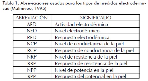
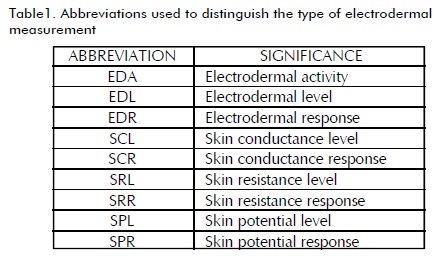
La respuesta electrodérmica (RED) es un fenómeno que está hoy en día bien relacionado con la respuesta de las glándulas sudoríparas (Fowles, 1986). Para ésta existen dos medidas principales: a partir de la resistencia o conductancia (exosomática) y mediante la detección de voltajes (endosomática) (Féré, 1888). Las mediciones pueden también ser tónicas (nivel de profundidad-L) o fásica (respuesta variable en el tiempo-R). La tabla 1 presenta las abreviaciones usadas para los tipos de medidas.
La medición es generalmente realizada sobre la palma con electrodos. Cuando el tipo de electrodo es reversible la polarización y los potenciales sesgados son minimizados. Las señales registradas se caracterizan por tener una recuperación lenta, con una duración alrededor de 40 s, amplitudes de fase alrededor de 2µS para la conductividad y 10-20 mV para el potencial, dependiendo del área del electrodo (Venables et al., 1980).
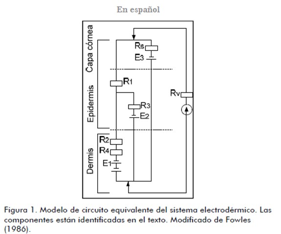
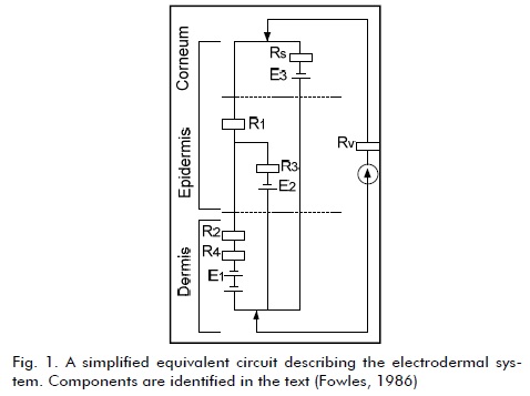
Fowles (1974) desarrolló un modelo global sobre RED (figura 1), útil sólo cualitativamente. La parte superior en el modelo representa la superficie de la piel y la inferior la interfaz entre la hipodermis y la dermis. R1 y R2 representan la resistencia al flujo de corriente en la epidermis y dermis, respectivamente. E1 y R4 corresponden al acceso a los de la dermis, y E2 y R3 el acceso a la epidermis. Los potenciales de transducción E1 y E2 surgen como resultado de la desigualdad de concentraciones iónicas por medio de los conductos, la resistencia R5 es de la superficie y E3 es el potencial.
Mecanismos fisiológicos
Entre los mecanismos fisiológicos de la estimulación eléctrica se encuentran: incremento en la circulación, diminución del edema, aumento en la migración de las células epiteliales, los neutrófilos y los macrófagos; inhibición de los mastocitos, estimulación de la síntesis de DNA e incremento de los factores de crecimiento. También se sugieren mayor producción de fibroblastos y niveles de colagenasa, inhibición bacterial, aumento en el debridamiento y la restauración del potencial bioeléctrico de cicatrización; además ha sido utilizada como bactericida y para incrementar el flujo sanguíneo y promover la cicatrización.
Rowley y colegas (1974) con extra alto voltaje (EAV) de polaridad negativa aplicado a heridas infectadas observaron disminución de la proliferación bacterial. Barranco y colegas (1974) inhibieron el crecimiento de estafilococos con corrientes de 0,4- 400 mA. Kincaid y Lavoie (1989), en un estudio in vitro con 150, 200, 250 y 300 V de polaridad negativa obtuvieron la destrucción de tres microorganismos presentes en heridas humanas. Los análisis mostraron mayor inhibición de los microorganismos a mayor intensidad y tiempo de aplicación.
La estimulación EAV también acelera el debridamiento y la trombosis de los tejidos, una corriente de polaridad negativa permite la reperfusión rápida de los tejidos y la disolución de hematomas (Nelson et al., 1999). Eberhardt y colegas (1986) realizaron un estudio sobre sujetos con lesión en la dermis del antebrazo tratada con un pulso rectangular de 100 Hz, 1 ms y 3- 35 mA, observando incrementos de los granulocitos en un 63,5% de quienes recibieron máxima estimulación, comparados con un 44,7% de células en quienes no recibieron estimulación. Mohr y colegas (1987) emplearon EAV en el flujo sanguíneo del miembro posterior en ratas, con cuatro tipos de frecuencias diferentes (2,20; 80 y 120 pps) y ambas polaridades. Para cada pulso en sus dos polaridades se halló aumento significativo de la velocidad del flujo sanguíneo y los mayores incrementos fueron para 20 pps y los pulsos negativos. Goldman y colegas (2001) aplicaron voltajes desde 80-330 V a 100 pps en pacientes diabéticos con isquemia inframaleolar, incrementando la microcirculación de las heridas isquémicas críticas y la promoción de la cicatrización de éstas.
Estimulación eléctrica para promover el proceso de cicatrización
Brown y colegas (1987) usando estimulación eléctrica pulsada monofásica de alto voltaje para la cicatrización de heridas en conejos, encontraron que en los primeros cuatro días la polaridad positiva fue contraindicada, pero posteriormente ésta aceleró el proceso de cicatrización mejor que la polaridad negativa. Chi-Sing y colegas (1996), con corriente directa (4-40 mA) hallaron efectos positivos en el proceso de cicatrización en edemas y quemaduras en ratas. Kloth y Feedar (1988) estudiaron pacientes con úlceras entre 1 mes a 2,5 años de evolución y utilizando EAV (100-175 V y 105 Hz) constataron que en todos los pacientes estimulados se cerraron sus heridas en un promedio de 7,3 semanas, con una tasa de cicatrización de 45% por semana, mientras que en el grupo control las úlceras se incrementaron en un porcentaje de 11,6% por semana.
Griffin y colegas (1991) evaluaron úlceras en un ensayo clínico con corriente de alto voltaje (200 V, 100 pps). El porcentaje de cierre de la úlcera en el grupo estimulado fue mayor después del día 5, al compararlo con el grupo control. Hounghton y colegas (2003) evaluaron úlceras crónicas por diabetes, insuficiencia arterial o insuficiencia venosa (150 V, 100 ms, 100 Hz, polaridad negativa), donde apreciaron reducción en 50% del área inicial de la herida, dos veces mayor que lo visto en el grupo placebo. Baker y colegas (1997) evaluaron la cicatrización a partir de estimulación eléctrica en pacientes diabéticos, encontrando incremento del 60% en la cicatrización del grupo estimulado con onda bifásica asimétrica.
Gentzkow y colegas (1993) hicieron un estudio de pacientes con úlceras por diabetes, enfermedad cardiovascular o renal y alteraciones motoras, sensitivas y con diversos grados de cronicidad. El 60,7% de las úlceras mejoraron después de 2 semanas de tratamiento, 80,4% luego de 4 semanas, 82% luego de la semana 7 y 23% se curaron completamente en un tiempo de 8,4 semanas. En otro de sus estudios las úlceras curaron más del doble con respecto al grupo control; la cicatrización total para el grupo estimulado se dio en un promedio de 9 semanas, y para el grupo control en 11,8 semanas (Gentzkow et al., 1991). Franek y colegas (2000), empleando EAV, notaron una reducción significativa en el tamaño de las úlceras venosas crurales y disminución de la presión de la úlcera.
Modelos de la actividad electrodérmica
El potencial de la piel se encuentra entre la superficie externa de la piel y el ambiente interno del cuerpo, lo que puede ser considerado como una respuesta ante un estímulo SPR o como un nivel de potencial de la piel, NPP. La respuesta de resistencia de la piel (RPP) puede ser monofásica, bifásica u ocasionalmente trifásica, y se atribuye esencialmente a la actividad de la glándula de sudor (Fowles, 1974). El NPP es comúnmente negativo en el exterior (Christie, 1981) e involucra mecanismos epidermales en mayor medida que en la RPP. Edelberg sugirió que las medidas eléctricas de la piel pueden reflejar el nivel de actividad de glándulas sudoríparas, el estado de los vasos sanguíneos locales y el estado de una o más capas de células vivas.
Modelo de hidratación de la piel
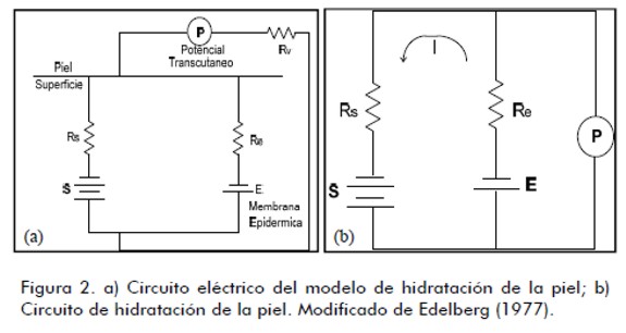
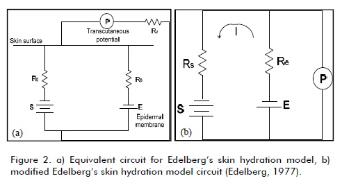
Martin y Venables (1966) encontraron que ante la ausencia de actividad sudorípara no se presentaba RRP. Edelberg (1968) construyó un modelo de hidratación considerando dos fuentes de potencial: el generador de glándulas sudoríparas y el generador de membrana de la epidermis (figura 2). En la figura 2, S es la batería de glándulas sudoríparas y E la batería de la membrana epidérmica. Rs y Re corresponden a la combinación de las resistencias internas y las baterías. P es el potencial transcutáneo y Rv la resistencia del medidor. Con S más negativo que E, circulará una corriente i, y con Rv constante e infinita resulta la figura 2b (Edelberg, 1977).
La piel como una batería
En la regeneración anfibia se estableció que los potenciales de amputación fueron derivados de la piel como generador de corriente (Borgens, 1982). El movimiento de iones positivos de sodio genera el potencial eléctrico a través de la epidermis (alrededor de 50 mV) y se demostró también la existencia de potencial entre dos sitios de la piel inferior a 1 mV (Barker et al., 1982; Foulds, 1983). La variación del potencial de batería de la piel depende de la concentración de glándulas sudoríparas y la densidad de las vellosidades y no parece estar relacionada con la distribución del dermatoma. Burr y colegas en los años treinta, y Becker y colegas en los sesenta, conceptualizaron los potenciales de la piel como un sistema de control DC. Becker (1962) demostró la existencia un patrón de líneas equipotenciales de campo eléctrico complejo en anfibios y en el hombre.
Potenciales de herida
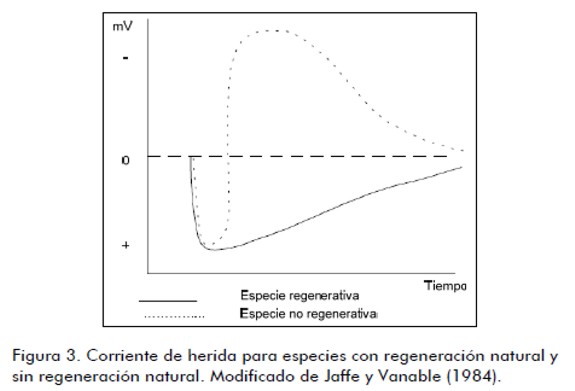
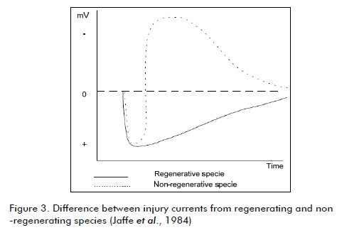
La corriente de herida es en esencia la diferencia de potencial y el subsecuente flujo de corriente entre el tejido herido y el normal debido a membranas dañadas, potenciales alterados o movimiento de partículas cargadas (Thakor et al., 1978). En el borde de la herida se encuentra un voltaje de más de 200 mV/mm y una corriente de 1-1,5 mA (Charman, 1990), lo que origina la migración y proliferación de células de fibroblastos y queratinocitos (Nishimura et al., 1996), aumentando el grosor de la cicatriz. En una especie no regeneradora la corriente de la herida es inicialmente positiva, mientras que en las especies regeneradoras cambia a negativa fuertemente después de una fase inicial positiva (figura 3) hasta volver a los niveles de preherida (Becker, 1967; 1974). De acuerdo con Becker, el sistema de control de DC es el responsable de la detección de la herida y del control del proceso de reparación (Nishimura et al., 1996). O’Leary y Goldring (1964) plantearon que el potencial de herida se desarrolla en los nervios, músculo o piel heridos y cualquier célula puede ser vulnerable a la despolarización de la membrana dada por el trauma.
Potenciales de herida en los tejidos musculoesqueléticos
Barker y colegas (1982), y Jaffe y Vanable (1984), trabajando en mamíferos, demostraron la existencia de una corriente en la piel herida y midieron en cero el potencial en el sitio de la herida. Illingworth y Barker (1980) obtuvieron densidades de corriente asociadas con amputación de dedos en niños con valores de 22µcm-2. Los resultados mostraron que la herida de la piel activó una batería epidermal con un voltaje en circuito abierto de 17-42 mV y la herida en el hueso activó una batería endosteal de 15-56 mV. La capa celular del periostio no presentó propiedades electrogénicas y la batería del hueso se asoció con la capa endosteal. Posteriormente se concluye que la herida muscular fue el factor dominante para generar el voltaje detectado. Las corrientes más grandes fueron de 10-32 µA en la superficie del tejido suave.
Aplicaciones en psicología
La AED es uno de los principales índices psicofisiológicos empleado como correlato de procesos psicológicos y está relacionada con la emoción, el arousal y la atención. Su medición es de alta sensibilidad (Wieland et al., 1970) y sus variaciones son hoy relacionadas con cambios en el estado cognitivo o emocional del individuo (Hugdahl, 1995). La AED se ha utilizado como indicador de estados de estrés (Clements y Turpin, 2000) y como índice clínico en el estudio de diversos trastornos psicofisiológicos relacionados con el estrés (Hugdahl, 1995) y la neurosis (Norris et al., 2007).
Algunos de los estudios sobre la neurosis muestran en los pacientes una mayor sintomatología depresiva después de acontecimientos estresantes, como el desempleo (Creed et al., 2001), cuidado del cónyuge (Gallant et al., 2003) y cirugía del cáncer de mama (Millar et al., 2005). Otros estudios han registrado mayores valores de conductancia de la piel en personas neuróticas, al igual que valores reducidos en la RCP ante estimulación sensorial repetitiva (Akdag et al., 2003) y una lenta habituación de RCP ante la angustia, el estado de ansiedad y la “excitabilidad” (Horan et al., 2005; Mardaga et al., 2006).
Conclusión
La existencia de señales eléctricas en el tejido biológico fue investigada hace muchos años y suspendida durante al menos una centuria. En la actualidad la aplicación y lectura de corrientes y potenciales eléctricos comienzan a ser aceptadas como alternativas a tratamientos farmacológicos, tal es el caso de heridas de piel, principalmente úlceras (Tomonori et al., 2004; Sakarai et al., 2008), patologías óseas como la no unión o pseudoartorisis, entre otras (Aaron et al., 2004; Anglen, 2003). Se incluyen también técnicas de diagnóstico más antiguas ya aceptadas y aplicadas, tal como la ECG, además del análisis de patologías psicológicas a partir de la RED (Norris, 2007; Horan et al., 2005). Esta revisión presentó evidencia teórica, clínica y experimental de trabajos alrededor del estudio de la respuesta electrodermal, dejando la bioelectricidad como un área aún muy joven en la que es indispensable más investigación que ahonde en el conocimiento del cuerpo humano y que lleve a generar nuevas y mejores alternativas a tratamientos.
Agradecimientos
A Tim Watson for School of Health & Emergency Professions University of Hertfordshire, UK, por la valiosa información suministrada.
Referencias
Aaron, R.K., Boyan, B.D., Ciombor, D.M., Schwartz, Z., Simon, B.J., Stimulation of growth factor synthesis by electric and electromagnetic fields., Clin Orthop Relat Res., 419, 2004, pp. 30-37.
Anglen, J., The clinical use of bone stimulators., J South Orthop Assoc., 12, 2003, pp. 46-54.
Baker, L., Chambers, R., DeMuth, S.K., Villar, F., Effects of electrical stimulation on wound healing in patients with diabetic ulcers., Diabetes Care, 20(3), 1997, pp. 405-412.
Barceló, F., Martínez-Selva, J.M., Palmero, F., Correlatos fisiológicos de la ansiedad a la entrevista en una muestra de mujeres jóvenes., Ansiedad y estrés, 2 (1), 1996, pp. 43-54.
Barker, A.T., Jaffe, L.E., Vanable, J.W., The glabrous epidermis of Cavies contains a powerful battery., Am J Physiol., 242, 1982, pp. R358-R366.
Barranco, S., Spadaro, J., In vitro effect of weak direct current on staphylococcus aureus., Clinical Orthopaedic, 100, 1974, pp. 250-255.
Becker, R., The significance of bioelectric potentials., Bioelectrochemistry and Bioenergetics, 1, 1974, pp. 187-199.
Becker, R., The electrical control of growth processes., Medical Times., 95, 1967, pp. 657-669.
Becker, R., The direct current control system: A link between environment and organism., New York State Journal of Medicine, 62, 1962, pp. 1169-1176.
Borgens, R., Endogenous ionic currents traverse intact and damaged bone., Science, 225, 1984, pp. 478-482.
Borgens, R., What is the role of naturally produced electric current in vertebrate regeneration and healing?, International Review of Cytology, 76, 1982, pp. 245-298.
Boucsein, W., Electrodermal Activity., Nueva York: Plenum Press, 1992.
Brown, M., Gogia, P.P., Effects of high voltage stimulation on cutaneous wound healing in rabbits., Physical Therapy, 67, 1987, pp. 662-667.
Charman, R.A., Part 3: Bioelectric potentials and tissue currents. ,Physiotherapy, 76, 10, 1990, pp. 643- 653.
Chi-Sing, C., Marylevich, N.P., McManus, A., Mason, A.D., Pruitt, B.A., Direct current reduces wound edema after full-thickness burn injury in rats., The Journal of Trauma: Injury,Infection and Critical Care, 40(5),1996, pp. 738-742.
Christie, M.J., Venables, P.H., Psychophysiology, Japanese Journal of Physiology, 8,1971, pp. 779-786.
Christie, M., Electrodermal activity in the 1980´s- A review., J Royal Soc Med., 74, 1981, pp. 616-622.
Clemens, K., Turpin, G., Life event exposure, physiological reactivity, and psychological strain., Journal of Behavioral Medicine, 23(1), 2000, pp. 73-94.
Creed, P.A., Muller, J., Machin, M.A., The role of satisfaction with occupational status, neuroticism, financial strain and categories of experience in predicting mental health in the unemployed, Personality and Individual Differences, 30, 2001, pp. 435-447.
Edelberg, R., Biopotentials from the skin surface: The hydration effect., Ann. N.Y. Acad. Sci, 148, 1968, pp. 252-62.
Edelberg, R., Relation of electrical properties of the skin to structure and physiologic state., J Invest Dermatol, 69, 1977, pp.324-327.
Eysenck, H.J., The biological basis of personality., Springfield, IL: Thomas, 1967.
Féré, C., Note sur les modifications de la resistance electrique sous l´influence des excitations sensorielles et des emotions., C.R. Soc. Biol., 5, 1888, pp. 217-9.
Foulds, I.S., Barker, A.T., Human skin battery potential and their possible rule in would healing., Br. J Dermatol, 109, 1983, pp. 515-522.
Fowles, D.C., Mechanisms of electrodermal activity: Methods in Physiological Psychology., Bioelectric Recording Techniques, Vol. 1, Ed. RF Thompson, MM Patterson. Academic Press, New York, 1974, pp. 231-71.
Fowles, D.C., The ecrine system and electrodermal activity in Psychophysiology., ed. MGH Coles, E Donchin, SW Porges. Guilford Press, New York, 1986, pp. 51-96.
Franek, A., Polak, A., Kucharzewski, M., Modern application of high voltage stimulation for enhanced healing of venous crural ulceration., Medical Engineering and Physics, 22, 2000, pp. 647-655.
Gallant, M.P., Connell, C. M., Neuroticism and depressive symptoms among spouse caregivers: Do health behaviors mediate this relationship?, Psychology and Aging., 18, 2003, pp. 587 -592.
Gentzkow, G.D., Alon, G., Taler, G.A., Eltorai, I.M., Montroy, R.E., Healing of refractory stage III and IV pressure ulcers by a new electrical stimulation device., Wounds, 5.3, 1993, pp. 160-172.
Gentzkow, G.D., Pollack, S.V., Kloth, L.C., Stubbs, H.A., Improved healing of pressure ulcers using dermapulse, a new electrical stimulation device., Wounds, 3(5), 1991, pp. 158- 170.
Goldman, R., Brewley, B., Goleen, M., Electrotherapy Reoxygenates inframalleolar ischemic wounds on diabetic patients., Advances in Skin & Wound Care, 15, 2001, pp. 112-120.
Griffin, W., Tooma, R.E., Mendius R.A., Clift J.K., Zwaag E.Z., Efficacy of high voltage pulsed current for healing of pressure ulcers in patients with spinal cord injury., Physical Therapy, 71, 1991, pp. 433- 442.
Horan, W.P., Subotnik, K.L., Reise, S.P., Ventura, J., Nuechterlein, K.H., Stability and clinical correlates of personality characteristics in recent-onset schizophrenia, Psychological Medicine, 35, 2005, pp. 995-1005.
Houghton, P.E., Kincaid, C.B., Lovell, M., Campbell, K.E., Keast, D.H., Woodbury, M.G., Harris, K.A., Effect of electrical stimulation on chronic leg ulcer size and appearance., Physical Terapy, 83,1, 2003, pp. 17-28.
Hugdahl, K., Psychophysiology: The mind-body perspective., United States: Harvard University Press, 1995.
Illingworth, C.M., Barker, A.T., Measurement of electrical currents emerging during the regeneration of amputated fingers tips in children., Clin. Phys. Physiol. Meas., 1(1), 1980, pp. 87-89.
Im, M.J., Lee, W., Hoopes, J., Effect of Electrical stimulation on survival of skin flaps in pigs., Physical Therapy, 70,1990, pp. 37-40.
Jaffe, L., Vanable, J., Electric fields and wound healing., Clinics in Dermatology, 2(3),1984, pp. 34-44.
Kincaid, C.B., Lavoie K.H., Inhibition of bacterial growth in vitro following stimulation with high voltage, monophasic, pulsed current., Physical Therapy, 69, 1989, pp. 29-33.
Klot, L., McCulloch, J.M., Promotion of wound healing with electrical stimulation., ADV Wound Care, 9(5), 1996, pp. 42-45.
Kloth, L., Feedar, J., Aceleration of wound healing with high voltage monophasic pulsed current., Physical Therapy, 68(5), 1988, pp. 503-508.
Malmivuo, J., Plonsey, R., Bioelectromagnetims: Principles and Applications of Bioelectric and Biomagnetic Fields., New York, Oxford University Press, Cap 27, 1995.
Mardaga, S., Laloyaux, O., Hansenne, M., Personality traits modulate skin conductance response to emotional pictures: An investigation with Cloninger´s model of personality., Personality and Individual Differences, 40, 1996, pp. 1603-1614.
Martin, I., Venables, P.H., Mechanisms of palmar skin resistance and skinpotential., Psychological Bulletin, 65, 1966, pp. 347 -357.
Millar, K., Purushotham, A.D., McLatchie, E., George, W.D., Murrary, G.D., A 1-year prospective study of individual variation in distress, and illness perceptions, after treatment for breast cancer., Journal of Psychosomatic Research, 58, 2006, pp. 335-342.
Mohr, T., Akers, T., Wessman H., Effect of high voltage stimulation on blood flow in the rat hind limb., Physical Therapy, 67, 1987, pp. 526-533.
Moncada, M.E., Martínez, A., Pinedo, C.R., Desarrollo experimental y computacional para estimar variables eléctricas inducidas en muestras de fémur bovino estimuladas por campos magnéticos de baja frecuencia., Revista Cubana de Investigaciones Biomédica, 27 (2), 2008, pp. 1-9.
Moncada M.E., Martínez A., Pinedo C.R., Cadavid H., Desarrollo e implementación de una técnica para la medición de impedancia en muestras de hueso húmedo de bovino., Rev. fac. ing. Univ. Antioquia, No 44, 2008, pp. 75-82.
Moncada, M.E., Cadavid, H., Metodología de construcción de muslos 3D para estimar variables eléctricas inducidas por campos magnéticos de baja frecuencia., Revista Brasilehir de Engenharia Biomédica, 23, 2007, pp. 123-130.
Nelson, R.M., Hayes, K.W., Currier. Clinical Electrotherapy., 3ed. Appleton and Lange, 1999.
Nishimura, K.Y., Isseroff, R., Nuccitelli, R., Human keratinocytes migrate to the negative pole in direct current electric fields comparable to those measured in mammalian wounds., Journal of Cell Science, 109, 1996, pp.199-207.
Norris, C.J., Larsen, J.T, Cacioppo, J.T., Neuroticism is associated with larger and more prolonged electrodermal responses to emotionally evocative pictures., Psychophysiology, 44, 2007, pp. 823-826.
O’Leary, J.L., Goldring, S., D-C Potentials of the Brain., Physiol Rev, 44(1), 1964, pp. 91-125.
Poltawski, L., Edwards, H., Todd, A., Watson, T., Lees, A., James, C., Cutaneous side effects of infused apomorphine: The patient and care experience., Brit J Neuroscience Nursing, 4 (12), 2008a, pp. 576-580.
Poltawski, L., Edwards, H., Todd, A., Watson, T., Lees, A., James, C., Ultrasound treatment of cutaneous side-effects of infused apomorphine: A randomised controlled pilot study., Mov Disord, 24, 2008 b, pp.115-118.
Poltawski, L., Watson, T., Bioelectricity and microcurrent therapy for tissue healing-a narrative review., Physical Therapy Reviews, 14(2), 2009, pp.104-114.
Rowley, B.A., McKenna, J., Chase, G.R., The influence of electrical current on an infecting microorganism in wounds., Ann NY Academic Scientific, 238, 1974, pp. 543-551.
Sakarai, T., Yoshimoto, M., Koyama, S., Miyakoshi, J., Exposure to extremely low frequency magnetic fields affects insulinsecreting cells., Bioelectromagnetics, 29(2), 2008, pp.118- 24.
Sandoval, M.C., Herrera, E., Efectos de la estimulación eléctrica sobre el proceso de cicatrización de las heridas., Salud UIS, 39, 2007, pp. 54-61.
Schwebel, D.C., Suls, J., Cardiovascular reactivity and neuroticism: Results from a laboratory and controlled ambulatory stress protocol., Journal of Personality, 67, 1999, pp. 67-92.
Selva, J.M., García-Sánchez, F., Gómez-Amor, J., Individual differences in psychophysiological and subjective correlates of speech anxiety., Journal of Psychophysiology, 3, 1989, pp.75-81.
Siddle, D.A., Lipp, O.V., Dall, P., The effects of task type and task requirements on the dissociation of skin conductance responses and secondary task probe reaction time., Psychophysiology, 33, 1996, pp. 73-83.
Thakor, N., Webster, J., The origin of skin potential and its variations., 31st ACEMB, Atlanta, Georgia, USA, 1978.
Tomonori, S., Akira, S., Shoichiro, S., Kazutomo, I., Junji, M., An Extremely Low Frequency Magnetic Field Attenuates Insulin Secretion From the Insulinoma Cell Line,RIN-m, Bioelectromagnetics, 2004, 25:160-166.
Venables, P.H., Christie, M.J., Techniques in Psychophysiology., Ed. I Martin, 1980, pp. 3-67.
Wieland, B.A., Mefferd, R.D., Systematic changes in levels of physiological activity during a fourth-month period., Psychophysiology, 6,1970, pp. 669-689.
Electrodermal activity - a review
María Elena Moncada1, Jorge de la Cruz2
1 Electrician Engineer, Universidad de Antioquia. PhD in Engineering , Universidad del Valle. Research Professor, Instituto Tecnológico Metropolitano, Colombia. mariamoncada@itm.edu.co
2 Electronic Engineering Student,, Universidad del Valle. jodelacruz@hotmail.com
ABSTRACT
the electricity in living tissue was widely studied around the 19th century. Such study was suspended for many years but has then been started again during recent decades. New research into bioelectricity is creating alternatives in the health field; one of them is an electrodermal response associated with the wound healing, cell stimulation and psychopathology diagnostic. This article presents some of the first responses and models concerning electrodermal activity. Theoretical, clinical and review papers were studied and classified to show the amplitude and variety of bioelectrical responses. Electrodermal activity is only one of many applications having an abundant amount of evidence regarding diagnosis and treatment starting from bioelectrical signals. Electrical tissue response requires more experimental, theoretical and clinical research in many fields involving an organism' s behaviour to ascertain, propose and create new treatment alternatives for different pathologies.
Keywords: bioelectricity, wound healing, skin, electric potential.
Received: February 25th 2010
Accepted: July 6th 2011
Introduction
The skin is one of our body' s most important organs. If more than 40% of the skin is lacking, then this is often incompatible with life. The skin has a selective protective function preventing the entry of harmful substances and its presence forms part of the body' s immune system. It has metabolism regulatory functions as it prevents the exit of substances, regulates the body’s temperature and converts sunshine into vitamin D.
Electric signal treatment has been studied since the 19th century. Researchers have demonstrated the presence of bioelectric endogenous systems (Klotet et al., 1996; Charman, 1990). References about the effects of electrical stimulation concerning ulcers generated by pressure, vascular insufficiency, trauma, diabetes, surgery abound in the literature (Sandoval et al., 2007; Poltawski, 2008 a, b; 2009). Electrical stimulation of the skin has been used as a bactericide (Nelson et al., 1999; Kincaid et al., 1989) to increase blood flow (Mohr et al., 1987; Goldman et al., 2001) and to promote healing (Im et al., 1990; Brown et al., 1987, Chi-Sing, 1996). Electrodermal activity (EDA) has also been studied and widely used in psychology as a stress level indicator (Clements and Turpin, 2000), including neurosis (Norris, 2007). It has high sensitivity when being measured; its variations are related to a person' s cognitive emotional state (Hugdahl, 1995).
The existence of electrical signals in biological tissue is a fact; however, studies to date have been unrelated, thereby preventing overall conclusions being reached. Developments in diagnosis, measurement and therapy have been achieved through the evidence reported to date. However, there is still much to explore and the need for new experimental, clinical and mathematical studies leading to knowledge about the body' s bioelectrical aspects has become apparent. Studies in this field and some clinical, experimental and computational work presenting the electrodermal system as a tissue component have been started in Colombia (Moncada et al., 2007; 2008 a, b; Sandoval et al., 2007) and it is expected that support alternatives to current medicine will be created in the future.
Electrodermal measurement
The electrodermal response (EDR) is a phenomenon which is very connected with sweat gland response nowadays (Fowles, 1986). Two main EDR measurements have traditionally involved resistance or conductance (Féré, 1888)] (exosomatic) and voltage detection (Tarchanoff, 1889) (endosomatic). These measurements can also be tonic (depth-L) or phasic (variable response-R). Table 1 shows the abbreviations used for types of measurement.
Measurement is usually done with electrodes on the palm; when an electrode is reversible, polarisation and slanted potentials become minimised. The recorded signals are characterised because they present slow recovery around 40s, phase amplitude for conductivity around 2μS, and 10-20 mV potency (depending on the electrode area (Venables et al., 1980).
Fowles (1974) developed a global EDR model (Fig. 1) which is a useful qualitatively. The top of the model represents the skin surface and the bottom the interface between hypodermis and dermis. R1 and R2 represent, respectively, resistance to current flow in epidermis and dermis, E1 and R4 represent access to the dermis and E2 and R3 access to the epidermis. Transduction potentials E1 and E2 emerge as a result of the inequality of ion concentration through the ducts. R5 is surface resistance and E3 is the potential.
Physiological mechanisms
Physiological electrical stimulation mechanisms would include increased circulation, decreased oedema, increased epithelial cell migration, neutrophils and macrophages, mast inhibition, DNA synthesis stimulation and increased growth factors. Increased fibroblast production, collagenase levels, bacterial inhibition, debridement and the restoration of healing bioelectric potential has also been suggested. Electrical stimulation has also been used as a bactericide and for increasing blood flow and promoting healing.
Rowley et al., (Rowley, 1974) observed a proliferation of bacterial decrease having negative polarity extra high voltage (EHV) applied to infected wounds. Barranco and et al., (1974) inhibited staphylococci growth using current from 0.4 to 400 mA. Kincaid and Lavoie (1989) manage to destroy three microorganisms present in human wounds in an in vitro study with 150, 200, 250 and 300 V negative polarity. Analysis revealed that more microorganisms were inhibited with greater voltage intensity and longer application time.
EHV stimulation also accelerates tissue debridement and thrombosis; negative polarity current allows fast tissue reperfusion and hematomas dissolution (Nelson, 1999). Eberhardt et al., (1986) did a study about people suffering forearm dermis wounds which had been treated with 100 Hz, 1 ms and 3 to 35 mA rectangular pulse. They observed that granulocytes increased by 63.5% in people who received maximum stimulation compared to 44.7% of cells which did not receive stimulation. Mohr et al., (1987) used EHV for rats' hind limb blood flow, using three frequencies (2.20, 80 and 120 pps) and both polarities. A significant increase in blood flow speed was found for each pulse at both polarities. The highest increases were at 20 pps and with negative pulse. Goldman et al., (2001) applied 80 to 330 V at 100 pps voltages to diabetic patients suffering inframaleolar ischemic wounds with increased microcirculation and promotion of their wound healing.
Electrical stimulation for promoting wound healing
Brown et al., (1987) researched high voltage pulsed monophasic electrical stimulation regarding wound healing in rabbits. They found that positive polarity was contraindicated during the first four days but later accelerated healing better than negative polarity. Chi-Sing et al., (1996) found positive effects in rats' oedema and burn healing when using direct current (4 and 40 mA). Kloth and Feedar (1988) studied patients suffering ulcers having 1 month to 2.5 years' evolution using EHV (100-175 V and 105 Hz), finding that stimulated patients' wounds closed in about 7.3 weeks, giving 45% per week healing rate compared to 11.6% per week increase for control group ulcers.
Griffin et al., (1991) evaluated ulcers in a clinical trial using high voltage current (200 V, 100 pps). The closing percentage for ulcers in the stimulated group compared to the control group was greater after the 5th day. Hounghton et al., (2003) evaluated chronic ulcers formed by diabetes and arterial or venous insufficiency (150 V, 100 ms, 100 Hz, negative polarity) where they found 50% reduction in initial wound area, twice more than in the placebo group. Baker et al., (1997) evaluated healing from electrical stimulation in diabetic patients, finding 60% increase in the healing of the group receiving biphasic asymmetrical stimulation.
Gentzkow et al., (1993) carried out a study on patients with diabetes-related ulcers, cardiovascular or renal illness, impaired sensory-motor function and varying degrees of chronicity; 60.7% of ulcers improved after two weeks treatment, 80.4% after four weeks, 82% after seven weeks and 23% healed completely in 8.4 weeks. In another of their studies, the ulcers healed more than twice as fast as those of the control group, total healing for the stimulated group being 9 weeks on average and 11.8 weeks for the control group (Gentzkow et al, 1991). Franek et al., (2000) found a significant reduction in the femoral venous ulcer size and pressure ulcer when using EHV.
Electrodermal activity models
The skin’s potential is located between the external surface of skin and the internal body' s environment which can be considered as skin potential response (SPR) to a stimulus or skin potential level (SPL). SPR can be monophasic, biphasic or occasionally triphasic and is mainly attributed to sweat gland activity (Fowles, 1974). SPL is usually negative on the surface (Christie, 1981) and involves more epidermal mechanisms than SPR. Edelberg has suggested that skin electrical measurements can reflect sweat gland activity level, the local blood vessels' state and the state of one or more living cell layers.
Skin hydration model
Martin and Venables (1966) found that there was no skin resistance response (SRR) in the absence of sweat activity. Edelberg (1968) built a hydration model considering two sources of potential: sweat gland generator and epidermis membrane generator (Fig. 2). In Figure 2, S is the sweat gland battery and E the epidermal membrane battery, Rs and Re are a combination of internal resistance and batteries, P is transcutaneous potential and Rv is measured resistance. An i current circulateds with S being more negative than E. Fig. 2b shows Rv being constant and infinite (Edelberg, 1977).
The skin battery
It has been established in amphibian regeneration that amputation potential was derived from the skin as a current generator (Borgens, 1982). Electrical potential through the epidermis (in the 50mV range) is generated by the movement of positive sodium ions; potential existence between two skin sites lower than 1 mV has also been demonstrated (Barker, 1982; Foulds, 1983). Skin battery potential variation depends on sweat gland concentration, speed and density but apparently is not related to dermatome distribution. Burr et al., in the 1930s and Becker et al., in the 1960s conceptualised skin potential as a DC control system. Becker (1962) demonstrated the existence of an equipotential pattern of lines for complex electric field in amphibians and humans.
Wound potential
Wound current is essentially the potential difference and subsequent current flow between injured and normal tissue due to damaged membranes, altered potential or charged particle movement (Thakor et al., 1978). There is a voltage of over 200 mV / mm at the edge of an injury and a 1 to 1.5 mA current (Charman, 1990); this causes fibroblast and keratinocyte cell migration and proliferation (Nishimura et al., 1996) thereby increasing scar thickness. Injury current is initially positive in non-regenerative species while it frequently changes to negative after an initial positive phase in regenerative species (Fig. 3) until returning again to pre-wound levels (Becker, 1967; 1974). According to Becker, a DC control system is responsible for detecting injury and controlling repair (Nishimura et al, 1996). O' Leary and Goldring (1964) suggested that injury potential is developed in nerves, muscle or skin which are wounded and that any cell can be vulnerable to membrane depolarisation caused by trauma.
Wound potential in muscle-skeletal tissues
Barker et al., (1982) and Jaffe and Vanable (1984) have done work on mammals. They showed the existence of a current in wounded skin and measured zero potential at the site of the wound. Illingworth and Barker (1980) obtained finger amputation- associated 22μAcm-2 current density in children; the results showed that the wound in the skin activated an epidermal battery having 17-42mV open circuit voltage and bone injury activated a 15-56mV endosteal battery. The periosteum cell layer did not present electrogenic properties and the bone battery was associated with the endosteal layer. It was later concluded that muscle wound was the dominant factor in generating the detected voltage; the larger currents were 10-32 μA on soft tissue surface.
Applications in psychology
EDA is one of the main psychophysiological indexes used to correlate psychological processes and is related to emotion, arousal and attention. Its measurement is highly sensitive (Wieland et al, 1970) and its variations related to changes in cognitive or emotional status in an individual (Hugdahl, 1995). EDA has been used as a stress state indicator (Clements and Turpin, 2000) and as clinical index in studying various stressrelated psychophysiological disorders (Hugdahl, 1995) and neuroticism (Norris, 2007).
Some neurosis studies have shown higher depressive symptomatology in patients after experiencing stressful events, such as unemployment (Creed et al., 2001), caring for a spouse (Gallant et al., 2003) and breast cancer surgery (Millar et al., 2005). Other studies have reported higher skin conductance values in neurotic people, as well as reduced values in SCR to repetitive sensory stimulation (Akdag et al., 2003) and slow SPR habituation to anguish, anxiety and "excitability" (Horan et al, 2005; Mardaga et al., 2006).
Conclusion
The existence of electrical signals in biological tissue was investigated many years ago and was suspended at least one hundred years ago. Nowadays, the application and reading current and electrical potential has begun to be accepted as alternatives to pharmacological treatment; this is the case of skin injuries, mainly ulcers (Tomonori et al., 2004; Sakarai et al., 2008) and bone pathologies such as non-union or pseudarthrosis (Aaron et al., 2004; Anglen, 2003). Older diagnostic techniques which are already accepted and which have been implemented would include ECG and analysis of psychological disorders by EDR (Norris, 2007, Horan et al, 2005). This review has presented theoretical, clinical and experimental evidence about work concerning the study of an electrodermal response, revealing bioelectricity to be a very young area where more research is needed to deepen knowledge about the human body and lead to generating new and better treatment alternatives.
Acknowledgments
We would like to thank Tim Watson from the School of Health & Emergency Professions, University of Hertfordshire, UK, for the valuable information provided.
References
Aaron, R.K., Boyan, B.D., Ciombor, D.M., Schwartz, Z., Simon, B.J., Stimulation of growth factor synthesis by electric and electromagnetic fields., Clin Orthop Relat Res., 419, 2004, pp. 30-37.
Anglen, J., The clinical use of bone stimulators., J South Orthop Assoc., 12, 2003, pp. 46-54.
Baker, L., Chambers, R., DeMuth, S.K., Villar, F., Effects of electrical stimulation on wound healing in patients with diabetic ulcers., Diabetes Care, 20(3), 1997, pp. 405-412.
Barceló, F., Martínez-Selva, J.M., Palmero, F., Correlatos fisiológicos de la ansiedad a la entrevista en una muestra de mujeres jóvenes., Ansiedad y estrés, 2 (1), 1996, pp. 43-54.
Barker, A.T., Jaffe, L.E., Vanable, J.W., The glabrous epidermis of Cavies contains a powerful battery., Am J Physiol., 242, 1982, pp. R358-R366.
Barranco, S., Spadaro, J., In vitro effect of weak direct current on staphylococcus aureus., Clinical Orthopaedic, 100, 1974, pp. 250-255.
Becker, R., The significance of bioelectric potentials., Bioelectrochemistry and Bioenergetics, 1, 1974, pp. 187-199.
Becker, R., The electrical control of growth processes., Medical Times., 95, 1967, pp. 657-669.
Becker, R., The direct current control system: A link between environment and organism., New York State Journal of Medicine, 62, 1962, pp. 1169-1176.
Borgens, R., Endogenous ionic currents traverse intact and damaged bone., Science, 225, 1984, pp. 478-482.
Borgens, R., What is the role of naturally produced electric current in vertebrate regeneration and healing?, International Review of Cytology, 76, 1982, pp. 245-298.
Boucsein, W., Electrodermal Activity., Nueva York: Plenum Press, 1992.
Brown, M., Gogia, P.P., Effects of high voltage stimulation on cutaneous wound healing in rabbits., Physical Therapy, 67, 1987, pp. 662-667.
Charman, R.A., Part 3: Bioelectric potentials and tissue currents. ,Physiotherapy, 76, 10, 1990, pp. 643- 653.
Chi-Sing, C., Marylevich, N.P., McManus, A., Mason, A.D., Pruitt, B.A., Direct current reduces wound edema after full-thickness burn injury in rats., The Journal of Trauma: Injury,Infection and Critical Care, 40(5),1996, pp. 738-742.
Christie, M.J., Venables, P.H., Psychophysiology, Japanese Journal of Physiology, 8,1971, pp. 779-786.
Christie, M., Electrodermal activity in the 1980' s- A review., J Royal Soc Med., 74, 1981, pp. 616-622.
Clemens, K., Turpin, G., Life event exposure, physiological reactivity, and psychological strain., Journal of Behavioral Medicine, 23(1), 2000, pp. 73-94.
Creed, P.A., Muller, J., Machin, M.A., The role of satisfaction with occupational status, neuroticism, financial strain and categories of experience in predicting mental health in the unemployed, Personality and Individual Differences, 30, 2001, pp. 435-447.
Edelberg, R., Biopotentials from the skin surface: The hydration effect., Ann. N.Y. Acad. Sci, 148, 1968, pp. 252-62.
Edelberg, R., Relation of electrical properties of the skin to structure and physiologic state., J Invest Dermatol, 69, 1977, pp.324-327.
Eysenck, H.J., The biological basis of personality., Springfield, IL: Thomas, 1967.
Féré, C., Note sur les modifications de la resistance electrique sous l’influence des excitations sensorielles et des emotions., C.R. Soc. Biol., 5, 1888, pp. 217-9.
Foulds, I.S., Barker, A.T., Human skin battery potential and their possible rule in would healing., Br. J Dermatol, 109, 1983, pp. 515-522.
Fowles, D.C., Mechanisms of electrodermal activity: Methods in Physiological Psychology., Bioelectric Recording Techniques, Vol. 1, Ed. RF Thompson, MM Patterson. Academic Press, New York, 1974, pp. 231-71.
Fowles, D.C., The ecrine system and electrodermal activity in Psychophysiology., ed. MGH Coles, E Donchin, SW Porges. Guilford Press, New York, 1986, pp. 51-96.
Franek, A., Polak, A., Kucharzewski, M., Modern application of high voltage stimulation for enhanced healing of venous crural ulceration., Medical Engineering and Physics, 22, 2000, pp. 647-655.
Gallant, M.P., Connell, C. M., Neuroticism and depressive symptoms among spouse caregivers: Do health behaviors mediate this relationship?, Psychology and Aging., 18, 2003, pp. 587 -592.
Gentzkow, G.D., Alon, G., Taler, G.A., Eltorai, I.M., Montroy, R.E., Healing of refractory stage III and IV pressure ulcers by a new electrical stimulation device., Wounds, 5.3, 1993, pp. 160-172.
Gentzkow, G.D., Pollack, S.V., Kloth, L.C., Stubbs, H.A., Improved healing of pressure ulcers using dermapulse, a new electrical stimulation device., Wounds, 3(5), 1991, pp. 158- 170.
Goldman, R., Brewley, B., Goleen, M., Electrotherapy Reoxygenates inframalleolar ischemic wounds on diabetic patients., Advances in Skin & Wound Care, 15, 2001, pp. 112-120.
Griffin, W., Tooma, R.E., Mendius R.A., Clift J.K., Zwaag E.Z., Efficacy of high voltage pulsed current for healing of pressure ulcers in patients with spinal cord injury., Physical Therapy, 71, 1991, pp. 433- 442.
Horan, W.P., Subotnik, K.L., Reise, S.P., Ventura, J., Nuechterlein, K.H., Stability and clinical correlates of personality characteristics in recent-onset schizophrenia, Psychological Medicine, 35, 2005, pp. 995-1005.
Houghton, P.E., Kincaid, C.B., Lovell, M., Campbell, K.E., Keast, D.H., Woodbury, M.G., Harris, K.A., Effect of electrical stimulation on chronic leg ulcer size and appearance., Physical Terapy, 83,1, 2003, pp. 17-28.
Hugdahl, K., Psychophysiology: The mind-body perspective., United States: Harvard University Press, 1995.
Illingworth, C.M., Barker, A.T., Measurement of electrical currents emerging during the regeneration of amputated fingers tips in children., Clin. Phys. Physiol. Meas., 1(1), 1980, pp. 87-89.
Im, M.J., Lee, W., Hoopes, J., Effect of Electrical stimulation on survival of skin flaps in pigs., Physical Therapy, 70,1990, pp. 37-40.
Jaffe, L., Vanable, J., Electric fields and wound healing., Clinics in Dermatology, 2(3),1984, pp. 34-44.
Kincaid, C.B., Lavoie K.H., Inhibition of bacterial growth in vitro following stimulation with high voltage, monophasic, pulsed current., Physical Therapy, 69, 1989, pp. 29-33.
Klot, L., McCulloch, J.M., Promotion of wound healing with electrical stimulation., ADV Wound Care, 9(5), 1996, pp. 42-45.
Kloth, L., Feedar, J., Aceleration of wound healing with high voltage monophasic pulsed current., Physical Therapy, 68(5), 1988, pp. 503-508.
Malmivuo, J., Plonsey, R., Bioelectromagnetims: Principles and Applications of Bioelectric and Biomagnetic Fields., New York, Oxford University Press, Cap 27, 1995.
Mardaga, S., Laloyaux, O., Hansenne, M., Personality traits modulate skin conductance response to emotional pictures: An investigation with Cloninger’s model of personality., Personality and Individual Differences, 40, 1996, pp. 1603-1614.
Martin, I., Venables, P.H., Mechanisms of palmar skin resistance and skinpotential., Psychological Bulletin, 65, 1966, pp. 347 -357.
Millar, K., Purushotham, A.D., McLatchie, E., George, W.D., Murrary, G.D., A 1-year prospective study of individual variation in distress, and illness perceptions, after treatment for breast cancer., Journal of Psychosomatic Research, 58, 2006, pp. 335-342.
Mohr, T., Akers, T., Wessman H., Effect of high voltage stimulation on blood flow in the rat hind limb., Physical Therapy, 67, 1987, pp. 526-533.
Moncada, M.E., Martínez, A., Pinedo, C.R., Desarrollo experimental y computacional para estimar variables eléctricas inducidas en muestras de fémur bovino estimuladas por campos magnéticos de baja frecuencia., Revista Cubana de Investigaciones Biomédica, 27 (2), 2008, pp. 1-9.
Moncada M.E., Martínez A., Pinedo C.R., Cadavid H., Desarrollo e implementación de una técnica para la medición de impedancia en muestras de hueso húmedo de bovino., Rev. fac. ing. Univ. Antioquia, No 44, 2008, pp. 75-82.
Moncada, M.E., Cadavid, H., Metodología de construcción de muslos 3D para estimar variables eléctricas inducidas por campos magnéticos de baja frecuencia., Revista Brasilehir de Engenharia Biomédica, 23, 2007, pp. 123-130.
Nelson, R.M., Hayes, K.W., Currier. Clinical Electrotherapy., 3ed. Appleton and Lange, 1999.
Nishimura, K.Y., Isseroff, R., Nuccitelli, R., Human keratinocytes migrate to the negative pole in direct current electric fields comparable to those measured in mammalian wounds., Journal of Cell Science, 109, 1996, pp.199-207.
Norris, C.J., Larsen, J.T, Cacioppo, J.T., Neuroticism is associated with larger and more prolonged electrodermal responses to emotionally evocative pictures., Psychophysiology, 44, 2007, pp. 823-826.
O’Leary, J.L., Goldring, S., D-C Potentials of the Brain., Physiol Rev, 44(1), 1964, pp. 91-125.
Poltawski, L., Edwards, H., Todd, A., Watson, T., Lees, A., James, C., Cutaneous side effects of infused apomorphine: The patient and care experience., Brit J Neuroscience Nursing, 4 (12), 2008a, pp. 576-580.
Poltawski, L., Edwards, H., Todd, A., Watson, T., Lees, A., James, C., Ultrasound treatment of cutaneous side-effects of infused apomorphine: A randomised controlled pilot study., Mov Disord, 24, 2008 b, pp.115-118.
Poltawski, L., Watson, T., Bioelectricity and microcurrent therapy for tissue healing-a narrative review., Physical Therapy Reviews, 14(2), 2009, pp.104-114.
Rowley, B.A., McKenna, J., Chase, G.R., The influence of electrical current on an infecting microorganism in wounds., Ann NY Academic Scientific, 238, 1974, pp. 543-551.
Sakarai, T., Yoshimoto, M., Koyama, S., Miyakoshi, J., Exposure to extremely low frequency magnetic fields affects insulinsecreting cells., Bioelectromagnetics, 29(2), 2008, pp.118- 24.
Sandoval, M.C., Herrera, E., Efectos de la estimulación eléctrica sobre el proceso de cicatrización de las heridas., Salud UIS, 39, 2007, pp. 54-61.
Schwebel, D.C., Suls, J., Cardiovascular reactivity and neuroticism: Results from a laboratory and controlled ambulatory stress protocol., Journal of Personality, 67, 1999, pp. 67-92.
Selva, J.M., García-Sánchez, F., Gómez-Amor, J., Individual differences in psychophysiological and subjective correlates of speech anxiety., Journal of Psychophysiology, 3, 1989, pp.75-81.
Siddle, D.A., Lipp, O.V., Dall, P., The effects of task type and task requirements on the dissociation of skin conductance responses and secondary task probe reaction time., Psychophysiology, 33, 1996, pp. 73-83.
Thakor, N., Webster, J., The origin of skin potential and its variations., 31st ACEMB, Atlanta, Georgia, USA, 1978.
Tomonori, S., Akira, S., Shoichiro, S., Kazutomo, I., Junji, M., An Extremely Low Frequency Magnetic Field Attenuates Insulin Secretion From the Insulinoma Cell Line,RIN-m, Bioelectromagnetics, 2004, 25:160-166.
Venables, P.H., Christie, M.J., Techniques in Psychophysiology., Ed. I Martin, 1980, pp. 3-67.
Wieland, B.A., Mefferd, R.D., Systematic changes in levels of physiological activity during a fourth-month period., Psychophysiology, 6,1970, pp. 669-689.
References
Aaron, R.K., Boyan, B.D., Ciombor, D.M., Schwartz, Z., Simon, B.J., Stimulation of growth factor synthesis by electric and electromagnetic fields., Clin Orthop Relat Res., 419, 2004, pp. 30-37. DOI: https://doi.org/10.1097/00003086-200402000-00006
Anglen, J., The clinical use of bone stimulators., J South Orthop Assoc., 12, 2003, pp. 46-54.
Baker, L., Chambers, R., DeMuth, S.K., Villar, F., Effects of electrical stimulation on wound healing in patients with diabetic ulcers., Diabetes Care, 20(3), 1997, pp. 405-412. DOI: https://doi.org/10.2337/diacare.20.3.405
Barceló, F., Martínez-Selva, J.M., Palmero, F., Correlatos fisiológicos de la ansiedad a la entrevista en una muestra de mujeres jóvenes., Ansiedad y estrés, 2 (1), 1996, pp. 43-54.
Barker, A.T., Jaffe, L.E., Vanable, J.W., The glabrous epidermis of Cavies contains a powerful battery., Am J Physiol., 242, 1982, pp. R358-R366. DOI: https://doi.org/10.1152/ajpregu.1982.242.3.R358
Barranco, S., Spadaro, J., In vitro effect of weak direct current on staphylococcus aureus., Clinical Orthopaedic, 100, 1974, pp. 250-255. DOI: https://doi.org/10.1097/00003086-197405000-00037
Becker, R., The significance of bioelectric potentials., Bioelectrochemistry and Bioenergetics, 1, 1974, pp. 187-199. DOI: https://doi.org/10.1016/0302-4598(74)85020-8
Becker, R., The electrical control of growth processes., Medical Times., 95, 1967, pp. 657-669.
Becker, R., The direct current control system: A link between environment and organism., New York State Journal of Medicine, 62, 1962, pp. 1169-1176.
Borgens, R., Endogenous ionic currents traverse intact and damaged bone., Science, 225, 1984, pp. 478-482. DOI: https://doi.org/10.1126/science.6740320
Borgens, R., What is the role of naturally produced electric current in vertebrate regeneration and healing?, International Review of Cytology, 76, 1982, pp. 245-298. DOI: https://doi.org/10.1016/S0074-7696(08)61793-3
Boucsein, W., Electrodermal Activity., Nueva York: Plenum Press, 1992. DOI: https://doi.org/10.1007/978-1-4757-5093-5
Brown, M., Gogia, P.P., Effects of high voltage stimulation on cutaneous wound healing in rabbits., Physical Therapy, 67, 1987, pp. 662-667. DOI: https://doi.org/10.1093/ptj/67.5.662
Charman, R.A., Part 3: Bioelectric potentials and tissue currents., Physiotherapy, 76, 10, 1990, pp. 643- 653. DOI: https://doi.org/10.1016/S0031-9406(10)63101-X
Chi-Sing, C., Marylevich, N.P., McManus, A., Mason, A.D., Pruitt, B.A., Direct current reduces wound edema after full-thickness burn injury in rats., The Journal of Trauma: Injury, Infection and Critical Care, 40(5),1996, pp. 738-742. DOI: https://doi.org/10.1097/00005373-199605000-00009
Christie, M.J., Venables, P.H., Psychophysiology, Japanese Journal of Physiology, 8,1971, pp. 779-786. DOI: https://doi.org/10.1111/j.1469-8986.1971.tb00515.x
Christie, M., Electrodermal activity in the 1980´s- A review., J Royal Soc Med., 74, 1981, pp. 616-622. DOI: https://doi.org/10.1177/014107688107400812
Clemens, K., Turpin, G., Life event exposure, physiological reactivity, and psychological strain., Journal of Behavioral Medicine, 23(1), 2000, pp. 73-94. DOI: https://doi.org/10.1023/A:1005472320986
Creed, P.A., Muller, J., Machin, M.A., The role of satisfaction with occupational status, neuroticism, financial strain and categories of experience in predicting mental health in the unemployed, Personality and Individual Differences, 30, 2001, pp. 435-447. DOI: https://doi.org/10.1016/S0191-8869(00)00035-0
Edelberg, R., Biopotentials from the skin surface: The hydration effect., Ann. N.Y. Acad. Sci, 148, 1968, pp. 252-62. DOI: https://doi.org/10.1111/j.1749-6632.1968.tb20353.x
Edelberg, R., Relation of electrical properties of the skin to structure and physiologic state., J Invest Dermatol, 69, 1977, pp.324-327. DOI: https://doi.org/10.1111/1523-1747.ep12507771
Eysenck, H.J., The biological basis of personality., Springfield, IL: Thomas, 1967.
Féré, C., Note sur les modifications de la resistance electrique sous l´influence des excitations sensorielles et des emotions., C.R. Soc. Biol., 5, 1888, pp. 217-9.
Foulds, I.S., Barker, A.T., Human skin battery potential and their possible rule in would healing., Br. J Dermatol, 109, 1983, pp. 515-522. DOI: https://doi.org/10.1111/j.1365-2133.1983.tb07673.x
Fowles, D.C., Mechanisms of electrodermal activity: Methods in Physiological Psychology., Bioelectric Recording Techniques, Vol. 1, Ed. RF Thompson, MM Patterson. Academic Press, New York, 1974, pp. 231-71.
Fowles, D.C., The eccrine system and electrodermal activity in Psychophysiology., ed. MGH Coles, E Donchin, SW Porges. Guilford Press, New York, 1986, pp. 51-96.
Franek, A., Polak, A., Kucharzewski, M., Modern application of high voltage stimulation for enhanced healing of venous crural ulceration., Medical Engineering and Physics, 22, 2000, pp. 647-655. DOI: https://doi.org/10.1016/S1350-4533(00)00077-1
Gallant, M.P., Connell, C. M., Neuroticism and depressive symptoms among spouse caregivers: Do health behaviors mediate this relationship?, Psychology and Aging., 18, 2003, pp. 587 -592. DOI: https://doi.org/10.1037/0882-7974.18.3.587
Gentzkow, G.D., Alon, G., Taler, G.A., Eltorai, I.M., Montroy, R.E., Healing of refractory stage III and IV pressure ulcers by a new electrical stimulation device., Wounds, 5.3, 1993, pp. 160-172.
Gentzkow, G.D., Pollack, S.V., Kloth, L.C., Stubbs, H.A., Improved healing of pressure ulcers using dermapulse, a new electrical stimulation device., Wounds, 3(5), 1991, pp. 158- 170.
Goldman, R., Brewley, B., Goleen, M., Electrotherapy Reoxygenates inframalleolar ischemic wounds on diabetic patients., Advances in Skin & Wound Care, 15, 2001, pp. 112-120. DOI: https://doi.org/10.1097/00129334-200205000-00006
Griffin, W., Tooma, R.E., Mendius R.A., Clift J.K., Zwaag E.Z., Efficacy of high voltage pulsed current for healing of pressure ulcers in patients with spinal cord injury., Physical Therapy, 71, 1991, pp. 433- 442. DOI: https://doi.org/10.1093/ptj/71.6.433
Horan, W.P., Subotnik, K.L., Reise, S.P., Ventura, J., Nuechterlein, K.H., Stability and clinical correlates of personality characteristics in recent-onset schizophrenia, Psychological Medicine, 35, 2005, pp. 995-1005. DOI: https://doi.org/10.1017/S003329170500440X
Houghton, P.E., Kincaid, C.B., Lovell, M., Campbell, K.E., Keast, D.H., Woodbury, M.G., Harris, K.A., Effect of electrical stimulation on chronic leg ulcer size and appearance., Physical Terapy, 83,1, 2003, pp. 17-28. DOI: https://doi.org/10.1093/ptj/83.1.17
Hugdahl, K., Psychophysiology: The mind-body perspective., United States: Harvard University Press, 1995.
Illingworth, C.M., Barker, A.T., Measurement of electrical currents emerging during the regeneration of amputated fingers tips in children., Clin. Phys. Physiol. Meas., 1(1), 1980, pp. 87-89. DOI: https://doi.org/10.1088/0143-0815/1/1/007
Im, M.J., Lee, W., Hoopes, J., Effect of Electrical stimulation on survival of skin flaps in pigs., Physical Therapy, 70,1990, pp. 37-40. DOI: https://doi.org/10.1093/ptj/70.1.37
Jaffe, L., Vanable, J., Electric fields and wound healing., Clinics in Dermatology, 2(3),1984, pp. 34-44. DOI: https://doi.org/10.1016/0738-081X(84)90025-7
Kincaid, C.B., Lavoie K.H., Inhibition of bacterial growth in vitro following stimulation with high voltage, monophasic, pulsed current., Physical Therapy, 69, 1989, pp. 29-33. DOI: https://doi.org/10.1093/ptj/69.8.651
Klot, L., McCulloch, J.M., Promotion of wound healing with electrical stimulation., ADV Wound Care, 9(5), 1996, pp. 42-45.
Kloth, L., Feedar, J., Acceleration of wound healing with high voltage monophasic pulsed current., Physical Therapy, 68(5), 1988, pp. 503-508. DOI: https://doi.org/10.1093/ptj/68.4.503
Malmivuo, J., Plonsey, R., Bioelectromagnetims: Principles and Applications of Bioelectric and Biomagnetic Fields., New York, Oxford University Press, Cap 27, 1995. DOI: https://doi.org/10.1093/acprof:oso/9780195058239.001.0001
Mardaga, S., Laloyaux, O., Hansenne, M., Personality traits modulate skin conductance response to emotional pictures: An investigation with Cloninger´s model of personality., Personality and Individual Differences, 40, 1996, pp. 1603-1614. DOI: https://doi.org/10.1016/j.paid.2005.12.006
Martin, I., Venables, P.H., Mechanisms of palmar skin resistance and skin-potential., Psychological Bulletin, 65, 1966, pp. 347 -357. DOI: https://doi.org/10.1037/h0023349
Millar, K., Purushotham, A.D., McLatchie, E., George, W.D., Murrary, G.D., A 1-year prospective study of individual variation in distress, and illness perceptions, after treatment for breast cancer., Journal of Psychosomatic Research, 58, 2006, pp. 335-342. DOI: https://doi.org/10.1016/j.jpsychores.2004.10.005
Mohr, T., Akers, T., Wessman H., Effect of high voltage stimulation on blood flow in the rat hind limb., Physical Therapy, 67, 1987, pp. 526-533. DOI: https://doi.org/10.1093/ptj/67.4.526
Moncada, M.E., Martínez, A., Pinedo, C.R., Desarrollo experimental y computacional para estimar variables eléctricas inducidas en muestras de fémur bovino estimuladas por campos magnéticos de baja frecuencia., Revista Cubana de Investigaciones Biomédica, 27 (2), 2008, pp. 1-9.
Moncada M.E., Martínez A., Pinedo C.R., Cadavid H., Desarrollo e implementación de una técnica para la medición de impedancia en muestras de hueso húmedo de bovino., Rev. fac. ing. Univ. Antioquia, No 44, 2008, pp. 75-82.
Moncada, M.E., Cadavid, H., Metodología de construcción de muslos 3D para estimar variables eléctricas inducidas por campos magnéticos de baja frecuencia., Revista Brasilehir de Engenharia Biomédica, 23, 2007, pp. 123-130.
Nelson, R.M., Hayes, K.W., Currier. Clinical Electrotherapy., 3ed. Appleton and Lange, 1999.
Nishimura, K.Y., Isseroff, R., Nuccitelli, R., Human keratinocytes migrate to the negative pole in direct current electric fields comparable to those measured in mammalian wounds., Journal of Cell Science, 109, 1996, pp.199-207. DOI: https://doi.org/10.1242/jcs.109.1.199
Norris, C.J., Larsen, J.T, Cacioppo, J.T., Neuroticism is associated with larger and more prolonged electrodermal responses to emotionally evocative pictures., Psychophysiology, 44, 2007, pp. 823-826. DOI: https://doi.org/10.1111/j.1469-8986.2007.00551.x
O’Leary, J.L., Goldring, S., D-C Potentials of the Brain., Physiol Rev, 44(1), 1964, pp. 91-125. DOI: https://doi.org/10.1152/physrev.1964.44.1.91
Poltawski, L., Edwards, H., Todd, A., Watson, T., Lees, A., James, C., Cutaneous side effects of infused apomorphine: The patient and care experience., Brit J Neuroscience Nursing, 4 (12), 2008a, pp. 576-580. DOI: https://doi.org/10.12968/bjnn.2008.4.12.31962
Poltawski, L., Edwards, H., Todd, A., Watson, T., Lees, A., James, C., Ultrasound treatment of cutaneous side-effects of infused apomorphine: A randomised controlled pilot study., Mov Disord, 24, 2008 b, pp.115-118. DOI: https://doi.org/10.1002/mds.22316
Poltawski, L., Watson, T., Bioelectricity and microcurrent therapy for tissue healing-a narrative review., Physical Therapy Reviews, 14(2), 2009, pp.104-114. DOI: https://doi.org/10.1179/174328809X405973
Rowley, B.A., McKenna, J., Chase, G.R., The influence of electrical current on an infecting microorganism in wounds., Ann NY Academic Scientific, 238, 1974, pp. 543-551. DOI: https://doi.org/10.1111/j.1749-6632.1974.tb26820.x
Sakarai, T., Yoshimoto, M., Koyama, S., Miyakoshi, J., Exposure to extremely low frequency magnetic fields affects insulinsecreting cells., Bioelectromagnetics, 29(2), 2008, pp.118- 24. DOI: https://doi.org/10.1002/bem.20370
Sandoval, M.C., Herrera, E., Efectos de la estimulación eléctrica sobre el proceso de cicatrización de las heridas., Salud UIS, 39, 2007, pp. 54-61.
Schwebel, D.C., Suls, J., Cardiovascular reactivity and neuroticism: Results from a laboratory and controlled ambulatory stress protocol., Journal of Personality, 67, 1999, pp. 67-92. DOI: https://doi.org/10.1111/1467-6494.00048
Selva, J.M., García-Sánchez, F., Gómez-Amor, J., Individual differences in psychophysiological and subjective correlates of speech anxiety., Journal of Psychophysiology, 3, 1989, pp.75-81.
Siddle, D.A., Lipp, O.V., Dall, P., The effects of task type and task requirements on the dissociation of skin conductance responses and secondary task probe reaction time., Psychophysiology, 33, 1996, pp. 73-83. DOI: https://doi.org/10.1111/j.1469-8986.1996.tb02110.x
Thakor, N., Webster, J., The origin of skin potential and its variations., 31st ACEMB, Atlanta, Georgia, USA, 1978.
Tomonori, S., Akira, S., Shoichiro, S., Kazutomo, I., Junji, M., An Extremely Low Frequency Magnetic Field Attenuates Insulin Secretion from the Insulinoma Cell Line, RIN-m, Bioelectromagnetics, 2004, 25:160-166. DOI: https://doi.org/10.1002/bem.10181
Venables, P.H., Christie, M.J., Techniques in Psychophysiology., Ed. I Martin, 1980, pp. 3-67.
Wieland, B.A., Mefferd, R.D., Systematic changes in levels of physiological activity during a fourth-month period., Psychophysiology, 6,1970, pp. 669-689. DOI: https://doi.org/10.1111/j.1469-8986.1970.tb02255.x
How to Cite
APA
ACM
ACS
ABNT
Chicago
Harvard
IEEE
MLA
Turabian
Vancouver
Download Citation
CrossRef Cited-by
1. Zuzana Visnovcova, Michal Mestanik, Michal Gala, Andrea Mestanikova, Ingrid Tonhajzerova. (2016). The complexity of electrodermal activity is altered in mental cognitive stressors. Computers in Biology and Medicine, 79, p.123. https://doi.org/10.1016/j.compbiomed.2016.10.014.
2. Shoya Shirai, Yoshihiko Takahashi. (2019). Psychological Effects of Visual Field Information on Wheelchair Users. 2019 19th International Conference on Control, Automation and Systems (ICCAS). , p.845. https://doi.org/10.23919/ICCAS47443.2019.8971749.
3. I. V. Boytsov, T. E. Belousova. (2015). Interrelations between electrodermal activity and internal diseases. Human Physiology, 41(6), p.659. https://doi.org/10.1134/S0362119715040064.
4. Irini Logothetis, Ignacio Gil, Xungai Wang, Joselito Razal. (2021). Comparison of silver-plated nylon (Ag/PA66) e-textile and Ag/AgCl electrodes for bioelectrical impedance analysis (BIA). Biomedical Physics & Engineering Express, 7(3), p.035011. https://doi.org/10.1088/2057-1976/abf2a0.
5. Sandra Cano, Leandro García, César A. Collazos, Sergio Albiol. (2019). Towards the design of a system to measure emotions using physiological signals as Biofeedback. Proceedings of the 5th Workshop on ICTs for improving Patients Rehabilitation Research Techniques. , p.122. https://doi.org/10.1145/3364138.3364164.
6. Matthew Fish, Carmen Russoniello, Dominiquie M. Clemmons‐James. (2018). The Use of Biofeedback in College Counseling. Journal of College Counseling, 21(3), p.274. https://doi.org/10.1002/jocc.12109.
Dimensions
PlumX
Article abstract page views
Downloads


License
Copyright (c) 2011 María Elena Moncada, Jorge de la Cruz

This work is licensed under a Creative Commons Attribution 4.0 International License.
The authors or holders of the copyright for each article hereby confer exclusive, limited and free authorization on the Universidad Nacional de Colombia's journal Ingeniería e Investigación concerning the aforementioned article which, once it has been evaluated and approved, will be submitted for publication, in line with the following items:
1. The version which has been corrected according to the evaluators' suggestions will be remitted and it will be made clear whether the aforementioned article is an unedited document regarding which the rights to be authorized are held and total responsibility will be assumed by the authors for the content of the work being submitted to Ingeniería e Investigación, the Universidad Nacional de Colombia and third-parties;
2. The authorization conferred on the journal will come into force from the date on which it is included in the respective volume and issue of Ingeniería e Investigación in the Open Journal Systems and on the journal's main page (https://revistas.unal.edu.co/index.php/ingeinv), as well as in different databases and indices in which the publication is indexed;
3. The authors authorize the Universidad Nacional de Colombia's journal Ingeniería e Investigación to publish the document in whatever required format (printed, digital, electronic or whatsoever known or yet to be discovered form) and authorize Ingeniería e Investigación to include the work in any indices and/or search engines deemed necessary for promoting its diffusion;
4. The authors accept that such authorization is given free of charge and they, therefore, waive any right to receive remuneration from the publication, distribution, public communication and any use whatsoever referred to in the terms of this authorization.