


Publicado
Mastoid foramen: morphometric study in dry skulls of Colombian population
Foramen mastoideo: estudio morfométrico en cráneos secos de población colombiana
DOI:
https://doi.org/10.15446/revfacmed.v72n1.106986Palabras clave:
Mastoid, Anatomy, Comparative, Prevalence (en)Proceso mastoides, Anatomía comparada, Prevalencia (es)
Descargas
Introduction: The mastoid foramen is an anatomically inconstant opening that transmits the mastoid emissary vein. It is located either near the occipitomastoid suture or at the posterior margin of the mastoid process of the temporal bone. Knowledge of the anatomic and morphologic variations of this foramen is essential to prevent complications during surgical procedures.
Objective: To describe the anatomical and morphometric characteristics of the mastoid foramen in a sample of skulls of Colombian population.
Materials and methods: A cross-sectional descriptive study was carried out in 103 dry skulls (206 hemiskulls) of adults (35-55 years old) owned by the human skeletal repositories of three universities in Manizales, Colombia. The mastoid part of the temporal bone and the occipitomastoid suture were observed macroscopically to determine the prevalence and number of mastoid foramina. The morphometric characteristics of the foramina were determined using a digital caliper and each foramen was probed using 0.35mm diameter nylon. In addition, a 100mL syringe was used to filter water through the foramina in order to verify the intracranial connection of each foramen.
Results: The prevalence of mastoid foramen was 94.17% (right half: 95.15%; left half: 93.20%). Moreover, 10.82%, 42.78% and 37.63% of the left and right skull halves had three, two, and one foramen, respectively. Most of the foramina (55.87%) communicated with the groove for sigmoid sinus.
Conclusions: In the Colombian population, the mastoid foramen is variable in size, may be absent or have one or more foramina in each skull half, and usually communicates with the groove for sigmoid sinus, although it may communicate with other parts of the endocranium.
Introducción. El foramen mastoideo es un detalle morfológico inconstante localizado cerca de la sutura occipitomastoidea o en el margen posterior del proceso mastoides del hueso temporal que transmite la vena emisaria mastoidea. El conocimiento de las variaciones anatómicas y morfológicas de este foramen es fundamental para prevenir complicaciones en procedimientos quirúrgicos.
Objetivo. Describir las características anatómicas y morfométricas del foramen mastoideo en una muestra de cráneos de población colombiana.
Materiales y métodos. Estudio descriptivo transversal realizado en 103 cráneos secos (206 hemicráneos) de adultos (35-55 años), pertenecientes a las osteotecas de tres universidades de Manizales, Colombia. Se observaron macroscópicamente la porción mastoidea y la sutura occipitomastoidea para determinar la prevalencia y el número de forámenes mastoideos. Las características morfométricas de los forámenes se determinaron utilizando un calibrador digital y cada foramen se sondeó con nailon de 0.35mm de diámetro. Además, se utilizó una jeringa de 100mL de capacidad para filtrar agua a través de los forámenes con el fin de verificar la conexión intracraneal de cada foramen.
Resultados. La prevalencia del foramen mastoideo fue 94.17% (lado derecho: 95.15%; lado izquierdo: 93.20%). Además, 10.82%, 42.78% y 37.63% de los hemicráneos tenía tres, dos y un foramen, respectivamente. La mayoría de los forámenes (55.87%) se comunicó con el surco del seno sigmoideo.
Conclusiones. En población colombiana, el foramen mastoideo es de tamaño variable; puede estar ausente o haber uno o más forámenes en cada hemicráneo, y suele comunicarse con el surco del seno sigmoideo, aunque puede comunicarse con otras partes del endocráneo.
Original research
Mastoid foramen: morphometric study in dry skulls of Colombian population
Foramen mastoideo: estudio morfométrico en cráneos secos de población colombiana
Jorge Eduardo Duque-Parra1![]() Jhony Alejandro Díaz-Vallejo2,3
Jhony Alejandro Díaz-Vallejo2,3![]() Eduardo Londoño-Garzón2
Eduardo Londoño-Garzón2![]()
1 Universidad de Caldas - Faculty of Health Sciences - Department of Basic Sciences - Manizales - Colombia.
2 Universidad de Caldas - School of Health Sciences - Basic Sciences Department - Manizales - Colombia.
3 Universidad de Caldas - Faculty of Agricultural Sciences - Nutrition, Metabolism and Food Safety Research Group - Manizales - Colombia.
Open access
Received: 27/01/2023
Accepted: 15/01/2024
Corresponding author: Jorge Eduardo Duque-Parra. Departamento de Ciencias Básicas, Facultad de Ciencias para la Salud, Universidad de Caldas. Manizales. Colombia. Email: jorge.duque_p@ucaldas.edu.co.
Keywords: Mastoid; Anatomy, Comparative; Prevalence (MeSH).
Palabras clave: Proceso mastoides; Anatomía comparada; Prevalencia (DeCS).
How to cite: Duque-Parra JE, Díaz-Vallejo JA, Londoño-Garzón E. Mastoid foramen: morphometric study in dry skulls of Colombian population. Rev. Fac. Med. 2024;72(1):e106986. English. doi: https://doi.org/10.15446/revfacmed.v72n1.106986.
Cómo citar: Duque-Parra JE, Díaz-Vallejo JA, Londoño-Garzón E. [Foramen mastoideo: estudio morfométrico en cráneos secos de población colombiana]. Rev. Fac. Med. 2024;72(1):e106986. English. doi: https://doi.org/10.15446/revfacmed.v72n1.106986.
Copyright: Copyright: ©2024 Universidad Nacional de Colombia. This is an open access article distributed under the terms of the Creative Commons Attribution License, which permits unrestricted use, distribution, and reproduction in any medium, as long as the original author and source are credited.
Abstract
Introduction: The mastoid foramen is an anatomically inconstant opening that transmits the mastoid emissary vein. It is located either near the occipitomastoid suture or at the posterior margin of the mastoid process of the temporal bone. Knowledge of the anatomic and morphologic variations of this foramen is essential to prevent complications during surgical procedures.
Objective: To describe the anatomical and morphometric characteristics of the mastoid foramen in a sample of skulls of Colombian population.
Materials and methods: A cross-sectional descriptive study was carried out in 103 dry skulls (206 hemiskulls) of adults (35-55 years old) owned by the human skeletal repositories of three universities in Manizales, Colombia. The mastoid part of the temporal bone and the occipitomastoid suture were observed macroscopically to determine the prevalence and number of mastoid foramina. The morphometric characteristics of the foramina were determined using a digital caliper and each foramen was probed using 0.35mm diameter nylon. In addition, a 100mL syringe was used to filter water through the foramina in order to verify the intracranial connection of each foramen.
Results: The prevalence of mastoid foramen was 94.17% (right half: 95.15%; left half: 93.20%). Moreover, 10.82%, 42.78% and 37.63% of the left and right skull halves had three, two, and one foramen, respectively. Most of the foramina (55.87%) communicated with the groove for sigmoid sinus.
Conclusions: In the Colombian population, the mastoid foramen is variable in size, may be absent or have one or more foramina in each skull half, and usually communicates with the groove for sigmoid sinus, although it may communicate with other parts of the endocranium.
Resumen
Introducción. El foramen mastoideo es un detalle morfológico inconstante localizado cerca de la sutura occipitomastoidea o en el margen posterior del proceso mastoides del hueso temporal que transmite la vena emisaria mastoidea. El conocimiento de las variaciones anatómicas y morfológicas de este foramen es fundamental para prevenir complicaciones en procedimientos quirúrgicos.
Objetivo. Describir las características anatómicas y morfométricas del foramen mastoideo en una muestra de cráneos de población colombiana.
Materiales y métodos. Estudio descriptivo transversal realizado en 103 cráneos secos (206 hemicráneos) de adultos (35-55 años), pertenecientes a las osteotecas de tres universidades de Manizales, Colombia. Se observaron macroscópicamente la porción mastoidea y la sutura occipitomastoidea para determinar la prevalencia y el número de forámenes mastoideos. Las características morfométricas de los forámenes se determinaron utilizando un calibrador digital y cada foramen se sondeó con nailon de 0.35mm de diámetro. Además, se utilizó una jeringa de 100mL de capacidad para filtrar agua a través de los forámenes con el fin de verificar la conexión intracraneal de cada foramen.
Resultados. La prevalencia del foramen mastoideo fue 94.17% (lado derecho: 95.15%; lado izquierdo: 93.20%). Además, 10.82%, 42.78% y 37.63% de los hemicráneos tenía tres, dos y un foramen, respectivamente. La mayoría de los forámenes (55.87%) se comunicó con el surco del seno sigmoideo.
Conclusiones. En población colombiana, el foramen mastoideo es de tamaño variable; puede estar ausente o haber uno o más forámenes en cada hemicráneo, y suele comunicarse con el surco del seno sigmoideo, aunque puede comunicarse con otras partes del endocráneo.
Introduction
The mastoid foramen is an inconstant opening with variable size, asymmetry and location. It may be located near the occipitomastoid suture or at the posterior margin of the mastoid part of the temporal bone and contains the mastoid emissary vein and a meningeal branch of the occipital artery.1 The mastoid emissary vein communicates the occipital (or posterior auricular) vein and the sigmoid sinus and is involved in blood cooling and the release of intracranial pressure.1
Unintentional injury to the mastoid emissary vein poses a major health concern, not only because of the difficulties related to hemostasis, but also because of its bidirectional flow and its proximity to the sigmoid sinus. In this situation, thromboembolism may occur, so proper intraoperative identification of these structures by the neurosurgeon is critical, especially in patients with craniosynostosis in whom this vein may be the only dominant drainage route to the brain.2,3 Therefore, preoperative knowledge of the anatomical and morphological variations of the mastoid foramen and its vascular elements is essential to prevent possible perioperative complications such as profuse bleeding, tinnitus, thrombosis, infections, and air embolism.3,4
In view of the above, the objective of the present study was to describe the anatomical and morphometric characteristics of the mastoid foramen of a sample of dry skulls of Colombian population.
Materials and methods
Cross-sectional descriptive observational study conducted in 2022 in 103 dry skulls (206 hemiskulls) of adults, with intact mastoid region, owned by the human skeletal repositories of the health sciences faculties of the Universidad de Caldas, Universidad Autónoma de Manizales, and Universidad de Manizales, all located in Manizales, Colombia. The morphometric characteristics of the foramina were established and a photographic record of the skulls was obtained.
The mastoid part of the temporal bone and the occipitomastoid suture were observed macroscopically in each skull to determine the prevalence and number of mastoid foramina. All foramina found, even if they were small in size, were probed with 0.35mm diameter nylon. A 10mL syringe was also used to filter water through the foramina to verify its flow into the intracranial region (Figure 1). The diameter and distance between the occipitomastoid suture and the foramina were evaluated with a digital metal caliper with a measurement accuracy of 0.02mm.
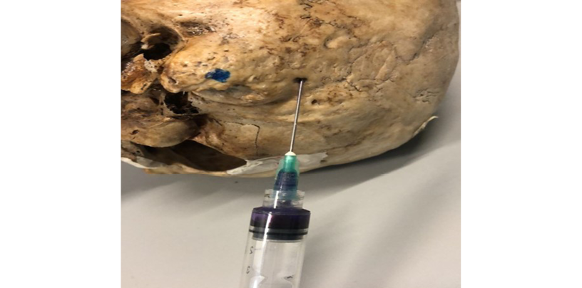
Figure 1. Example of the verification of mastoid foramen flow into the intracranial region by means of water filtration.
Statistical analysis
Data were entered into a spreadsheet created in Microsoft Excel for analysis and described using absolute frequencies, percentages, means, and ranges (minimum and maximum).
Results
The mastoid foramen was found in 94.17% (n=194) of the analyzed hemiskulls (right half: 95.15%; left half: 93.20%) (Table 1). Moreover, 10.82%, 42.78% and 37.63% of the hemiskulls had three foramina, two foramina, and one foramen, respectively.
Table 1. Frequency of mastoid foramen in the hemiskulls analyzed (n=206).
|
Frequency of mastoid foramen in the hemiskulls analyzed |
||||||
|
Right half (n=103) |
Left half (n=103) |
Total halves (n=206) |
||||
|
n |
% |
n |
% |
n |
% |
|
|
No foramen |
5 |
4.85 |
7 |
6.80 |
12 |
5.83 |
|
With a foramen |
98 |
95.15 |
96 |
93.20 |
194 |
94.17 |
|
Distribution of foramina |
||||||
|
Right half (n=98) |
Left half (n=96) |
Total halves (n=194) |
||||
|
1 foramen |
37 |
37.76 |
36 |
37.5 |
73 |
37.63 |
|
2 foramina |
43 |
43.88 |
40 |
41.67 |
83 |
42.78 |
|
3 foramina |
8 |
8.16 |
13 |
13.54 |
21 |
10.82 |
|
4 foramina |
6 |
6.12 |
4 |
4.16 |
10 |
5.15 |
|
5 foramina |
3 |
3.06 |
0 |
0 |
3 |
1.55 |
|
6 foramina |
1 |
1.02 |
2 |
2.08 |
3 |
1.55 |
|
7 foramina |
0 |
0 |
0 |
0 |
0 |
0 |
|
8 foramina |
0 |
0 |
1 |
1.04 |
1 |
0.52 |
Regarding morphometric characteristics, the average diameter and distance to the occipitomastoid suture were 1.82mm (range: 0.28-7.3) and 4.08mm (range: 0-27.16), respectively (Table 2).
Table 2. Variations in the mastoid foramen.
|
Parameter |
Total number of foramina found |
Minimum |
Maximum |
Median |
|
Diameter (mm) |
383 |
0.28 |
7.3 |
1.82 |
|
Distance to the occipitomastoid suture (mm) |
383 |
0 |
27.16 |
4.08 |
A total of 55.87% of the foramina communicated with the groove for sigmoid sinus (Table 3). In addition, in one case, a foramen communicated with the groove for greater petrosal nerve at the level of the middle cranial fossa (Figure 2) and, in another, a possible communication with other emissary veins within the diploe of the temporal bone was observed (Figure 3).
Table 3. Mastoid foramen communication.
|
Communication |
Number of foramina (n=383) |
Percentage |
|
None |
148 |
38.64% |
|
Groove for sigmoid sinus |
214 |
55.87% |
|
Lateral part of the cerebellar fossa |
14 |
3.66% |
|
At the transverse sigmoid junction |
5 |
1.31% |
|
Groove for greater petrosal nerve, in the middle cranial fossa |
1 |
0.26% |
|
Emissary veins within the diploe of the temporal bone |
1 |
0.26% |
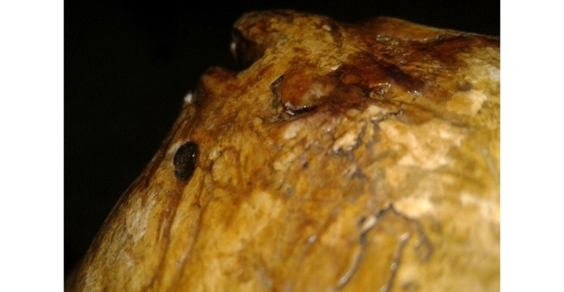
Figure 2. Mastoid foramen communicating with the groove for greater petrosal nerve in the middle cranial fossa.
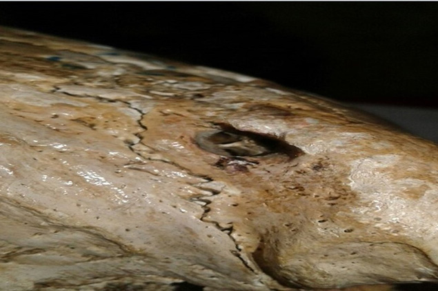
Figure 3. Possible communication of the mastoid foramen with other emissary veins within the diploe of the temporal bone.
Discussion
Morphometric variations of the mastoid process have been studied since the early twentieth century, mainly in dried skulls and cadavers.5 However, in vivo studies have also been performed using different technologies such as CT scans, X-rays, and image analysis software.6 Based on the knowledge obtained from these studies and those carried out in other species, it has been hypothesized that the presence of the mastoid foramen allows a greater cerebral blood flow in humans, which has been interpreted as a trait of human evolution.5,6
There are multiple openings in the skull through which important neural and vascular structures run. Therefore, identifying these structures is essential not only to understand the neurovascular anatomy of the region, but also to differentiate between normal structures and possible anomalies, as misinterpretation of these variations can lead to complications during surgery.7
In the present study, the prevalence of the mastoid foramen was 94.17% (n=194), with a presence of between 1 and 8 foramina per hemiskull. This figure is higher than that reported by Kim et al.2 in a study conducted in Australia on 80 dry skulls, in which 83.4% of the skulls were found to have at least one foramen. It is also higher than the figure reported by Turgut et al.8 in a study of 586 dry skulls from Anatolia, in which they found a prevalence of mastoid foramen of 78.5%. Finally, Yurdabakan et al.6 also reported a prevalence of mastoid foramen of 82% (0-4 foramina) in a study of 472 patients in Turkey.
Furthermore, in the present study the prevalence of mastoid foramen per hemiskull was 95.15% for the right half and 93.20% for the left half. These results are partially similar to those reported by Louis et al.9 in a work performed in the United States on 200 skulls (100 cadaver heads and 100 dried human skulls), where the prevalence of mastoid foramina (1 to 4) was 72% and 98% for the left and right halves, respectively.
The average mastoid foramen diameter in our study was 1.82mm, with a minimum of 0.28mm and a maximum of 7.3mm.This value is higher than the one reported by Kim et al.2 (average diameter of 1.64mm, with a maximum of 7mm), but lower than the one described by Yurdabakan et al.6 (3.39±1.48mm).
The mastoid foramen and some bone canals on the external surface of the mastoid region are important not only as an epigenetic variation, but also because of the vessels that run through them.10 In this regard, it should be noted that, first, the structure of the canal is not described in classical anatomy textbooks, and second, the information on this foramen and the mastoid emissary vein is not very extensive.11
Based on the results obtained in the present study, as well as those reported worldwide, it is possible to assume that there is a great variety in the presence and dimensions of mastoid foramina in the dry skulls of the adult population, since they may be absent or even have multiple mastoid foramina with different diameters in one or both halves of the skull. Furthermore, these figures probably vary depending on the place of origin of the skulls.
In this sense, proper intraoperative identification of the anatomical variations of these structures, especially by neurosurgeons and maxillofacial surgeons, is crucial, since unawareness of these variations can lead to misinterpretations during surgery and, consequently, to complications.3,4
It should be pointed out that a limitation of this research was the failure to make inferences due to the use of a sample of dry skulls from non-comparable populations.
Conclusion
In the Colombian population, the mastoid foramen is of variable size. It may be absent or there may be one or more foramina in each half of the skull, and it generally communicates with the groove for sigmoid sinus, although it may communicate with other parts of the endocranium.
Conflicts of interest
None stated by the authors.
Funding
None stated by the authors.
Acknowledgments
None stated by the authors.
References
1.Hampl M, Kachlik D, Kikalova K, Riemer R, Halaj M, Novak V, et al. Mastoid foramen, mastoid emissary vein and clinical implications in neurosurgery. Acta Neurochir (Wien). 2018;160(7):1473-82. https://doi.org/gdrfws.
2.Kim LK, Ahn CS, Fernandes AE. Mastoid emissary vein: anatomy and clinical relevance in plastic & reconstructive surgery. J Plast Reconstr Aesthet Surg. 2014;67(6):775-80. https://doi.org/f54q6j.
3.Zhou W, Di G, Rong J, Hu Z, Tan M, Duan K, et al. Clinical applications of the mastoid emissary vein. Surg Radiol Anat. 2023;45(1):55-63. https://doi.org/mgs9.
4.Pekçevik R, Öztürk A, Pekçevik Y, Toka O, Güçlü-Aslan G, Çukurova İ. Mastoid Emissary Vein Canal Incidence and Its Relationship with Jugular Bulb and Sigmoid Sulcus Anatomical Variations. Turk Arch Otorhinolaryngol. 2021;59(4):244-52. https://doi.org/mgtb.
5.Pereira GAM, Lopes PTC, Santos AMPV, Pozzobon A. Study of landmarks in dried skulls in a Brazil population. J Morphol Sci. 2013;30(2):94-7.
6.Yurdabakan ZZ, Okumuş Ö, Orhan K. The morphometric analysis of mastoid foramen and mastoid emissary canal on cone-beam computed tomography (CBCT). Surg Radiol Anat. 2023;45(3):303-14. https://doi.org/mgtc.
7.Freire AR, Rossi AC, de Oliveira VCS, Prado FB, Caria PHF, Botacin PR. Emissary foramens of the human skull: anatomical characteristics and its relations with clinical neurosurgery. Int J Morphol. 2013;31(1):287-92. https://doi.org/mgtd.
8.Turgut HB, Anil A, Peker T, Pelin C, Sevim A. The incidence and localization of mastoid foramen and superficial parietomastoid canal and their relations with each other. Kaibogaku Zashi. 1998;73(3):223-31.
9.Louis RG Jr, Loukas M, Wartmann CT, Tubbs RS, Apaydin N, Gupta AA, et al. Clinical anatomy of the mastoid and occipital emissary veins in a large series. Surg Radiol Anat. 2009;31(2):139-44. https://doi.org/dq82wp.
10.Koesling S, Kunkel P, Schul T. Vascular anomalies, sutures and small canals of the temporal bone on axial CT. Eur J Radiol. 2005;54(3):335-43. https://doi.org/dh77w8.
11.Hernández-Rodríguez AN, Galindo-de León S, Morales-Avalos R, Theriot-Girón MC, de la Garza-Castro O, Elizondo-Omaña RE, et al. Prevalencia y Características Morfométricas del Foramen Mastoideo y Vena Emisaria Mastoidea en Población Mexicana. Int. J. Morphol. 2014;32(2):395-98. https://doi.org/mgtf.
Referencias
Hampl M, Kachlik D, Kikalova K, Riemer R, Halaj M, Novak V, et al. Mastoid foramen, mastoid emissary vein and clinical implications in neurosurgery. Acta Neurochir (Wien). 2018;160(7):1473-82. https://doi.org/gdrfws.
Kim LK, Ahn CS, Fernandes AE. Mastoid emissary vein: anatomy and clinical relevance in plastic & reconstructive surgery. J Plast Reconstr Aesthet Surg. 2014;67(6):775-80. https://doi.org/f54q6j.
Zhou W, Di G, Rong J, Hu Z, Tan M, Duan K, et al. Clinical applications of the mastoid emissary vein. Surg Radiol Anat. 2023;45(1):55-63. https://doi.org/mgs9.
Pekçevik R, Öztürk A, Pekçevik Y, Toka O, Güçlü-Aslan G, Çukurova İ. Mastoid Emissary Vein Canal Incidence and Its Relationship with Jugular Bulb and Sigmoid Sulcus Anatomical Variations. Turk Arch Otorhinolaryngol. 2021;59(4):244-52. https://doi.org/mgtb.
Pereira GAM, Lopes PTC, Santos AMPV, Pozzobon A. Study of landmarks in dried skulls in a Brazil population. J Morphol Sci. 2013;30(2):94-7. DOI: https://doi.org/10.4067/S0717-95022012000200006
Yurdabakan ZZ, Okumuş Ö, Orhan K. The morphometric analysis of mastoid foramen and mastoid emissary canal on cone-beam computed tomography (CBCT). Surg Radiol Anat. 2023;45(3):303-14. https://doi.org/mgtc.
Freire AR, Rossi AC, de Oliveira VCS, Prado FB, Caria PHF, Botacin PR. Emissary foramens of the human skull: anatomical characteristics and its relations with clinical neurosurgery. Int J Morphol. 2013;31(1):287-92. https://doi.org/mgtd.
Turgut HB, Anil A, Peker T, Pelin C, Sevim A. The incidence and localization of mastoid foramen and superficial parietomastoid canal and their relations with each other. Kaibogaku Zashi. 1998;73(3):223-31.
Louis RG Jr, Loukas M, Wartmann CT, Tubbs RS, Apaydin N, Gupta AA, et al. Clinical anatomy of the mastoid and occipital emissary veins in a large series. Surg Radiol Anat. 2009;31(2):139-44. https://doi.org/dq82wp.
Koesling S, Kunkel P, Schul T. Vascular anomalies, sutures and small canals of the temporal bone on axial CT. Eur J Radiol. 2005;54(3):335-43. https://doi.org/dh77w8.
Hernández-Rodríguez AN, Galindo-de León S, Morales-Avalos R, Theriot-Girón MC, de la Garza-Castro O, Elizondo-Omaña RE, et al. Prevalencia y Características Morfométricas del Foramen Mastoideo y Vena Emisaria Mastoidea en Población Mexicana. Int. J. Morphol. 2014;32(2):395-98. https://doi.org/mgtf.
Cómo citar
APA
ACM
ACS
ABNT
Chicago
Harvard
IEEE
MLA
Turabian
Vancouver
Descargar cita
Licencia
Derechos de autor 2024 Revista de la Facultad de Medicina

Esta obra está bajo una licencia Creative Commons Reconocimiento 3.0 Unported.
-


