


Publicado
El verdadero origen aparente de los nervios glosofaríngeo, vago y accesorio
The true apparent origin of glossopharyngeal, vagus and accessory nerves
DOI:
https://doi.org/10.15446/revfacmed.v67n2.68096Palabras clave:
Neuroanatomía, Médula oblongada, Nervios craneales (es)Neuroanatomy, Medulla Oblongata, Cranial nerves (en)
Introducción. Existe un vacío conceptual asociado con los sitios precisos por donde emergen las raíces de los nervios glosofaríngeo, vago y accesorio, un conocimiento que es de suma importancia para los neurocirujanos.
Objetivo. Determinar el sitio preciso por donde las raíces de los nervios glosofaríngeo, vago y accesorio emergen como origen aparente en la médula oblongada.
Materiales y métodos. Se valoraron 67 troncos encefálicos humanos que con anterioridad habían sido fijados en solución de formalina al 10%. Mediante inspección directa, luego de retirar las meninges, se examinó y registró el sitio preciso por donde emergen las raíces de tales nervios y se comparó con lo registrado en la literatura.
Resultados. En el 100% de los troncos encefálicos estudiados se encontró que las raíces nerviosas emergen entre 2mm a 3mm por detrás del surco retro-olivar, distinto a lo reportado en la literatura consultada.
Conclusión. Hay disparidad de criterios en cuanto al origen aparente de los nervios glosofaríngeo, vago y accesorio, lo que amerita un estudio más amplio que permita llegar a un consenso generalizado sobre el sitio preciso por donde las raíces de tales nervios hacen su aparición.
Introduction: There is a conceptual gap associated with the sites where the roots of the glossopharyngeal, vagus and accessory nerves emerge, which is information of utmost importance for neurosurgeons.
Objective: To determine the exact site where the roots of the glossopharyngeal, vagus and accessory nerves emerge, considering the medulla oblongata as their apparent place of origin.
Materials and methods: 67 human brainstems that had previously been fixed in 10% formalin solution were examined. After removing the meninges, the precise site where the roots of these nerves emerged was analyzed by direct inspection and the results recorded and compared to the literature.
Results: In 100% of the brainstems studied, it was found that the nerve roots emerge between 2mm and 3mm behind the retro-olivary groove, which differs from the reports of the consulted literature.
Conclusion: There are different opinions about the apparent origin of the glossopharyngeal, vagus and accessory nerves. This requires a deeper study that allows reaching a general consensus on the precise place where the roots of these nerves originate.
Investigación original
DOI: https://doi.org/10.15446/revfacmed.v67n2.68096
El verdadero origen aparente de los nervios glosofaríngeo, vago y accesorio
The true apparent origin of glossopharyngeal, vagus and accessory nerves
Recibido: 03/10/2017. Aceptado: 26/12/2017.
Jorge Eduardo Duque-Parra1,2 • John Barco-Ríos1,2 • Johanna Alexandra Barco-Cano2,3
1 Universidad de Caldas - Facultad de Ciencias para la Salud - Departamento de Ciencias Básicas - Manizales - Colombia.
2 Universidad de Caldas - Grupo Neurociencia de Caldas - Manizales - Colombia.
3 Universidad de Caldas - Facultad de Ciencias para la Salud - Departamento Clínico - Manizales - Colombia.
Correspondencia: John Barco-Ríos. Departamento de Ciencias Básicas, Facultad de Ciencias para la Salud, Universidad de Caldas. Calle 65 No. 26-10, oficina: 203. Teléfono: +57 6 8781500. Manizales. Colombia. Correo electrónico: jhon.barco@ucaldas.edu.co.
| Resumen |
Introducción. Existe un vacío conceptual asociado con los sitios precisos por donde emergen las raíces de los nervios glosofaríngeo, vago y accesorio, un conocimiento que es de suma importancia para los neurocirujanos.
Objetivo. Determinar el sitio preciso por donde las raíces de los nervios glosofaríngeo, vago y accesorio emergen como origen aparente en la médula oblongada.
Materiales y métodos. Se valoraron 67 troncos encefálicos humanos que con anterioridad habían sido fijados en solución de formalina al 10%. Mediante inspección directa, luego de retirar las meninges, se examinó y registró el sitio preciso por donde emergen las raíces de tales nervios y se comparó con lo registrado en la literatura.
Resultados. En el 100% de los troncos encefálicos estudiados se encontró que las raíces nerviosas emergen entre 2mm a 3mm por detrás del surco retro-olivar, distinto a lo reportado en la literatura consultada.
Conclusión. Hay disparidad de criterios en cuanto al origen aparente de los nervios glosofaríngeo, vago y accesorio, lo que amerita un estudio más amplio que permita llegar a un consenso generalizado sobre el sitio preciso por donde las raíces de tales nervios hacen su aparición.
Palabras clave: Neuroanatomía; Médula oblongada; Nervios craneales (DeCS).
Duque-Parra JE, Barco-Ríos J, Barco-Cano JA. El verdadero origen aparente de los nervios glosofaríngeo, vago y accesorio. Rev. Fac. Med. 2019;67(2):217-20. Spanish. doi: https://doi.org/10.15446/revfacmed.v67n2.68096.
| Abstract |
Introduction: There is a conceptual gap associated with the sites where the roots of the glossopharyngeal, vagus and accessory nerves emerge, which is information of utmost importance for neurosurgeons.
Objective: To determine the exact site where the roots of the glossopharyngeal, vagus and accessory nerves emerge, considering the medulla oblongata as their apparent place of origin.
Materials and methods: 67 human brainstems that had previously been fixed in 10% formalin solution were examined. After removing the meninges, the precise site where the roots of these nerves emerged was analyzed by direct inspection and the results recorded and compared to the literature.
Results: In 100% of the brainstems studied, it was found that the nerve roots emerge between 2mm and 3mm behind the retro-olivary groove, which differs from the reports of the consulted literature.
Conclusion: There are different opinions about the apparent origin of the glossopharyngeal, vagus and accessory nerves. This requires a deeper study that allows reaching a general consensus on the precise place where the roots of these nerves originate.
Keywords: Neuroanatomy; Medulla Oblongata; Cranial Nerves (MeSH).
Duque-Parra JE, Barco-Ríos J, Barco-Cano JA. [The true apparent origin of glossopharyngeal, vagus and accessory nerves]. Rev. Fac. Med. 2019;67(2):217-20. Spanish. doi: https://doi.org/10.15446/revfacmed.v67n2.68096.
Introducción
En la Terminologia Anatomica se nominan algunos elementos neuroanatómicos que hacen parte de la médula oblongada, como la oliva, el surco retro-olivar, el surco posterolateral y el área retro-olivar (1,2), todos ellos asociados con el origen aparente de los nervios craneales glosofaríngeo, vago y accesorio. En diversos textos de neuroanatomía y artículos relacionados se observa un vacío conceptual asociado con los sitios precisos por donde emergen las raíces de tales nervios, mientras que en otros textos, que se citan más adelante, se manifiestan algunos errores de apreciación y de nominación de algunos elementos que no son reconocidos por la Terminologia Anatomica (1).
En la literatura consultada es evidente la disparidad de criterios relacionados con el sitio por donde emergen las raíces de los nervios glosofaríngeo, vago y accesorio, además de una serie de inconsistencias en la descripción neuroanatómica de algunas estructuras relacionadas con dicho origen: lateral a la oliva bulbar queda el surco posterolateral, el cual se extiende anterior al fascículo cuneiforme y la terminación anterior del tubérculo trigeminal (2); a lo largo de este surco, en la región del bulbo, se encuentra el origen aparente de estos tres nervios, en línea con las raíces posteriores de los nervios espinales (3). Otros autores refieren que en el surco retro-olivar (4-6) o fisura post-olivar (7) se encuentra el nervio vago, el cual emerge detrás de la oliva, igual que lo hacen el glosofaríngeo y el accesorio (2,4-6,8,9). Asimismo, se describe que a lo largo del surco retro-olivar, una ranura poco profunda ubicada entre la oliva y la superficie posterolateral de la médula, emergen estos tres nervios como una línea de raicillas (10). Para Tubbs et al. (11), los nervios glosofaríngeo y vago surgen del tercio superior de la cara posterior de la oliva. Como se puede apreciar, no existe una descripción unificada que indique el lugar exacto por donde tales nervios se hacen visibles como origen aparente, lo que lleva a confusión y a errores de interpretación; esto es evidente en las siguientes descripciones:
Nervio glosofaríngeo: está formado por 3 a 5 raicillas que salen del surco dorsolateral de la médula oblongada, por encima del vago y por debajo del vestíbulo coclear (12). También se menciona que surge por la parte posterior de la oliva como una o raramente dos raicillas, caudal al origen del nervio facial (10). Otros autores estiman que emerge entre la oliva inferior y el pedúnculo cerebeloso inferior (13,14); incluso se cita que emerge del surco lateral de la médula, caudal al nervio facial (15). También se señala que tiene su origen en la parte superior del surco colateral posterior (16,17), o que se ubica en el surco retro-olivar (5,6,18,19) o surco post-olivar (20). Asimismo, se afirma, sin dar mayores detalles, que emerge detrás de la oliva (21). Tubbs et al. (11) señalan que emerge por la cara posterior de la oliva, en su tercio superior, pero se quedan cortos al no mencionar ningún surco de salida.
Nervio vago: está conformado por 10 a 15 raíces procedentes de la medula oblonga, por detrás de la oliva (21,22) o lateral a ella (14,23,24) e incluso en el tercio superior de su cara lateral (10,11). Chatain & Bustamante (12) le dan una ubicación más específica a este nervio al señalar que emerge con 16 filetes radiculares del surco dorsolateral de la médula oblongada, por debajo del nervio glosofaríngeo, mientras que otros autores describen que las fibras del vago emergen del surco retro-olivar (5,6,18,19) o fisura post-olivar (7,9). También se señala que la emergencia de este ocurre por el surco colateral del bulbo (16,17), entre los nervios glosofaríngeo y accesorio (16); Pansky (15) lo ubica en el surco lateral de la médula y Escobar & Pimienta (25) indican que se origina de la médula oblongada, sin especificar el sitio exacto.
Nervio accesorio: hace su aparición mediante 4 a 6 raíces (22) por detrás de la oliva (2,4,5,8,9,21,22), pero no se especifica el lugar preciso de su aparición. También se describe sin mayores detalles que sus raíces craneales emergen por la parte lateral de las olivas (14,23,26), en sus dos tercios inferiores (10); Sarrazin et al. (18) lo ubican en el surco retro-olivar (18). Algunos autores hacen una descripción un poco más detallada, pues señalan que se hace visible por el surco posterolateral (2,3) o dorsolateral de la médula oblongada, con 4 a 5 filetes radiculares que emergen a continuación de las raíces del vago (12); para Spalteholz (27), emerge con 3 a 6 filetes nerviosos por un surco que recorre el bulbo por detrás de la oliva bulbar, inmediatamente por debajo del nervio vago. También se menciona al surco colateral posterior como el sitio de origen del accesorio (16,17). Testut & Latarjet (28) señalan que presenta 4 a 5 raíces bulbares que emergen por el surco lateral del bulbo, por debajo del vago. Finalmente, Lachman et al. (29) refieren que la raíz craneal del nervio emerge entre la oliva y el pedúnculo cerebeloso inferior, sin más detalle.
El objetivo del presente estudio fue determinar el sitio preciso en la médula oblongada por donde las raíces de estos tres nervios craneales emergen como origen aparente.
Materiales y métodos
Se realizó un estudio observacional cuantitativo en 75 troncos encefálicos humanos que se conservan en los laboratorios de anatomía de la Universidad de Caldas y la Universidad de Manizales con el propósito de identificar con precisión el sitio donde ocurre el origen aparente de los nervios glosofaríngeo, vago y accesorio. Los troncos encefálicos estaban previamente fijados con solución de formalina al 10%.
Como criterios de exclusión, se omitieron 8 piezas que presentaban algún tipo de deformación o destrucción de las raíces nerviosas por la manipulación indebida, quedando 67 piezas para el estudio.
Mediante disección, se removieron las meninges para dejar al descubierto las áreas de estudio, en específico las dos áreas retro-olivares. Se procedió a una inspección directa para verificar y registrar el sitio preciso por donde las raíces de los nervios glosofaríngeo, vago y accesorio hacen su aparición en la médula oblongada en sus lados derecho e izquierdo, correspondiente a sus orígenes aparentes, para confrontarlos luego con aquellos sitios de origen que aparecen descritos en la literatura. Posteriormente, con un calibrador digital se midió, en milímetros, la distancia entre el surco retro-olivar y el sitio por donde emergen las raíces nerviosas. Se consignó la información obtenida y se fotografiaron las piezas neuroanatómicas estudiadas.
Para esta investigación se siguieron todos los principios de la Declaración de Helsinki (30) para la investigación médica, respetando las partes obtenidas en el estudio y la confidencialidad de los datos del fallecido, su dignidad e integridad y los requerimientos consagrados en la Resolución 8430 del Ministerio de Salud que rige la investigación biomédica en Colombia (31).
El presente artículo y el estudio del que se deriva fueron presentados ante comité de ética institucional para verificar el cumplimiento de los aspectos éticos. Se obtuvo aprobación por parte del Comité de Bioética de la Universidad de Caldas mediante acta No. 012 del 16 de octubre de 2018.
Resultados
De los 67 troncos encefálicos estudiados, se encontró que en ninguno de ellos las raíces nerviosas de los nervios glosofaríngeo, vago y accesorio hacían su aparición como origen aparente en el surco
retro-olivar, tal como se describe en la literatura consultada. Se observó que, en el 100% de las piezas neuroanatómicas estudiadas, el origen aparente de estos tres nervios craneales forma una línea continua de raicillas que se localiza entre 2mm a 3mm por detrás del surco retro-olivar, en sus lados derecho e izquierdo, concretamente en las áreas retro-olivares, tal como se puede apreciar en las Figuras 1, 2, 3 y 4. Además, no se evidencia ningún surco en aquel sitio por donde emergen las raicillas de estos nervios craneales.
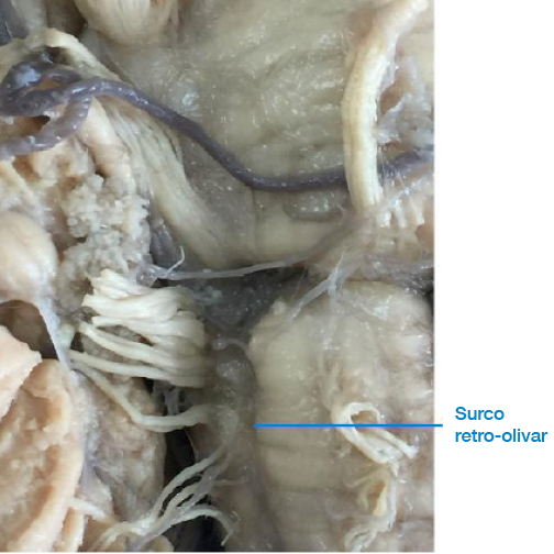
Figura 1. Surco retro-olivar donde las raíces nerviosas emergen por detrás del surco y no a través de él.
Fuente: Elaboración propia.

Figura 2. Raíces nerviosas de los nervios craneales glosofaríngeo, vago y accesorio.
Fuente: Elaboración propia.

Figura 3. Línea continua de raíces nerviosas que emergen por detrás del surco retro-olivar.
Fuente: Elaboración propia.

Figura 4. Tronco encefálico fresco donde una línea continua de raíces nerviosas emerge unos pocos milímetros por detrás del surco retro-olivar.
Fuente: Elaboración propia.
Discusión
Múltiples autores indican que el origen aparente de los nervios glosofaríngeo, vago y accesorio se encuentra en el surco retro-olivar (5,6,18,19) o surco post-olivar (7,9,20). Sin embargo, una inspección más detallada en las muestras estudiadas sobre dicho sitio de origen revela que tal descripción no es cierta y carece de precisión, por lo que se sugiere hacer más estudios en otros lugares geográficos y con una muestra mayor, esto con el fin de precisar dicho sitio de emergencia o revaluar este concepto. En los 67 troncos encefálicos utilizados para este trabajo, se encontró que en ninguno de ellos las raicillas nerviosas de estos nervios craneales emergen por los surcos retro-olivares. En el 100% de las piezas neuroanatómicas analizadas, se evidenció y registró fotográficamente que las raicillas nerviosas hacen su aparición entre 2mm a 3mm por detrás del surco retro-olivar en sus lados derecho e izquierdo, concretamente en las áreas retro-olivares; además, contrario a lo que se registra en la literatura, no se observó la presencia de ningún surco en aquel sitio por donde emergen las raicillas nerviosas.
Dos de los autores referenciados indican que estos tres nervios se originan en el surco colateral posterior del bulbo raquídeo (16,17); sin embargo, en el presente estudio no se encontró la presencia de tal surco, por otro lado, en la terminología anatómica tampoco se encuentra registrado este término neuroanatómico (1).
Puesto que el conocimiento neuroanatómico es de gran importancia en la formación de los neurocirujanos, es importante aventurarse hasta el borde del universo conocido de educación anatómica (32). Este conocimiento se enfatiza en los libros de texto durante la formación universitaria de pregrado y posgrado, pero una deficiente descripción de ciertos aspectos neuroanatómicos que requieren de gran precisión en estos profesionales dificulta la localización anatómica exacta de una lesión que afecta el tronco encefálico (33). El aprendizaje de conceptos erróneos, contradictorios o imprecisos durante la formación profesional genera en la mente un conocimiento falsamente preconcebido que pocos se atreven a cuestionar y que se va incorporando en el saber de manera permanente.
Conclusiones
Los nervios craneales glosofaríngeo, vago y accesorio no tienen su origen aparente en el surco retro-olivar, como tradicionalmente se describe en los textos y artículos referenciados. El verdadero origen aparente de estos nervios ocurre entre 2mm a 3mm por detrás del surco retro-olivar, lugar donde se aprecia una línea continua de raicillas nerviosas que se hacen visibles en ambos lados de la médula oblongada, en específico en las áreas retro-olivares. Tampoco se encontró evidencia de ningún surco en el sitio donde supuestamente emergen las raicillas de estos nervios craneales, distinto a lo que se describe en la literatura.
Debido a la dificultad para conseguir un número mayor de troncos encefálicos para que la muestra fuera aún más representativa, se sugiere la realización de otros estudios a gran escala y en otras latitudes con el fin de corroborar o rechazar estos hallazgos, lo que a su vez resolverá la disparidad de criterios que se observa en la literatura consultada. El conocimiento del verdadero origen aparente de estos tres nervios craneales puede resultar de gran utilidad a los neurocirujanos que realizan procedimientos en estas regiones, evitándose la tracción o sección involuntaria de estas raíces nerviosas.
Conflicto de intereses
Ninguno declarado por los autores.
Financiación
Ninguna declarada por los autores.
Agradecimientos
Ninguno declarado por los autores.
Referencias
1.Federativa Committee on Anatomical Terminology. Terminologia Anatomica = International anatomical terminology. Stuttgart: Thieme; 1998.
2.Dauber W. Pocket atlas of human anatomy. New York: Thieme; 2007.
3.Standring S, editor. Gray´s Anatomy. The Anatomical basis of clinical practice. 25th ed. Philadelphia: Churchill Livingstone; 2008.
4.Pró E. Anatomía Clínica. Buenos Aires: Editorial Médica Panamericana; 2012.
5.Iñiguez RA, Rebollo MA. Neuroanatomía. 7th ed. Buenos Aires: Interamericana; 1979.
6.Bustamante J. Neuroanatomía funcional y clínica. 4th ed. Bogotá: Editorial Médica Celsus; 2007.
7.Haines DE. Neuroanatomy: An Atlas of Structures, Sections, and Systems. 4th ed. Williams & Wilkins; 1995.
8.Khale W, Frotscher M. Nervous system and sensory organs. New York: Thieme; 2003.
9.Carpenter MB. Neuroanatomía. Fundamentos. 4th ed. Madrid: Editorial Médica Panamericana; 1994.
10.Rhoton AL Jr. The Cerebellopontine Angle and Posterior Fossa Cranial Nerves by the Retrosigmoid Approach. Neurosurgery. 2000;47(Suppl 3):93-129. http://doi.org/cvd2.
11.Tubbs RS, Mortazavi MM, Loukas M, Shoja MM, Cohen-Gadol AA. Intraoperative and anatomical descriptions of intracranial connections between the glossopharyngeal and vagus nerves: clinical implications. J Neurosurg. 2011;115(1):179-81. http://doi.org/ccn5xs.
12.Chatain I, Bustamante J. Anatomía macroscópica funcional y clínica. Wilmington D.C.: Addison Wesley Iberoamericana S.A.; 1986.
13.Rubin M, Safdieh J. Netter’s Concise Neuroanatomy. Philadephia: Saunders; 2007.
14.Snell RS. Neuroanatomía Clínica. 7th ed. Madrid: Lippincott Williams and Wilkins. Wolters Kluwer Health; 2009.
15.Pansky B. Review of Gross Anatomy. 6th ed. McGraw-Hill; 1996.
16.Coello CR, Coello SR. Anatomía Humana. Descripción por regiones. 2nd ed. Guayaquil: Universidad de Guayaquil; 2009.
17.Legros B, Fournier P, Chiaroni P, Ritz O, Fusciardi J. Basal fracture of the skull and lower (IX, X, XI, XII) cranial nerves palsy: four case reports including two fractures of the occipital Condyle-a literature reviews. J Trauma. 2000;48(2):342-8. http://doi.org/cfgtpq.
18.Sarrazin JL, Toulgoat F, Benoudiba F. The lower cranial nerves: IX, X, XI, XII. Diagn Interv Imaging. 2013;94(10):1051-62. http://doi.org/cvd3.
19.Policeni BA, Smoker WR. Pathologic conditions of the lower cranial nerves IX, X, XI, and XII. Neuroimaging Clin N Am. 2008;18(2):347-68. http://doi.org/bsdbns.
20.Surek CC, Van Ess M, Stephens R. Acousticofacial-glossopharyngeal triangle: an anatomic model for rapid surgical orientation. Skull Base. 2010;20(3):139-42. http://doi.org/bdhw8c.
21.Prives M, Lisenkov N, Bushkovich V. Anatomía Humana. Tomo III. Moscú: Editorial MIR Moscú; 1984.
22.Sinelnikov RD. Atlas de anatomía humana. Tomo III. Moscú: Editorial MIR Moscú; 1978.
23.Afifi AK, Bergman RA. Neuroanatomía Funcional. México D.F.: McGraw Hill Interamericana; 2006.
24.García-Porrero Pérez JA, Hurlé-González JM. Neuroanatomía humana. Madrid: Editorial Médica Panamericana; 2015.
25.Damodaran O, Risk E, Rodríguez J, Lee G. Cranial nerve assessment: a concise guide to clinical examination. Clin Anat. 2014;27(1):25-30. http://doi.org/f5k2kj.
26.Escobar M, Pimienta H. Sistema Nervioso: neuroanatomía funcional, neurohistología, neurotransmisores, receptores y clínica. Cali: Editorial Universidad del Valle; 2008.
27.Spalteholz W. Atlas de anatomía humana. Tomo III. Barcelona: Editorial Labor; 1990.
28.Testut L, Latarjet A. Tratado de anatomía humana. Tomo III. Barcelona: Salvat editores S.A.; 1973.
29.Lachman N, Acland RD, Rosse C. Anatomical evidence for the absence of a morphologically distinct cranial root of the accessory nerve in man. Clin Anat. 2002;15(1):4-10. http://doi.org/b725h5.
30.Asociación Médica Mundial. Declaración de Helsinki de la Asociación Médica Mundial. Principios éticos para las investigaciones médicas en seres humanos. Fortaleza: 64.a Asamblea General de la AMM; 2013.
31.Colombia. Ministerio de Salud. Resolución 8430 de 1993 (octubre 4): Por la cual se establecen las normas científicas, técnicas y administrativas para la investigación en salud. Bogotá D.C.; octubre 4 de 1993.
32.Paalman MH. News frontiers in anatomy education. The Anat Rec. 2000;261(2):47. http://doi.org/fsc6d3.
33.Fernández-Gil MA, Palacios-Bote R, Leo-Barahona M, Mora-Encinas JP. Anatomy of the brainstem: a gaze into the stem of life. Semin Ultrasound CT MR. 2010;31(3):196-219. http://doi.org/cs248t.
Referencias
Federativa Committee on Anatomical Terminology. Terminologia Anatomica = International anatomical terminology. Stuttgart: Thieme; 1998.
Dauber W. Pocket atlas of human anatomy. New York: Thieme; 2007.
Standring S, editor. Gray´s Anatomy. The Anatomical basis of clinical practice. 25th ed. Philadelphia: Churchill Livingstone; 2008.
Pró E. Anatomía Clínica. Buenos Aires: Editorial Médica Panamericana; 2012.
Iñiguez RA, Rebollo MA. Neuroanatomía. 7th ed. Buenos Aires: Interamericana; 1979.
Bustamante J. Neuroanatomía funcional y clínica. 4th ed. Bogotá: Editorial Médica Celsus; 2007.
Haines DE. Neuroanatomy: An Atlas of Structures, Sections, and Systems. 4th ed. Williams & Wilkins; 1995.
Khale W, Frotscher M. Nervous system and sensory organs. New York: Thieme; 2003.
Carpenter MB. Neuroanatomía. Fundamentos. 4th ed. Madrid: Editorial Médica Panamericana; 1994.
Rhoton AL Jr. The Cerebellopontine Angle and Posterior Fossa Cranial Nerves by the Retrosigmoid Approach. Neurosurgery. 2000;47(Suppl 3):93-129. http://doi.org/cvd2.
Tubbs RS, Mortazavi MM, Loukas M, Shoja MM, Cohen-Gadol AA. Intraoperative and anatomical descriptions of intracranial connections between the glossopharyngeal and vagus nerves: clinical implications. J Neurosurg. 2011;115(1):179-81. http://doi.org/ccn5xs.
Chatain I, Bustamante J. Anatomía macroscópica funcional y clínica. Wilmington D.C.: Addison Wesley Iberoamericana S.A.; 1986.
Rubin M, Safdieh J. Netter’s Concise Neuroanatomy. Philadephia: Saunders; 2007.
Snell RS. Neuroanatomía Clínica. 7th ed. Madrid: Lippincott Williams and Wilkins. Wolters Kluwer Health; 2009.
Pansky B. Review of Gross Anatomy. 6th ed. McGraw-Hill; 1996.
Coello CR, Coello SR. Anatomía Humana. Descripción por regiones. 2nd ed. Guayaquil: Universidad de Guayaquil; 2009.
Legros B, Fournier P, Chiaroni P, Ritz O, Fusciardi J. Basal fracture of the skull and lower (IX, X, XI, XII) cranial nerves palsy: four case reports including two fractures of the occipital Condyle-a literature reviews. J Trauma. 2000;48(2):342-8. http://doi.org/cfgtpq.
Sarrazin JL, Toulgoat F, Benoudiba F. The lower cranial nerves: IX, X, XI, XII. Diagn Interv Imaging. 2013;94(10):1051-62. http://doi.org/cvd3.
Policeni BA, Smoker WR. Pathologic conditions of the lower cranial nerves IX, X, XI, and XII. Neuroimaging Clin N Am. 2008;18(2):347-68. http://doi.org/bsdbns.
Surek CC, Van Ess M, Stephens R. Acousticofacial-glossopharyngeal triangle: an anatomic model for rapid surgical orientation. Skull Base. 2010;20(3):139-42. http://doi.org/bdhw8c.
Prives M, Lisenkov N, Bushkovich V. Anatomía Humana. Tomo III. Moscú: Editorial MIR Moscú; 1984.
Sinelnikov RD. Atlas de anatomía humana. Tomo III. Moscú: Editorial MIR Moscú; 1978.
Afifi AK, Bergman RA. Neuroanatomía Funcional. México D.F.: McGraw Hill Interamericana; 2006.
García-Porrero Pérez JA, Hurlé-González JM. Neuroanatomía humana. Madrid: Editorial Médica Panamericana; 2015.
Damodaran O, Risk E, Rodríguez J, Lee G. Cranial nerve assessment: a concise guide to clinical examination. Clin Anat. 2014;27(1):25-30. http://doi.org/f5k2kj.
Escobar M, Pimienta H. Sistema Nervioso: neuroanatomía funcional, neurohistología, neurotransmisores, receptores y clínica. Cali: Editorial Universidad del Valle; 2008.
Spalteholz W. Atlas de anatomía humana. Tomo III. Barcelona: Editorial Labor; 1990.
Testut L, Latarjet A. Tratado de anatomía humana. Tomo III. Barcelona: Salvat editores S.A.; 1973.
Lachman N, Acland RD, Rosse C. Anatomical evidence for the absence of a morphologically distinct cranial root of the accessory nerve in man. Clin Anat. 2002;15(1):4-10. http://doi.org/b725h5.
Asociación Médica Mundial. Declaración de Helsinki de la Asociación Médica Mundial. Principios éticos para las investigaciones médicas en seres humanos. Fortaleza: 64.a Asamblea General de la AMM; 2013.
Colombia. Ministerio de Salud. Resolución 8430 de 1993 (octubre 4): Por la cual se establecen las normas científicas, técnicas y administrativas para la investigación en salud. Bogotá D.C.; octubre 4 de 1993.
Paalman MH. News frontiers in anatomy education. The Anat Rec. 2000;261(2):47. http://doi.org/fsc6d3.
Fernández-Gil MA, Palacios-Bote R, Leo-Barahona M, Mora-Encinas JP. Anatomy of the brainstem: a gaze into the stem of life. Semin Ultrasound CT MR. 2010;31(3):196-219. http://doi.org/cs248t.
Cómo citar
APA
ACM
ACS
ABNT
Chicago
Harvard
IEEE
MLA
Turabian
Vancouver
Descargar cita
Licencia
Derechos de autor 2019 Revista de la Facultad de Medicina

Esta obra está bajo una licencia Creative Commons Reconocimiento 3.0 Unported.
-


