


Publicado
Fibronasolaringoscopia en el diagnóstico de síndrome de apnea-hipopnea obstructiva del sueño (SAHOS)
Fibronasolaringoscopy in the diagnosis of obstructive sleep apnea-hypopnea syndrome (OSAHS)
DOI:
https://doi.org/10.15446/revfacmed.v65n1Sup.59560Palabras clave:
Síndromes de la Apnea del Sueño, Endoscopia, Obstrucción de las vías aéreas, Ronquido. (es)Sleep Apnea Syndromes, Endoscopy, Airway Obstruction, Snoring. (en)
En los pacientes con diagnóstico de trastornos respiratorios del sueño (TRS) no se ha podido asociar ningún hallazgo anatómico a la severidad de la patología que el paciente presenta o a su éxito quirúrgico. Los avances actuales en la tecnología de video han permitido evaluar de manera más fidedigna las medidas de la vía aérea (VA) y trazar un mapa más exacto del sitio específico de obstrucción. La fibronasolaringoscopia es una técnica accesible y económica para la evaluación de la VA en múltiples posiciones en períodos de sueño y vigilia; esta requiere un amplio conocimiento por parte del examinador de la anatomía y fisiología de la vía aérea superior para determinar los sitios exactos de obstrucción y los patrones de colapso que podrían determinar la posibilidad del manejo quirúrgico o no, haciendo de este examen diagnóstico parte fundamental en el estudio de los pacientes con TRS.
In patients diagnosed with sleep-disordered breathing (SDB), no anatomical findings can be associated with the severity of the pathology or the surgical success. Current advances in video technology have allowed a more accurate assessment of airway measurements and a more accurate map of the specific site of obstruction.
Fibronasolaryngoscopy is an affordable and economical technique for evaluating the airway in multiple positions during sleep and wakeful periods; this requires a thorough understanding of the anatomy and physiology of the upper airway by the examiner to determine the exact sites of obstruction and patterns of collapse that could lead to a possible surgical management of the condition, making this diagnostic examination a fundamental part of the study of patients with SDB.
artículo de reflexión
DOI: https://doi.org/10.15446/revfacmed.v65n1Sup.59560
Fibronasolaringoscopia en el diagnóstico de síndrome de apnea-hipopnea obstructiva del sueño (SAHOS)
Fibronasolaringoscopy in the diagnosis of obstructive sleep apnea-hypopnea syndrome (OSAHS)
Recibido: 12/08/2016. Aceptado: 16/05/2017.
Sandra Irene Zabala-Parra1,2 Steve Amado-Galeano3 • Fritz Eduardo Gempeler-Rueda4
1 Cayre - Clínica Especializada en trastornos del sueño - Bogotá D.C. - Colombia.
2 Centro Medico Dalí - Bogotá D.C. - Colombia.
3 Clínica de la Policía - Servicio Otorrinolaringología - Bogotá D.C. - Colombia.
4 Pontificia Universidad Javeriana - Facultad de Medicina - Hospital Universitario San Ignacio - Servicio de Anestesiología
- Bogotá D.C. - Colombia.
Correspondencia: Sandra Irene Zabala-Parra. Centro Medico Dalí. Calle 97 No. 23-37, consultorio 512. Teléfono: +57 1 6425487. Bogotá D.C. Colombia. Correo electrónico: durmiendobien@yahoo.com.
| Resumen |
En los pacientes con diagnóstico de trastornos respiratorios del sueño (TRS) no se ha podido asociar ningún hallazgo anatómico a la severidad de la patología que el paciente presenta o a su éxito quirúrgico. Los avances actuales en la tecnología de video han permitido evaluar de manera más fidedigna las medidas de la vía aérea (VA) y trazar un mapa más exacto del sitio específico de obstrucción. La fibronasolaringoscopia es una técnica accesible y económica para la evaluación de la VA en múltiples posiciones en períodos de sueño y vigilia; esta requiere un amplio conocimiento por parte del examinador de la anatomía y fisiología de la vía aérea superior para determinar los sitios exactos de obstrucción y los patrones de colapso que podrían determinar la posibilidad del manejo quirúrgico o no, haciendo de este examen diagnóstico parte fundamental en el estudio de los pacientes con TRS.
Palabras clave: Síndromes de la Apnea del Sueño; Endoscopia; Obstrucción de las vías aéreas; Ronquido (DeCS).
Zabala-Parra SI, Amado-Galeano S, Gempeler-Rueda FE. Fibronasolaringoscopia en el diagnóstico de síndrome de apnea-hipopnea obstructiva del sueño (SAHOS). Rev. Fac. Med. 2017;65:S97-100. Spanish. doi: https://doi.org/10.15446/revfacmed.v65n1Sup.59560.
| Abstract |
In patients diagnosed with sleep-disordered breathing (SDB), no anatomical findings can be associated with the severity of the pathology or the surgical success. Current advances in video technology have allowed a more accurate assessment of airway measurements and a more accurate map of the specific site of obstruction.
Fibronasolaryngoscopy is an affordable and economical technique for evaluating the airway in multiple positions during sleep and wakeful periods; this requires a thorough understanding of the anatomy and physiology of the upper airway by the examiner to determine the exact sites of obstruction and patterns of collapse that could lead to a possible surgical management of the condition, making this diagnostic examination a fundamental part of the study of patients with SDB.
Keywords: Sleep Apnea Syndromes; Endoscopy; Airway Obstruction; Snoring (MeSH).
Zabala-Parra SI, Amado-Galeano S, Gempeler-Rueda FE. [Fibronasolaringoscopy in the diagnosis of obstructive sleep apnea-hypopnea syndrome (OSAHS)]. Rev. Fac. Med. 2017;65:S97-100. Spanish. doi: https://doi.org/10.15446/revfacmed.v65n1Sup.59560.
Introducción
En los pacientes con diagnóstico de trastornos respiratorios del sueño (TRS) no se ha podido asociar ningún hallazgo anatómico específico a la severidad de la patología que el paciente presenta o a su éxito quirúrgico, sea cual sea la anatomía de la vía aérea superior (VAS) o la impresión subjetiva de mayor grado de estrechez o de amplitud de la misma.
Muchas veces se encuentran pacientes longilíneos, con vías aéreas amplias y TRS severos, o pacientes obesos con trastornos craneofaciales y obstrucciones de la VAS, sin ningún tipo de patología. Estos hallazgos individuales, aunque son datos limitados, sugirieren que la apnea obstructiva del sueño es más frecuente en población adulta y que sus consecuencias son deletéreas sobre la salud de los pacientes (1).
Desarrollo
La fibronasolaringoscopia es una técnica accesible y económica para la evaluación de la vía aérea (VA) en múltiples posiciones en períodos de sueño y vigilia. Los avances en la tecnología de vídeo han permitido evaluar de manera más fidedigna las medidas de la VA y trazar un mapa más exacto del sitio específico de obstrucción. Es necesario un amplio conocimiento de la anatomía y fisiología (2).
Las diferencias significativas en la capacidad de cada examinador para detectar colapsos en cada nivel entre los observadores hace necesaria la realización de la videoendoscopia bajo sedación (DISE, Drug Induced Sleep Endoscopy); esta se indica en los casos en que los sitios de obstrucción y los patrones de colapso no se pueden identificar con la técnica convencional y, por lo tanto, tampoco se puede determinar la posibilidad del manejo quirúrgico o no (3,4). De igual forma, las diferencias de los hallazgos y la correlación entre una fibronasolaringoscopia convencional y un DISE dependen de la experiencia y el conocimiento del especialista que lo realice (5).
Fibronasolaringoscopia dinámica
Determinar la génesis anatómica exacta es de gran utilidad para definir posibles intervenciones en el nivel afectado y mejorar la permeabilidad de la VA (6). Es aún mejor si se correlacionan estos hallazgos estáticos de la morfología individual de cada paciente, por lo que se hace preciso conocer la anatomía y fisiología de un lugar determinado y ver las condiciones normales y cómo pueden variar con maniobras específicas, para así establecer diferentes escenarios de los resultados quirúrgicos (7).
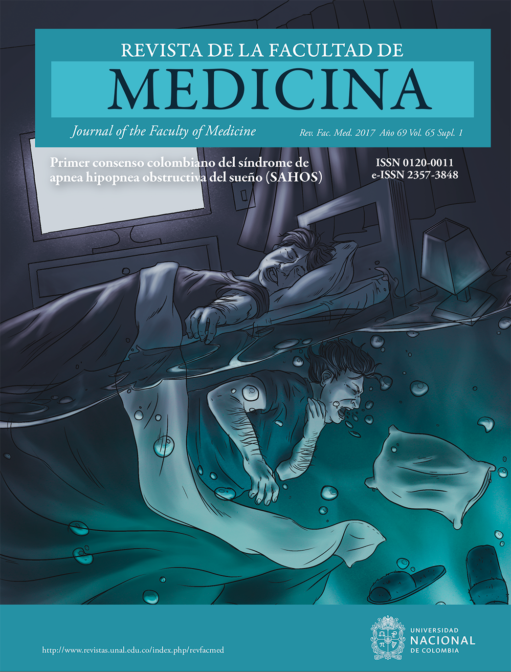
El protocolo comienza con una revisión fibroscópica estática con descripción de la morfología de cada paciente, la cual se inicia a nivel nasal, haciendo énfasis en el tamaño, forma e inserción de los cornetes, la anatomía septal y sus relaciones (Figura 1). Un sitio importante en la fisiología respiratoria es el área de la válvula nasal, el cual debe ser un sitio que el examinador evalúe de forma rutinaria para descartar vibración, colapso o estrechez a este nivel, pues su estrechez ocasiona problemas obstructivos considerables. Luego, se continúa con la evaluación del espacio nasofaríngeo, evidenciando la presencia de remanentes adenoideos o masas que pueden ser hallazgos frecuentes en pacientes con apnea obstructiva (8). Por último, se realiza una vista estática palatofaríngea superior —cuya presentación más usual es ovalada— y los hallazgos de cambios de esta morfología hacen suponer alteraciones en las estructuras anatómicas ubicadas a este nivel.
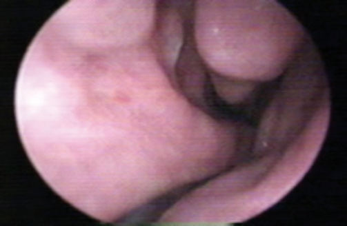
Figura 1. Vista endoscopia nasal de una septodesviación.
Fuente: Documento obtenido durante la realización del estudio.
El conocimiento de la anatomía permite realizar una evaluación exacta de los posibles sitios comprometidos; para esto, se toma la VA como un diagrama de hexágono donde se ubican cada una de las estructuras (Figura 2) y, al realizar las maniobras de ronquido e inspiración forzada —maniobra de Müller tradicional—, se evalúa el tipo de colapso presentado que puede ser anteroposterior (Figura 3), lateral (Figura 4) o mixto, lo que determina la posibilidad de manejo y el posible éxito quirúrgico.
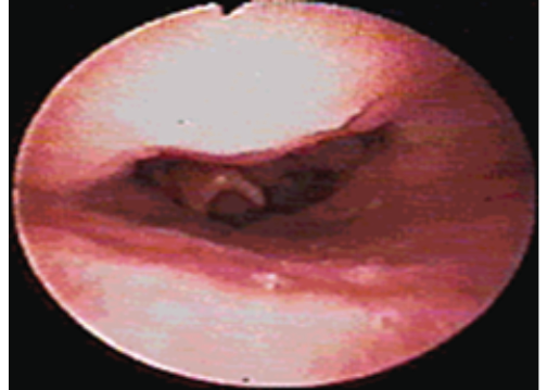
Figura 2. Hexágono diagnóstico palatofaringeo.
Fuente: Elaboración con base en Amado-Galeano (9).
Figura 3. Patrón de colapso anteroposterior.
Fuente: Documento obtenido durante la realización del estudio.

Figura 4. Patrón de colapso lateral.
Fuente: Documento obtenido durante la realización del estudio.
A nivel retroglósico, se realiza la misma evaluación teniendo en cuenta la morfología de base de lengua, así como su relación con estructuras hipofaríngeas y supraglóticas en estado de reposo (Figura 5).
Figura 5. Hexágono diagnóstico hipofaringeo.
Fuente: Elaboración con base en Amado-Galeano (9).
Una vez finalizada esta fase, se realiza un nuevo barrido donde se ven los cambios y patrones de movimientos en fonación, deglución, inspiración y espiración; se usa la maniobra de Müller para evaluar patrones de cierre, dando más importancia a las maniobras de presión negativa posterior a espiración forzada, que se denomina de presión critica de cierre. Tanto los individuos sanos como los que presentan TRS pueden presentar cambios de colapso positivo con esta maniobra.
La morfología del cierre en los pacientes con TRS es la que realmente interesa, pues muestra los vectores que se deben cambiar y corregir para lograr un aumento en el área del sitio de obstrucción. Luego, se deben buscar los sitios de vibración, haciendo maniobras que reproduzcan sonidos inducidos por el ronquido como los de absorción, espiración e inspiración nasal con boca abierta, con y sin hiperextensión, para realizar la evaluación del sitio o sitios anatómicos que están ocasionando este síntoma en los pacientes. La maniobra de protrusión mandibular permite valorar el compromiso de la posición de la mandíbula en el colapso retroglósico, el posible manejo quirúrgico maxilofacial o el éxito con uso de dispositivos de avance mandibular (DAM).
Por último, se realiza la valoración de los cambios posicionales en todos los pacientes repitiendo exactamente el mismo procedimiento con el paciente en decúbito supino o con un ángulo de inclinación no mayor de 45 grados para evaluar el efecto gravitacional sobre la anatomía. Se realizan valoraciones en decúbito lateral o prono solo cuando la clínica o posición del paciente al dormir dan indicios de que pueda encontrarse algún hallazgo con este cambio.
Fibronasolaringoscopia bajo sedación
La fibronasolaringoscopia inducida por medicamentos o bajo sedación se realiza solo cuando la técnica tradicional estándar no ha brindado la información suficiente para determinar el posible sitio de obstrucción y las posibilidades de manejo del paciente. Como se ha mencionado, el conocimiento de las zonas anatómicas donde ocurre el colapso faríngeo es indispensable para la toma de decisiones en el tratamiento médico o quirúrgico en los pacientes con síndrome de apnea-hipopnea obstructiva del sueño (SAHOS). La endoscopia durante sueño inducido por medicamentos permite la observación directa de la VAS durante el sueño.
El objetivo es llegar a un plano anestésico en el que el paciente se encuentre en respiración espontánea lo suficientemente activa para generar turbulencia, ronquido y colapso, pero lo suficientemente superficial para no generar depresión respiratoria ni apnea inducida por medicamentos (10). Es decir, inducir el sueño para crear una situación similar al sueño espontáneo y observar si se presentan ronquidos y patrones de apnea secundarios a obstrucción en diferentes partes de la VAS (11,12).
Se debe tener en cuenta que la sedación puede alterar significativamente la dinámica de las vías respiratorias superiores durante los eventos de apnea; por lo cual debe tenerse sumo cuidado en no inducir relajación muscular excesiva con los medicamentos, pues se puede llegar a patrones respiratorios inadecuados creando falsos positivos del colapso de la VAS. Antes del procedimiento es obligatorio realizar una adecuada valoración clínica, pues la apnea obstructiva del sueño y los trastornos del sueño pueden causar, de forma crónica, importantes cambios fisiológicos en los sistemas cardíaco y pulmonar, los cuales, a su vez, pueden tener implicaciones importantes en la forma de administrar la sedación que comprometen la seguridad durante el procedimiento.
Se recomienda que esta técnica de sedación sea ejecutada por un anestesiólogo con experiencia en este tipo de procedimiento y que se lleve a cabo en un espacio adecuado, donde se cuente con fuente de oxígeno, equipos y medicamentos necesarios en caso de paro respiratorio o cardíaco. Antes de iniciar el procedimiento, se realiza monitoreo fisiológico permanente con electrocardiografía, oximetría de pulso y medición de la presión arterial de forma no invasiva a intervalos regulares. En las instituciones donde se cuentan con el BIS (análisis biespectral electroencefalográfico, por sus siglas en inglés) o entropía, se recomienda su utilización para monitorear de forma más objetiva el plano de sedación —entre 60 y 80 unidades del BIS— (15).
Más adelante se aplica anestesia tópica con lidocaína en las fosas nasales y la orofaringe con el fin de disminuir los requerimientos de sedación durante la introducción del fibrolaringoscopio y se inicia la administración de oxígeno suplementario por medio de cánula o catéter nasal a necesidad, según la saturación de oxígeno. Algunos autores recomiendan la utilización previa de atropina o glicopirrolato con el objetivo de disminuir las secreciones en la VAS y así facilitar la endoscopia (13).
Desde 1991, cuando Croft & Pringle (16) propusieron por primera vez la técnica DISE, muchos agentes sedantes han sido considerados en diferentes dosis. En la actualidad, la sedación se puede lograr con múltiples medicamentos como Propofol, Midazolam, Dexmedetomidina, entre otros, que se administran combinados o de forma individual, a dosis diferentes, ya sea con administración manual de bolos, infusión continua o bombas de infusión Target Control Infusión (TCI).
El objetivo final es lograr una sedación con respiración espontánea —sueño inducido—, en la cual el paciente tolere la introducción de fibroscopio sin producir apnea inducida por los medicamentos. Los opioides y los anestésicos inhalados, por lo general, no se recomiendan dados los efectos depresores respiratorios. Es de anotar que no hay protocolos de sedación estandarizados ni ensayos clínicos aleatorios: solo existen reportes de casos.
Entre los agentes más utilizados en esta técnica está el midazolam; algunos autores recomiendan su administración intravenosa lenta de pequeños bolos (12). El propofol, que en la actualidad es uno de los más usados en esta técnica diagnóstica, tiene una fluctuación de la concentración en sangre bastante grande debido a la variación subjetiva en la administración de bolos manuales, con la consecuente fluctuación del efecto de sedación. La infusión continua de propofol induce de forma más fiable un sueño adecuado, al tiempo que permite una rápida recuperación cuando se detiene la infusión (11).
Es de anotar que el sistema TCI ha sido desarrollado para proporcionar una mayor comodidad y control durante la anestesia intravenosa. El principio básico es que se ajusta la dosis de la infusión para alcanzar cierta concentración plasmática y, por ende, la profundidad de sedación deseada. Con este sistema, las tasas de infusión se alteran de forma automática y de acuerdo a un modelo farmacocinético incluido en la bomba de infusión TCI. Para algunos autores, esta técnica debe ser la primera opción en la realización de la endoscopia del sueño debido a su precisión, estabilidad y seguridad (11).
Existe un nuevo agente alfa 2 agonista adrenérgico selectivo, la dexmedetomidina, que se utiliza para la sedación con la gran ventaja de que no induce apnea. Aunque no tiene un efecto directo sobre el corazón, puede presentarse una respuesta hemodinámica bifásica con la dosis de carga inicial (13). Este aumento transitorio de la presión arterial y la frecuencia cardíaca es seguido por una disminución gradual de las mismas. La principal desventaja de este medicamento es el prolongado tiempo de latencia, que puede oscilar entre 10 y 30 minutos, además su efecto de acción prolongado puede llegar hasta 4 o 6 horas, lo que no lo hace ideal para procedimientos ambulatorios.
Una vez alcanzado el plano de sedación adecuado con la técnica seleccionada, se introduce el fibronasolaringoscopio y se buscan los sitios de vibración y de colapso. Si durante el examen despierto el paciente presenta una predisposición posicional, en el examen bajo sedación se intenta reproducir dicha posición para evaluar los hallazgos (14). Todos estos hallazgos permiten identificar el o los sitios de obstrucción (15).
Es importante tener en cuenta que para todo diagnóstico se puede contar —además del examen endoscópico— con las imágenes diagnósticas con técnicas convencionales, las más utilizadas son: tomografía axial computada de senos paranasales, radiografías laterales, cefalometría y exámenes de reconstrucción tridimensional de VA, útiles en pacientes donde la obstrucción no ha sido determinada de forma clara o hay dudas en la misma.
Conclusiones
En la actualidad, realizar un adecuado diagnóstico polisomnográfico, clínico, radiológico y endoscópico permite al especialista tomar la mejor opción de tratamiento en el paciente con SAHOS: tratamiento con presión positiva sobre la vía aérea (PAP), manejos médicos, quirúrgicos, uso de dispositivos de avance mandibular o manejos combinados que tendrán una mejor tasa de efectividad si se está dando manejo al sitio o sitios específicos de obstrucción de la VAS.
Conflicto de intereses
Ninguno declarado por los autores.
Financiación
Ninguna declarada por los autores.
Agradecimientos
A la Asociación Colombiana de Medicina Interna (ACMI® - Médicos para adultos), la Asociación Colombiana de Neurología (ACN) y la Asociación Colombiana de Sociedades Científicas (ACSC) por permitir a los autores usar sus instalaciones como lugar de reunión de trabajo.
Referencias
1.Carrasco-Llatas M, Marcano-Acuña M, Zerpa-Zerpa V, Dalmau-Galofre J. Surgical results of different palate techniques to treat oropharyngeal collapse. Eur Arch Otorhinolaryngol. 2015;272(9):2535-40. http://doi.org/bmjg.
2.Zhang P, Ye J, Pan C, Xian J, Sun N, Li J, et al. Comparison of drug-induced sleep endoscopy and upper airway computed tomography in obstructive sleep apnea patients. Eur Arch Otorhinolaryngol. 2014;271(10):2751-6. http://doi.org/bnr8.
3.Koo SK, Choi JW, Myung NS, Lee HJ, Kim YJ, Kim YJ. Analysis of obstruction site in obstructive sleep apnea syndrome patients by drug induced sleep endoscopy. Am J Otolaryngol. 2013;34(6):626-30. http://doi.org/f2xrr9.
4.Aktas O, Erdur O, Cirik AA, Kayhan FT. The role of drug-induced sleep endoscopy in surgical planning for obstructive sleep apnea syndrome. Eur Arch Otorhinolaryngol. 2015;272(8):2039-43. http://doi.org/bnr9.
5.Yilmaz YF, Kum RO, Ozcan M, Gungor V, Unal A. Drug-induced sleep endoscopy versus Müller maneuver in patients with retropalatal obstruction. Laryngoscope. 2015;125(9):2220-5. http://doi.org/bmjh.
6.Zhang P, Ye J, Pan C, Sun N, Kang D. The Role of Obstruction Length and Height in Predicting Outcome of Velopharyngeal Surgery. Otolaryngol Head Neck Surg. 2015;153(1):144-9. http://doi.org/bnsb.
7.Lovato A, Kotecha B, Vianello A, Giacomelli L, Staffieri A, Marchese-Ragona R. Nasal and oral snoring endoscopy: novel and promising diagnostic tools in OSAS patients. Eur Arch Otorhinolaryngol. 2015;272(7):1793-9. http://doi.org/bnsc.
8.Aksoy EA, Serin GM, Polat S, Ünal OF, Tanyeri H. The morphology of the nasopharyngeal inlet in obstructive sleep apnea. Eur Arch Otorhinolaryngol. 2014;271(4):771-5. http://doi.org/bnsd.
9.Amado-Galeano S. Implante neuroestimulador del nervio hipogloso para el SAHOS. In: Plaza G, Baptista P, O’Connor C, editors. Diagnóstico y tratamiento de los trastornos respiratorios del sueño. Madrid: Hospital de la Zarzuela; 2015.
10.Carrasco-Llatas M, Agostini-Porras G, Cuesta-González MT, Rodrigo-Sanbartolomé A, Giner-Bayarri P, Gómez-Pajares F, et al. Drug-induced sleep endoscopy: a two drug comparison and simultaneous polysomnography. Eur Arch Otorhinolaryngol. 2014;271(1):181-7. http://doi.org/bnsf.
11.De Vito A, Agnoletti V, Berrettini S, Piraccini E, Criscuolo A, Corso R, et al. Drug-induced sleep endoscopy: conventional versus target controlled infusion techniques—a randomized controlled study. Eur Arch Otorhinolaryngol. 2011;268(3):457-62. http://doi.org/d7g46n.
12.Eichler C, Sommer JU, Stuck BA Hörmann K, Maurer JT. Does drug-induced sleep endoscopy change the treatment concept of patients with snoring and obstructive sleep apnea? Sleep Breath. 2013;17(1):63-8. http://doi.org/fzsj44.
13.Mathews AMV, Goh JPS, Teo LM. A Case Report on the Anaesthetic Management of Dexmedetomidine-induced Sleep Endoscopy and Transoral Robotic Surgery for the Treatment of Obstructive Sleep Apnoea. Proceedings of Singapore Healthcare. 2013;22(2):151-5. http://doi.org/bnsg.
14.Lan MC, Liu SY, Lan MY, Modi R, Capasso R. Lateral pharyngeal wall collapse associated with hypoxemia in obstructive sleep apnea. Laryngoscope. 2015;125(10):2408-12. http://doi.org/bmjj.
15.Aktas O, Erdur O, Cirik AA, Kayhan FT. The role of drug-induced sleep endoscopy in surgical planning for obstructive sleep apnea syndrome. Eur Arch Otorhinolaryngol. 2014;272(8):2039-43. http://doi.org/bnr9.
16.Croft CB, Pringle M. Sleep nasendoscopy: a technique of assessment in snoring and obstructive sleep apnoea. Clin Otolaryngol Allied Sci. 1991;16(5):504-9. http://doi.org/fmz77k.
Recibido: 12 de agosto de 2016; Aceptado: 16 de mayo de 2017
Resumen
En los pacientes con diagnóstico de trastornos respiratorios del sueño (TRS) no se ha podido asociar ningún hallazgo anatómico a la severidad de la patología que el paciente presenta o a su éxito quirúrgico. Los avances actuales en la tecnología de video han permitido evaluar de manera más fidedigna las medidas de la vía aérea (VA) y trazar un mapa más exacto del sitio específico de obstrucción. La fibronasolaringoscopia es una técnica accesible y económica para la evaluación de la VA en múltiples posiciones en períodos de sueño y vigilia; esta requiere un amplio conocimiento por parte del examinador de la anatomía y fisiología de la vía aérea superior para determinar los sitios exactos de obstrucción y los patrones de colapso que podrían determinar la posibilidad del manejo quirúrgico o no, haciendo de este examen diagnóstico parte fundamental en el estudio de los pacientes con TRS.
Palabras clave:
Síndromes de la Apnea del Sueño, Endoscopia, Obstrucción de las vías aéreas, Ronquido (DeCS).Abstract
In patients diagnosed with sleep-disordered breathing (SDB), no anatomical findings can be associated with the severity ofthe pathology or the surgical success. Current advances in video technology have allowed a more accurate assessment of airway measurements and a more accurate map of the specific site of obstruction.
Fibronasolaryngoscopy is an affordable and economical technique for evaluating the airway in multiple positions during sleep and wakeful periods; this requires a thorough understanding of the anatomy and physiology of the upper airway by the examiner to determine the exact sites of obstruction and patterns of collapse that could lead to a possible surgical management of the condition, making this diagnostic examination a fundamental part of the study of patients with SDB.
Keywords:
Sleep Apnea Syndromes, Endoscopy, Airway Obstruction, Snoring (MeSH).Introducción
En los pacientes con diagnóstico de trastornos respiratorios del sueño (TRS) no se ha podido asociar ningún hallazgo anatómico específico a la severidad de la patología que el paciente presenta o a su éxito quirúrgico, sea cual sea la anatomía de la vía aérea superior (VAS) o la impresión subjetiva de mayor grado de estrechez o de amplitud de la misma.
Muchas veces se encuentran pacientes longilíneos, con vías aéreas amplias y TRS severos, o pacientes obesos con trastornos craneofaciales y obstrucciones de la VAS, sin ningún tipo de patología. Estos hallazgos individuales, aunque son datos limitados, sugirieren que la apnea obstructiva del sueño es más frecuente en población adulta y que sus consecuencias son deletéreas sobre la salud de los pacientes 1.
Desarrollo
La fibronasolaringoscopia es una técnica accesible y económica para la evaluación de la vía aérea (VA) en múltiples posiciones en períodos de sueño y vigilia. Los avances en la tecnología de vídeo han permitido evaluar de manera más fidedigna las medidas de la VA y trazar un mapa más exacto del sitio específico de obstrucción. Es necesario un amplio conocimiento de la anatomía y fisiología 2.
Las diferencias significativas en la capacidad de cada examinador para detectar colapsos en cada nivel entre los observadores hace necesaria la realización de la videoendoscopia bajo sedación (DISE, Drug Induced Sleep Endoscopy); esta se indica en los casos en que los sitios de obstrucción y los patrones de colapso no se pueden identificar con la técnica convencional y, por lo tanto, tampoco se puede determinar la posibilidad del manejo quirúrgico o no 3,4. De igual forma, las diferencias de los hallazgos y la correlación entre una fibronasolaringoscopia convencional y un DISE dependen de la experiencia y el conocimiento del especialista que lo realice 5.
Fibronasolaringoscopia dinámica
Determinar la génesis anatómica exacta es de gran utilidad para definir posibles intervenciones en el nivel afectado y mejorar la permeabilidad de la VA 6. Es aún mejor si se correlacionan estos hallazgos estáticos de la morfología individual de cada paciente, por lo que se hace preciso conocer la anatomía y fisiología de un lugar determinado y ver las condiciones normales y cómo pueden variar con maniobras específicas, para así establecer diferentes escenarios de los resultados quirúrgicos 7.
El protocolo comienza con una revisión fibroscópica estática con descripción de la morfología de cada paciente, la cual se inicia a nivel nasal, haciendo énfasis en el tamaño, forma e inserción de los cornetes, la anatomía septal y sus relaciones (Figura 1). Un sitio importante en la fisiología respiratoria es el área de la válvula nasal, el cual debe ser un sitio que el examinador evalúe de forma rutinaria para descartar vibración, colapso o estrechez a este nivel, pues su estrechez ocasiona problemas obstructivos considerables. Luego, se continúa con la evaluación del espacio nasofaríngeo, evidenciando la presencia de remanentes adenoideos o masas que pueden ser hallazgos frecuentes en pacientes con apnea obstructiva 8. Por último, se realiza una vista estática palatofaríngea superior -cuya presentación más usual es ovalada- y los hallazgos de cambios de esta morfología hacen suponer alteraciones en las estructuras anatómicas ubicadas a este nivel.
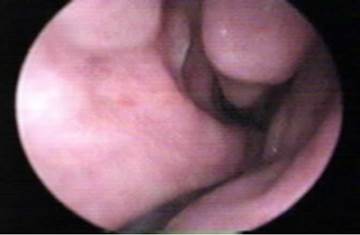
Figura 1: Vista endoscopia nasal de una septodesviación.
El conocimiento de la anatomía permite realizar una evaluación exacta de los posibles sitios comprometidos; para esto, se toma la VA como un diagrama de hexágono donde se ubican cada una de las estructuras (Figura 2) y, al realizar las maniobras de ronquido e inspiración forzada -maniobra de Müller tradicional-, se evalúa el tipo de colapso presentado que puede ser anteroposterior (Figura 3), lateral (Figura 4) o mixto, lo que determina la posibilidad de manejo y el posible éxito quirúrgico.

Figura 2: Hexágono diagnóstico palatofaringeo.
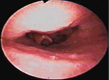
Figura 3: Patrón de colapso anteroposterior.

Figura 4: Patrón de colapso lateral.
A nivel retroglósico, se realiza la misma evaluación teniendo en cuenta la morfología de base de lengua, así como su relación con estructuras hipofaríngeas y supraglóticas en estado de reposo (Figura 5).
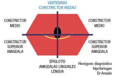
Figura 5: Hexágono diagnóstico hipofaringeo.
Una vez finalizada esta fase, se realiza un nuevo barrido donde se ven los cambios y patrones de movimientos en fonación, deglución, inspiración y espiración; se usa la maniobra de Müller para evaluar patrones de cierre, dando más importancia a las maniobras de presión negativa posterior a espiración forzada, que se denomina de presión critica de cierre. Tanto los individuos sanos como los que presentan TRS pueden presentar cambios de colapso positivo con esta maniobra.
La morfología del cierre en los pacientes con TRS es la que realmente interesa, pues muestra los vectores que se deben cambiar y corregir para lograr un aumento en el área del sitio de obstrucción. Luego, se deben buscar los sitios de vibración, haciendo maniobras que reproduzcan sonidos inducidos por el ronquido como los de absorción, espiración e inspiración nasal con boca abierta, con y sin hiperextensión, para realizar la evaluación del sitio o sitios anatómicos que están ocasionando este síntoma en los pacientes. La maniobra de protrusión mandibular permite valorar el compromiso de la posición de la mandíbula en el colapso retroglósico, el posible manejo quirúrgico maxilofacial o el éxito con uso de dispositivos de avance mandibular (DAM).
Por último, se realiza la valoración de los cambios posicionales en todos los pacientes repitiendo exactamente el mismo procedimiento con el paciente en decúbito supino o con un ángulo de inclinación no mayor de 45 grados para evaluar el efecto gravitacional sobre la anatomía. Se realizan valoraciones en decúbito lateral o prono solo cuando la clínica o posición del paciente al dormir dan indicios de que pueda encontrarse algún hallazgo con este cambio.
Fibronasolaringoscopia bajo sedación
La fibronasolaringoscopia inducida por medicamentos o bajo sedación se realiza solo cuando la técnica tradicional estándar no ha brindado la información suficiente para determinar el posible sitio de obstrucción y las posibilidades de manejo del paciente. Como se ha mencionado, el conocimiento de las zonas anatómicas donde ocurre el colapso faríngeo es indispensable para la toma de decisiones en el tratamiento médico o quirúrgico en los pacientes con síndrome de apnea-hipopnea obstructiva del sueño (SAHOS). La endoscopia durante sueño inducido por medicamentos permite la observación directa de la VAS durante el sueño.
El objetivo es llegar a un plano anestésico en el que el paciente se encuentre en respiración espontánea lo suficientemente activa para generar turbulencia, ronquido y colapso, pero lo suficientemente superficial para no generar depresión respiratoria ni apnea inducida por medicamentos 10. Es decir, inducir el sueño para crear una situación similar al sueño espontáneo y observar si se presentan ronquidos y patrones de apnea secundarios a obstrucción en diferentes partes de la VAS 11,12.
Se debe tener en cuenta que la sedación puede alterar significativamente la dinámica de las vías respiratorias superiores durante los eventos de apnea; por lo cual debe tenerse sumo cuidado en no inducir relajación muscular excesiva con los medicamentos, pues se puede llegar a patrones respiratorios inadecuados creando falsos positivos del colapso de la VAS. Antes del procedimiento es obligatorio realizar una adecuada valoración clínica, pues la apnea obstructiva del sueño y los trastornos del sueño pueden causar, de forma crónica, importantes cambios fisiológicos en los sistemas cardíaco y pulmonar, los cuales, a su vez, pueden tener implicaciones importantes en la forma de administrar la sedación que comprometen la seguridad durante el procedimiento.
Se recomienda que esta técnica de sedación sea ejecutada por un anestesiólogo con experiencia en este tipo de procedimiento y que se lleve a cabo en un espacio adecuado, donde se cuente con fuente de oxígeno, equipos y medicamentos necesarios en caso de paro respiratorio o cardíaco. Antes de iniciar el procedimiento, se realiza monitoreo fisiológico permanente con electrocardiografía, oximetría de pulso y medición de la presión arterial de forma no invasiva a intervalos regulares. En las instituciones donde se cuentan con el BIS (análisis biespectral electroencefalográfico, por sus siglas en inglés) o entropía, se recomienda su utilización para monitorear de forma más objetiva el plano de sedación -entre 60 y 80 unidades del BIS- 15.
Más adelante se aplica anestesia tópica con lidocaína en las fosas nasales y la orofaringe con el fin de disminuir los requerimientos de sedación durante la introducción del fibrolaringoscopio y se inicia la administración de oxígeno suplementario por medio de cánula o catéter nasal a necesidad, según la saturación de oxígeno. Algunos autores recomiendan la utilización previa de atropina o glicopirrolato con el objetivo de disminuir las secreciones en la VAS y así facilitar la endoscopia 13.
Desde 1991, cuando Croft & Pringle 16 propusieron por primera vez la técnica DISE, muchos agentes sedantes han sido considerados en diferentes dosis. En la actualidad, la sedación se puede lograr con múltiples medicamentos como Propofol, Midazolam, Dexmedetomidina, entre otros, que se administran combinados o de forma individual, a dosis diferentes, ya sea con administración manual de bolos, infusión continua o bombas de infusión Target Control Infusión (TCI).
El objetivo final es lograr una sedación con respiración espontánea -sueño inducido-, en la cual el paciente tolere la introducción de fibroscopio sin producir apnea inducida por los medicamentos. Los opioides y los anestésicos inhalados, por lo general, no se recomiendan dados los efectos depresores respiratorios. Es de anotar que no hay protocolos de sedación estandarizados ni ensayos clínicos aleatorios: solo existen reportes de casos.
Entre los agentes más utilizados en esta técnica está el midazolam; algunos autores recomiendan su administración intravenosa lenta de pequeños bolos 12. El propofol, que en la actualidad es uno de los más usados en esta técnica diagnóstica, tiene una fluctuación de la concentración en sangre bastante grande debido a la variación subjetiva en la administración de bolos manuales, con la consecuente fluctuación del efecto de sedación. La infusión continua de propofol induce de forma más fiable un sueño adecuado, al tiempo que permite una rápida recuperación cuando se detiene la infusión 11.
Es de anotar que el sistema TCI ha sido desarrollado piara proporcionar una mayor comodidad y control durante la anestesia intravenosa. El principio básico es que se ajusta la dosis de la infusión para alcanzar cierta concentración plasmática y, por ende, la profundidad de sedación deseada. Con este sistema, las tasas de infusión se alteran de forma automática y de acuerdo a un modelo farmacocinético incluido en la bomba de infusión TCI. Para algunos autores, esta técnica debe ser la primera opción en la realización de la endoscopia del sueño debido a su precisión, estabilidad y seguridad 11.
Existe un nuevo agente alfa 2 agonista adrenérgico selectivo, la dexmedetomidina, que se utiliza para la sedación con la gran ventaja de que no induce apnea. Aunque no tiene un efecto directo sobre el corazón, puede presentarse una respuesta hemodinámica bifásica con la dosis de carga inicial 13. Este aumento transitorio de la presión arterial y la frecuencia cardíaca es seguido por una disminución gradual de las mismas. La principal desventaja de este medicamento es el prolongado tiempo de latencia, que puede oscilar entre 10 y 30 minutos, además su efecto de acción prolongado puede llegar hasta 4 o 6 horas, lo que no lo hace ideal para procedimientos ambulatorios.
Una vez alcanzado el plano de sedación adecuado con la técnica seleccionada, se introduce el fibronasolaringoscopio y se buscan los sitios de vibración y de colapso. Si durante el examen despierto el paciente presenta una predisposición posicional, en el examen bajo sedación se intenta reproducir dicha posición para evaluar los hallazgos 14. Todos estos hallazgos permiten identificar el o los sitios de obstrucción 15.
Es importante tener en cuenta que para todo diagnóstico se puede contar -además del examen endoscópico- con las imágenes diagnósticas con técnicas convencionales, las más utilizadas son: tomografía axial computada de senos paranasales, radiografías laterales, cefalometría y exámenes de reconstrucción tridimensional de VA, útiles en pacientes donde la obstrucción no ha sido determinada de forma clara o hay dudas en la misma.
Conclusiones
En la actualidad, realizar un adecuado diagnóstico polisomnográfico, clínico, radiológico y endoscópico permite al especialista tomar la mejor opción de tratamiento en el paciente con SAHOS: tratamiento con presión positiva sobre la vía aérea (PAP), manejos médicos, quirúrgicos, uso de dispositivos de avance mandibular o manejos combinados que tendrán una mejor tasa de efectividad si se está dando manejo al sitio o sitios específicos de obstrucción de la VAS.
Conflicto de intereses
Ninguno declarado por los autores.
Financiación
Ninguna declarada por los autores.
Agradecimientos
A la Asociación Colombiana de Medicina Interna (ACMI® - Médicos para adultos), la Asociación Colombiana de Neurología (ACN) y la Asociación Colombiana de Sociedades Científicas (ACSC) por permitir a los autores usar sus instalaciones como lugar de reunión de trabajo.
Referencias
Referencias
Carrasco-Llatas M, Marcano-Acuña M, Zerpa-Zerpa V, Dalmau-Galofre J. Surgical results of different palate techniques to treat oropharyngeal collapse. Eur Arch Otorhinolaryngol. 2015;272(9):2535-40. http://doi.org/bmjg.
Zhang P, Ye J, Pan C, Xian J, Sun N, Li J, et al. Comparison of drug-induced sleep endoscopy and upper airway computed tomography in obstructive sleep apnea patients. Eur Arch Otorhinolaryngol. 2014;271(10):2751-6. http://doi.org/bnr8.
Koo SK, Choi JW, Myung NS, Lee HJ, Kim YJ, Kim YJ. Analysis of obstruction site in obstructive sleep apnea syndrome patients by drug induced sleep endoscopy. Am J Otolaryngol. 2013;34(6):626-30. http://doi.org/f2xrr9.
Aktas O, Erdur O, Cirik AA, Kayhan FT. The role of drug-induced sleep endoscopy in surgical planning for obstructive sleep apnea syndrome. Eur Arch Otorhinolaryngol. 2015;272(8):2039-43. http://doi.org/bnr9.
Yilmaz YF, Kum RO, Ozcan M, Gungor V, Unal A. Drug-induced sleep endoscopy versus Müller maneuver in patients with retropalatal obstruction. Laryngoscope. 2015;125(9):2220-5. http://doi.org/bmjh.
Zhang P, Ye J, Pan C, Sun N, Kang D. The Role of Obstruction Length and Height in Predicting Outcome of Velopharyngeal Surgery. Otolaryngol Head Neck Surg. 2015;153(1):144-9. http://doi.org/bnsb.
Lovato A, Kotecha B, Vianello A, Giacomelli L, Staffieri A, Marchese-Ragona R. Nasal and oral snoring endoscopy: novel and promising diagnostic tools in OSAS patients. Eur Arch Otorhinolaryngol. 2015;272(7):1793-9. http://doi.org/bnsc.
Aksoy EA, Serin GM, Polat S, Ünal OF, Tanyeri H. The morphology of the nasopharyngeal inlet in obstructive sleep apnea. Eur Arch Otorhinolaryngol. 2014;271(4):771-5. http://doi.org/bnsd.
Amado-Galeano S. Implante neuroestimulador del nervio hipogloso para el SAHOS. In: Plaza G, Baptista P, O’Connor C, editors. Diagnóstico y tratamiento de los trastornos respiratorios del sueño. Madrid: Hospital de la Zarzuela; 2015.
Carrasco-Llatas M, Agostini-Porras G, Cuesta-González MT, Rodrigo-Sanbartolomé A, Giner-Bayarri P, Gómez-Pajares F, et al. Drug-induced sleep endoscopy: a two drug comparison and simultaneous polysomnography. Eur Arch Otorhinolaryngol. 2014;271(1):181-7. http://doi.org/bnsf.
De Vito A, Agnoletti V, Berrettini S, Piraccini E, Criscuolo A, Corso R, et al. Drug-induced sleep endoscopy: conventional versus target controlled infusion techniques—a randomized controlled study. Eur Arch Otorhinolaryngol. 2011;268(3):457-62. http://doi.org/d7g46n.
Eichler C, Sommer JU, Stuck BA Hörmann K, Maurer JT. Does drug-induced sleep endoscopy change the treatment concept of patients with snoring and obstructive sleep apnea? Sleep Breath. 2013;17(1):63-8. http://doi.org/fzsj44.
Mathews AMV, Goh JPS, Teo LM. A Case Report on the Anaesthetic Management of Dexmedetomidine-induced Sleep Endoscopy and Transoral Robotic Surgery for the Treatment of Obstructive Sleep Apnoea. Proceedings of Singapore Healthcare. 2013;22(2):151-5. http://doi.org/bnsg.
Lan MC, Liu SY, Lan MY, Modi R, Capasso R. Lateral pharyngeal wall collapse associated with hypoxemia in obstructive sleep apnea. Laryngoscope. 2015;125(10):2408-12. http://doi.org/bmjj.
Aktas O, Erdur O, Cirik AA, Kayhan FT. The role of drug-induced sleep endoscopy in surgical planning for obstructive sleep apnea syndrome. Eur Arch Otorhinolaryngol. 2014;272(8):2039-43. http://doi.org/bnr9.
Croft CB, Pringle M. Sleep nasendoscopy: a technique of assessment in snoring and obstructive sleep apnoea. Clin Otolaryngol Allied Sci. 1991;16(5):504-9. http://doi.org/fmz77k.
Cómo citar
APA
ACM
ACS
ABNT
Chicago
Harvard
IEEE
MLA
Turabian
Vancouver
Descargar cita
CrossRef Cited-by
1. Antoinette am Zehnhoff-Dinnesen, Sevtap Akbulut, Eugenia Chávez Calderón, Muhittin Demir, Dirk Deuster, Michael Fuchs, Ahmed Geneid, Thomas Murry, Tadeus Nawka, Christiane Neuschaefer-Rube, Ewa Niebudek-Bogusz, Andrzej Obrębowski, Haldun Oguz, Arno Olthoff, Anders Overgård Jønsson, Mette Pedersen, Bernhard Richter, John Rubin, Berit Schneider-Stickler, Kevin Shields, Mariola Śliwińska-Kowalska, Bożena Wiskirska-Woźnica, Virginie Woisard, Waldemar Wojnowski. (2020). Phoniatrics I. European Manual of Medicine. , p.239. https://doi.org/10.1007/978-3-662-46780-0_5.
Dimensions
PlumX
Visitas a la página del resumen del artículo
Descargas
Licencia
Derechos de autor 2017 Revista de la Facultad de Medicina

Esta obra está bajo una licencia Creative Commons Reconocimiento 3.0 Unported.
-


