


Publicado
Nanopartículas de CuO y su propiedad antimicrobiana en cepas intrahospitalarias
CuO nanoparticles and their antimicrobial activity against nosocomial strains
DOI:
https://doi.org/10.15446/rev.colomb.quim.v46n3.62386Palabras clave:
CuO, cepas nosocomiales, nanopartículas (es)Antimicrobial activity, plate diffusion method, nosocomial strains, CuO nanoparticles (en)
Using a prototype reactor, CuO nanoparticles (NPs) were synthetized through the precipitation method, starting from CuSO2·5H2O and Cu(CH3COO)2·H2O. The obtained NPs were characterized by XDR, FT-IR, SEM, and TEM. The antimicrobial activity of the NPs was determined by the plate diffusion method, placing 20 mg of NPs onto four nosocomial strains obtained from north Lima national hospital Intensive-Care Unit (Staphylococcus epidermidis, Aerococcus viridans, Ochrobactrum anthropic, and Micrococcus lylae). NPs characterization revealed that those synthetized from acetate (CuO–Acet) shown pure CuO phase, while those synthetized from sulphate CuO–Sulf shown two phases where CuO was the predominant one, having more than 84%. The crystal domains for CuO–Acet and CuO–Sulf were 15 and 19 nm, respectively. The inhibition halos for the studied strains were larger for CuO–Sulf NPs than CuO–Acet NPs, only Ochrobactrum anthropi displayed similar inhibition halos for both types of NPs.
Red de Revistas Científicas de América Latina y el Caribe, España y Portugal
Aplicada y Analítica
Nanopartículas de CuO y su propiedad antimicrobiana en cepas intrahospitalarias
CuO nanoparticles and their antimicrobial activity against nosocomial strains
Nanopartículas de CuO e sua propriedade antimicrobiana frente a cepas intrahospitalares
Nanopartículas de CuO y su propiedad antimicrobiana en cepas intrahospitalarias
Revista Colombiana de Química, vol. 46, núm. 3, 2017
Universidad Nacional de Colombia
Recepción: 02 Febrero 2017
Aprobación: 26 Abril 2017
Resumen: Empleando un prototipo de reactor, se sintetizaron nanopartículas (NPs) de CuO a través del método de precipitación a partir de CuSO2·5H2O y de Cu(CH3COO)2·H2O. Las NPs obtenidas fueron caracterizadas mediante XRD, FT-IR, TEM y SEM. La actividad antimicrobiana de las NPs se determinó mediante el método de difusión en placa, colocando 20 mg de NPs de CuO sobre cuatro cepas intrahospitalarias o nosocomiales aisladas de la Unidad de Cuidados Intensivos de un hospital nacional de Lima norte (Staphylococcus epidermidis, Aerococcus viridans, Ochrobactrum anthropi y Micrococcus lylae). La caracterización de las NPs de CuO demostró que las sintetizadas a partir de acetato (CuO–Acet) presentaron una fase pura de CuO, mientras que las sintetizadas a partir de sulfato (CuO–Sulf) presentaron dos fases, donde la de CuO representó más del 84%. Los dominios cristalinos del CuO–Acet y CuO–Sulf fueron 15 y 19 nm, respectivamente. Los halos de inhibición de las cepas estudiadas fueron mayores para las NPs de CuO–Sulf que para las NPs de CuO–Acet; solo para la cepa Ochrobactrum anthropi se presentaron halos similares para ambos tipos de NPs.
Palabras clave: Actividad antimicrobiana, método de difusión en placa, cepas nosocomiales, nanopartículas de CuO.
Abstract: Using a prototype reactor, CuO nanoparticles (NPs) were synthetized through the precipitation method, starting from CuSO2·5H2O and Cu(CH3COO)2·H2O. The obtained NPs were characterized by XDR, FT-IR, SEM, and TEM. The antimicrobial activity of the NPs was determined by the plate diffusion method, placing 20 mg of NPs onto four nosocomial strains obtained from north Lima national hospital Intensive-Care Unit (Staphylococcus epidermidis, Aerococcus viridans, Ochrobactrum anthropic, and Micrococcus lylae). NPs characterization revealed that those synthetized from acetate (CuO–Acet) shown pure CuO phase, while those synthetized from sulphate CuO–Sulf shown two phases where CuO was the predominant one, having more than 84%. The crystal domains for CuO–Acet and CuO–Sulf were 15 and 19 nm, respectively. The inhibition halos for the studied strains were larger for CuO–Sulf NPs than CuO–Acet NPs, only Ochrobactrum anthropi displayed similar inhibition halos for both types of NPs.
Keywords: Antimicrobial activity, plate diffusion method, nosocomial strains, CuO nanoparticles.
Resumo: Usando um protótipo de reator, sintetizaram-se nano partículas (NPs) de CuO pelo método de precipitação a partir de CuSO2·5H2O e de Cu(CH3COO)2·H2O. As NPs obtidas foram caracterizadas por meio das seguintes técnicas: XRD, FT-IR, TEM e SEM. A atividade antimicrobiana das NPs foi determinada pelo método de difusão em placa, colocando 20 mg de NPs de CuO sobre quatro cepas nosocomiais isoladas de uma Unidade de Terapia Intensiva no hospital nacional de Lima norte (Staphylococcus epidermidis Aerococcus viridans, Ochrobactrum anthropic and Micrococcus lylae). A caracterização das NPs de CuO mostrou que as nanopartículas sintetizadas a partir do acetato (CuO–Acet) apresentavam uma fase pura de CuO, enquanto que as sintetizadas a partir do sulfato (CuO–Sulf), apresentavam duas fases sendo a fase maior de CuO, com mais de 84%. Os domínios cristalinos de CuO–Acet y CuO–Sulf foram 15 e 19 nm, respectivamente. Os halos de inibição das cepas estudadas foram maiores para as NPs de CuO–Sulf do que as NPs de CuO–Acet somente para a cepa Ochrobactrum anthropi, apresentaram-se halos de inibição parecidos.
Palavras-chave: Atividade antimicrobiana, método de difusão em placa, cepas nosocomiais, nanoparticulas de CuO.
Introducción
En la actualidad, se han estudiado las NPs de CuO debido a sus promisorias propiedades antimicrobianas ( 1 , 2 ). Así lo demuestra su efectividad frente a bacterias como Escherichia coli ( 3 - 5 ), Pseudomona aeruginosa ( 6 ), Klebsiella pneumoniae ( 5 ), Enterococcus faecalis, Shigella flexneri, Salmonella typhimurium ( 7 ), Proteus vulgaris y Staphylococcus aureus. Además, se ha evidenciado que las NPs de CuO, frente a bacterias como Micrococcus luteus, Staphylococcus aureus, Escherichia coli, Klebsiella pneumoniae y Pseudomonas aeruginosa, muestran una mayor actividad inhibitoria comparada con el halo de inhibición presentado por los hongos como Aspergillus flavus, Aspergillus niger y Candida albicans ( 9 ). Otras investigaciones corroboran el efecto inhibitorio de las NPs de CuO frente a bacterias Gram positivas y Gram negativas, donde el tamaño de las nanopartículas esta correlacionado con su actividad antimicrobiana ( 10 ).
El CuO es usado en la industria cosmética y en prendas textiles por sus propiedades antimicrobianas. Las fibras impregnadas con NPs de CuO presentan un amplio espectro antimicrobiano y antifúngico ( 11 , 12 ), por tanto, su uso está siendo desarrollado en diferentes partes del mundo en el ámbito de la salud pública ( 10 , 13 ).
Investigaciones realizadas sobre telas recubiertas con NPs de CuO mostraron una efectiva actividad antimicrobiana frente a los microorganismos E. coli, K. pneumoniae, S. aureus y S. aureus Meticilina Resistente (MRSA, por sus siglas en inglés), reduciendo significativamente la presencia de estos microorganismos ( 11 , 14 ). También, se han demostrado las propiedades de las NPs de CuO como agente bactericida frente a bacterias estándares ( 8 , 15 - 17 ), usándose como soporte en materiales inertes, sin embargo, no existen referencias de su actividad bactericida frente a las bacterias intrahospitalarias.
Ahora bien, la Organización Mundial de la Salud (WHO, por sus siglas en inglés) reportó que las infecciones nosocomiales o intrahospitalarias (IIH) actualmente llamadas Infecciones Asociadas a la Atención de Salud (IAAS) ( 18 ), son una importante causa de morbilidad y mortalidad, lo que demuestra una calidad deficiente en la atención del sector salud ( 19 ).
A finales de 1998, el Ministerio de Salud del Perú promovió la vigilancia, prevención y control de las IAAS con la finalidad de mejorar la calidad de atención y, por consiguiente, disminuir la morbilidad, mortalidad y costos hospitalarios ( 18 ). Según estudios de la Dirección General de Epidemiología del Ministerio de Salud del Perú, se reportaron tasas de prevalencia de IAAS de 4,8% para el 2014 y 3,9% para el 2015 ( 20 ). Por tanto, la toma de medidas de bioseguridad en un medio donde se cuenta con pocos recursos económicos y con alta presencia de las IAAS tiene vigencia e importancia en la actualidad ( 20 ), teniendo en cuenta que el gasto por paciente que reingresa al hospital es de alrededor de 150000 nuevos soles ( 21 ) (aproximadamente 45000 USD), cantidad que podría ahorrarse si se evitan los reingresos y por ende las complicaciones de los pacientes.
Por otro lado, actualmente el Perú es el segundo productor de cobre a nivel mundial y, en el mercado nacional, es viable acceder a sales de cobre de grado técnico para uso industrial a muy bajos costos. El tratamiento de estas sales, para darles un valor agregado, es un aspecto de desarrollo tecnológico importante a nivel nacional. Por tanto, en el presente trabajo se estudió la síntesis de CuO, utilizando un prototipo de reactor diseñado para producir hasta 100 g de NPs. Adicionalmente, se evaluó la impregnación de estas NPs en textiles de algodón, como respuesta a la problemática nacional de salud relacionada a las IAAS.
Materiales y métodos
Síntesis de las nanopartículas de CuO
La síntesis se realizó mediante el método de precipitación ( 16 ): se preparon soluciones acuosas de 0,02 M a partir de sales precursoras de acetato de cobre monohidratado (Cu(CH3COO)2∙H2O) de grado químicamente puro, Merck Millipore, Alemania y sulfato de cobre pentahidratado (CuSO2∙5H2O) de grado técnico suministrado por la empresa J.A. Elmer, Perú. Luego cada solución de sal precursora preparada se vertió en un reactor piloto de acero inoxidable (33” de altura, 15” de diámetro, motor de 1720 revoluciones por minuto (rpm) y 0,37 kW de potencia, calentador por inmersión Rotkappe® de marca MAZURCZAK) tal como se muestra en la Figura 1. La reacción se inició en 100 °C con agitación vigorosa de 500 rpm, luego se agregó una solución de 0,1 M de hidróxido de sodio (NaOH) con 99,0% de pureza de la marca Merck Millipore, Alemania. Inmediatamente, se observó la formación de un precipitado marrón y para completar la reacción se mantuvo el sistema en agitación por 5 min. Posteriormente, el reactor fue vaciado y las NPs de CuO fueron lavadas 3 veces con agua destilada y secadas a 80 °C. Las NPs de CuO obtenidas a partir de las sales precursoras de Cu(CH3COO)2∙H2O y CuSO2∙5H2O fueron denominadas de CuO-Acet y CuO-Sulf, respectivamente.

Difracción de rayos X
El estudio estructural de las NPS de CuO se realizó por difracción de rayos X empleando un difractómetro PANalytical X'Pert Pro con una radiación CuK α (λ = 1,5406 Å), equipado con un contador de centelleo y un monocromador de grafito a la salida del haz difractado. Los datos se recolectaron en un rango de 20 ° £ 2q £ 120 º con 0,02 ° de ancho de paso y una tasa de conteo no menor de 15 s por paso. El ajuste del difractograma se realizó por el método de Rietveld, utilizando el programa FullProf ( 22 ).
Microscopía electrónica
La caracterización morfológica de las NPS de CuO se realizó por microscopía electrónica de barrido de emisión de campo (FEGSEM por sus siglas en inglés), utilizando el microscopio electrónico Zeiss LEO 982 GEMINI, operado a 3 kV. Del mismo modo, se utilizó microscopía electrónica de barrido y transmisión (STEM/TEM) empleando un microscopio electrónico de emisión de campo de alta resolución JEOL JEM-2200FS operado a 200 kV con corrector de aberración esférica en modo STEM.
Espectroscopía infrarroja con transformada de Fourier
Otra caracterización de vibración molecular de las NPs de CuO se llevó a cabo por espectroscopia infrarroja con Transformada de Fourier (FTIR), empleando un espectrofotómetro IR Prestige-21 Shimadzu utilizando la técnica de reflectancia atenuada total (ATR) con rango de análisis de 4000 a 400 cm-1.
Evaluación de la actividad antimicrobiana
Para la evaluación de la actividad antimicrobiana se realizó el muestreo de 432 superficies correspondientes a tres áreas de UCI: Emergencia (12 muestras), Quirúrgico (12 muestras) y Neonatología (12 muestras). En cada unidad se realizó el hisopado de tres superficies diferentes tomando cuatro ejemplares de cada una de ellas (manijas, barandas, incubadoras e historias clínicas). Las muestras obtenidas fueron sembradas en medios de cultivo Agar Tripticasa de Soya (TSA) y Agar MacConkey e incubadas a 37 °C por 24 h. Finalmente, se realizó el recuento bacteriano en Unidades Formadoras de Colonias (UFC) y cada una de las colonias diferentes fue repicada (acción de trasplantar de un medio de cultivo a otro) en TSA para su posterior identificación.
La identificación de los microorganismos se realizó por pruebas bioquímicas convencionales. Para la identificación de las bacterias Gram positivas se realizó la búsqueda de las enzimas oxidasa, catalasa, coagulasa y DNasa, tipos de hemolisis, fermentación del manitol, susceptibilidad a la bacitracina (0,04 UI – Unidad Internacional) y optoquina (clorhidrato de etilhidrocupreína) 5 µg, logrando determinar el género bacteriano.
En el caso de las bacterias Gram negativas se realizaron pruebas para determinar el tipo de fermentación a los carbohidratos como glucosa, lactosa y sacarosa a través del medio bioquímico Agar Triple Azúcar Hierro (TSI), presencia de la enzima lisina descarboxilasa (LIA), presencia de motilidad y producción de indol (medio SIM), utilización de citrato como única fuente de carbono y presencia de acetil-metil-carbinol (acetoína) e hidrolisis de la urea ( 23 ).
Finalmente se aplicó el sistema de identificación multipruebas Analytical Profile Index (API) de la empresa Biomérieux: API Staph, API Strep para identificar bacterias Gram positivos, API 20E para la Familia Enterobacteriaceae y API NE para las bacterias no fermentadoras ( 24 ).
Para determinar la actividad antimicrobiana de las NPs de CuO se utilizó el método de difusión en placa empleado en el Instituto de Normas Clínicas y de Laboratorio (CLSI por sus siglas en inglés,) ( 25 ). Este método es una modificación del método cilindro placa de cultivo de la Farmacopea Americana USP 32 ( 26 ), donde la inhibición bacteriana, mediante la difusión del principio activo de la muestra a probar en un medio de cultivo sólido, es evidenciada con la formación de halos de inhibición ( 25 , 27 ). Se utiliza, además, un cilindro de acero de 6 mm de diámetro que se ubica al centro de la placa, y dentro del cual se vierte un peso determinado de las nanopartículas (para este trabajo fue 20 mg), permitiendo que estas se distribuyan únicamente en el diámetro del cilindro ( 28 ).
Para cada una de las cepas intrahospitalarias aisladas e identificadas (Staphylococcus epidermidis, Aerococcus viridans, Ochrobactrum anthropi y Micrococcus lylae), se preparó un inóculo con una turbidez equivalente a 0,5 de la escala de Mac Farland ( 29 ) (1,5x10⁸ UFC/ml) y se sembró por duplicado sobre la superficie del medio de cultivo TSA, con ayuda de un hisopo estéril. Posteriormente, en el centro de la placa con TSA se colocó el cilindro de acero para inmediatamente añadir 0,02 g de NPs de CuO. Al finalizar, las placas se dejaron incubar a 37 °C por 24 h.
Aquellas cepas que presentaron halos de inhibición fueron medidas en mm con ayuda de un Vernier de marca Stanley Hardened. Se realizó el mismo procedimiento con los controles: como control positivo se tenían discos impregnados con 100 µL de desinfectante a base de amonio cuaternario (dilución 1/30), utilizado actualmente en el sector salud; como control negativo, se emplearon discos impregnados con 100 µL de agua destilada estéril. Todas las siembras fueron realizadas por duplicado para cada uno de los controles.
Resultados y discusión
Difracción de rayos X
La Figura 2 muestra los patrones de difracción de rayos X de las NPs CuO-Acet y CuO-Sulf. CuO-Acet solamente muestra los picos de difracción correspondiente al CuO, mientras que CuO-Sulf, muestra una segunda fase, identificada como Cu4SO4(HO)6 (16%). La fase de CuO en ambas muestras presentó una ligera orientación preferencial del plano (001), cuya intensidad es mayor que la que corresponde a una muestra de CuO sin orientación preferencial.

Los parámetros de red a, b, c y β de la celda monoclínica del CuO fueron determinados por el refinamiento Rietveld de los datos, usando el modelo y estructura inicial según Åsbrink y Norrby ( 30 ). Se tuvo en cuenta la contribución de los efectos del tamaño y la tensión interna al ensanchamiento del pico de difracción; ambos parámetros fueron refinados empleando un modelo isotrópico. Los parámetros estructurales y microestructurales obtenidos del refinamiento Rietveld son mostrados en la Tabla 1.
| a (Å) | b (Å) | c (Å) | β (°) | D nm | e * | |
| CuO-Acet | 4,6902 (1) | 3,4282(1) | 5,1384(1) | 99,34 (1) | 15,76 (1) | 50,53(4) |
| CuO-Sulf | 4,6944 (1) | 3,4247(1) | 5,1431(1) | 99,40 (1) | 19,20 (1) | 64,41(4) |
Estos picos, indexados respecto al sistema cristalino monoclínico (grupo espacial C2/c ), archivo N° 45 – 0937 de la base de datos del International Centre for Diffraction Data (ICDD, por sus siglas en inglés), son característicos de las NPs de CuO ( 8 ). La medida del tamaño del cristalito fue estimado del refinamiento de Rietveld y la fórmula de Scherrer, con valores de 15 y 19 nm para las NPs de CuO-Acet y CuO-Sulf, respectivamente.
Microscopia Electrónica
En la Figura 3 se muestra la caracterización morfológica por SEM de las NPs sintetizadas a partir de dos tipos de sales precursoras. En las Figuras 3a y 3b se muestran imágenes a baja (200 kX) y alta (400 kX) magnificación, obtenidas con electrones secundarios de las NPs de CuO-Acet, donde se observan conglomerados de partículas cristalinas de apariencia redondeada con tamaños relativamente heterogéneos que varían entre 20 y 100 nm. Las micrografías de las Figuras 3c y 3d muestran la morfología de las NPs de CuO-Sulf. Estos cristales forman conglomerados laminares, cada uno de ellos tiene partículas alargadas de varios tamaños que van entre 10 y 110 nm.

Las imágenes de la Figura 3 muestras dos morfologías que diferencian las NPs de CuO-Acet y CuO-Sulf. Con la primera sal precursora los aglomerados de CuO son más redondeados que con las obtenidas por la segunda, y presentan aglomerados laminares de CuO alargados (forma de varillas).
Para determinar con mayor detalle las formas y la cristalinidad de las partículas que conforman los conglomerados, se estudiaron las muestras mediante STEM. Las imágenes se adquirieron en modo de campo claro y contraste Z. La Figura 4 muestra las NPs de CuO-Acet, las imágenes en campo claro (Figura 4a) y contraste Z (Figura 4b) muestran la forma de las partículas que conforman los aglomerados de tamaño uniforme, en forma prismática, cuyo ancho es ~15 nm y largo de ~20 nm.

La imagen de la Figura 4c muestra a mayor magnificación la cristalinidad de las partículas. En esta imagen se ven distancias interplanares que corresponden al CuO, cuyos valores están marcados en la imagen. El patrón de difracción de electrones (Figura 4d) corrobora la cristalinidad de las partículas, cuyas intensidades indexan con CuO. Se indican los índices de Miller en la imagen. Esta información es concordante con lo encontrado por XRD.
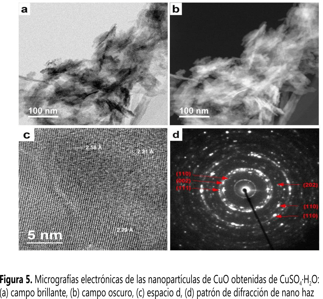
La Figura 5 muestra en detalle la morfología y la cristalinidad de las NPs de CuO-Sulf; también la imagen en campo claro (Figura 5a) y contraste Z (Figura 5b) muestran el crecimiento alargado de las partículas de CuO-Sulf, con diámetro de ~10 nm y longitud de ~80 nm. Del mismo modo, la Figura 5c muestra las distancias interplanares de las partículas, cuyos planos cristalinos pertenecen al CuO. El patrón de difracción de electrones muestra intensidades semi-continuas a lo largo del círculo que muestran un crecimiento textural de las NPs, debido a la monocristalinidad de cada una de las partículas alargadas.
Además de estas partículas, la Figura 6 muestra la imagen de una partícula laminar que no fue encontrada en las imágenes por SEM. La imagen de campo claro (Figura 6a) y campo oscuro (Figura 6b) muestran que se encuentra con las partículas alargadas; la intensidad uniforme en la partícula laminar muestra que tiene un espesor uniforme. En el patrón de difracción de electrones (Figura 6c) se observan intensidades puntuales bien marcadas, las cuales corresponden a la fase de CuSO4·H2O, cuyos índices de Miller están en color verde; las otras intensidades provienen de las partículas alargadas.

A partir de estas imágenes por microscopia electrónica de transmisión, puede observarse que la síntesis de NPs prismáticas se forma con el precursor acetato y, en el caso de las partículas alargadas y laminares monocristalinas, con el precursor sulfato. Usando diferentes tipos de precursores fue posible obtener partículas con morfologías diferentes.
Espectroscopia infrarroja con transformada de Fourier
En la Figura 7 se observa el espectro de transmisión de las NPs (a) CuO-Acet y (b) CuO-Sulf. Tanto para las Figuras 7a y 7b las bandas formadas entre 2500 a 4000 cm-1 son principalmente atribuidas al modo stretching del enlace O-H- en la superficie de las NPs de CuO. Además de ello, para ambos espectros la banda formada alrededor de 1640 cm-1 es correspondiente al modo bending del agua ( 12 , 31 ). En la Figura 7a las bandas en 1550 cm-1 y 1410 cm-1se pueden atribuir al modo stretching del –COO y modo bending del –CH3 respectivamente ( 12 , 31 ). Las bandas entre 800 a 1250 cm-1, específicamente en 1122 cm-1 y 1085 cm-1 de la Figura 7b pueden ser correspondientes a los modos de vibración del CuSO4·H2O remanente sin reaccionar ( 32 ). Las bandas de absorción infrarroja en valores menores que 700 cm-1 muestran los modos de vibración característicos de las NPs de CuO. Las bandas en 473 cm-1 y 597 cm-1 de la Figura 7a y las bandas en 477 cm-1 y 592 cm-1 de la Figura 7b corresponden al modo de vibración stretching del enlace Cu-O ( 12 , 33 ).

Actividad Antimicrobiana
De la evaluación de la actividad antimicrobiana, se observó que los halos de inhibición para el control positivo para Staphylococcus epidermidis, Aerococcus viridans, Ochrobactrum anthropi y Micrococcus lylae fueron de 21, 18, 7 y 19 mm, respectivamente, mientras que para el control negativo fue de 6 mm para todas las bacterias estudiadas. En la Figura 8 se muestra la formación de halos de inhibición (delimitados por líneas punteadas de color rojo) para las nanopartículas de CuO-Acet para bacterias: (a) Staphylococcus epidermidis (12 mm), (b) Aerococcus viridans (10 mm), (c) Ochrobactrum anthropi (16 mm) y (d) Micrococcus lylae (14 mm). Las nanopartículas de CuO-Sulf presentaron los siguientes halos de inhibición: (e) Staphylococcus epidermidis (17 mm), (f) Aerococcus viridans (13 mm), (g) Ochrobactrum anthropi (16 mm) y (h) Micrococcus lylae (16 mm); la Tabla 2 muestra un resumen de estos datos.
También se visualiza claramente la presencia de un color azulado oscuro alrededor de las NPs, lo que indica un proceso de formación de complejos de Cu2+ en el medio agar. Los iones del Cu2+ para el caso de las NPs CuO-Acet, probablemente corresponden a un proceso de disolución de los átomos externos de las NPs. Para el caso de las NPs CuO-Sulf, se puede esperar que los Cu2+ provengan también de la fase Cu4SO4(HO)6.

| Cepa intrahospitalaria | CuO-Acet (mm) | CuO-Sulf (mm) | Control positivo (mm) | Control negativo (mm) |
| Staphylococcus epidermidis | 12 | 17 | 21 | 6 |
| Aerococcus viridans | 10 | 13 | 18 | 6 |
| Ochrobactrum anthropi | 16 | 16 | 7 | 6 |
| Micrococcus lylae | 14 | 16 | 19 | 6 |
Una posible explicación a la inhibición bacteriana por parte de las nanopartículas de CuO podría ser la interacción directa que se estaría dando con la superficie de la membrana externa de la bacteria. En este contexto, estudios realizados con óxidos metálicos sugieren que la actividad antimicrobiana estaría dándose por las especies reactivas del oxígeno (ROS, por sus siglas en inglés) producidas por la presencia de estos óxidos. Allí, las especies de oxígeno estarían interactuando con la membrana celular de la bacteria y permitiendo el ingreso de las nanopartículas a la célula ( 34 ). Otros estudios realizados observan que el efecto antimicrobiano de estas nanopartículas aumenta con la concentración utilizada, por lo que probablemente esta inhibición se deba al aumento de interrupciones generadas por las nanopartículas de CuO en la superficie de la membrana de la célula generando así un mal funcionamiento de los componentes de la célula tales como el ADN, lípidos, peptidoglicanos y proteínas ( 34 , 35 ).
Muchos investigadores afirman que el mecanismo esencial para la citotoxicidad que generan las nanopartículas es la liberación de iones Cu2+ que reaccionan con los grupos tiol (SH) de las proteínas presentes en la superficie de la membrana de la célula bacteriana. Estas proteínas sobresalen de la membrana celular, permitiendo el transporte de nutrientes a través de la pared celular. Las nanopartículas estarían inactivando estas proteínas, reduciendo la permeabilidad de la membrana y llevando a la célula a la muerte ( 36 , 37 ).
En este trabajo, la liberación de los iones Cu2+ podría proceder más fácilmente de las NPs de CuO-Sulf, dado que en ellas se encuentra también una pequeña cantidad de Cu4SO4(HO)6 el cual, por su forma hidratada, interactúa más fácilmente con el medio agar, a diferencia de las NPs de CuO-Acet.
Otros trabajos de investigación han demostrado que las células bacterianas, luego de la fijación inicial a una superficie, se mantienen unidas por un polímero extracelular y forman una biopelícula. Entonces, suprimir la formación de la biopelícula es una forma de prevenir o evitar el crecimiento de las bacterias ( 38 ). Así, se ha observado que recubrimientos delgados de ZnO que tienen una superficie compuesta de agujas de tamaño nanométrico, pueden penetrar en la membrana celular cuando están en contacto. Además, tales características de la estructura superficial de la película de ZnO afecta la última etapa del desarrollo de la biopelícula. Por lo anterior, el efecto combinado de las características nanoscópicas de la aguja y las propiedades antibacterianas del ZnO son los responsables del menor número de bacterias en una superficie ( 39 , 40 ).
Se puede decir entonces, de manera análoga, que la morfología alargada y laminar de las NPs de CuO-Sulf favorece la interacción de estas con la membrana celular de las bacterias a diferencia de las NPs de CuO-Acet que presentaron forma globular.
Conclusiones
La caracterización de las NPs de CuO demostró que cuando son sintetizadas a partir de Cu(CH3COO)2∙H2O presentan una fase de CuO pura, mientras que si se sintetizan a partir de CuSO2∙5H2O presentan dos fases, siendo la fase mayoritaria de CuO. Lo anterior puede deberse a la pureza grado técnico de la sal precursora. Sin embargo, se puede observar que ambos tipos de NPs tienen un efecto antimicrobiano frente a las diferentes cepas intrahospitalarias de un hospital nacional de Lima norte.
Los halos de inhibición de las cepas estudiadas fueron mayores para las NPs de CuO obtenidas a partir de CuSO2∙5H2O que para las obtenidas a partir de Cu(CH3COO)2∙H2O; solo para la cepa Ochrobactrum anthropi presentan halos similares. Este hecho se sustentaría, no solo en la pureza del CuO, sino también debido a que la morfología de las NPs de CuO de formas alargadas y laminares, obtenidas a partir de CuSO2∙5H2O, podrían dañar con mayor facilidad la membrana celular de las bacterias, lo que conllevaría a la inactivación de las mismas.
Agradecimientos
M.M.G.L. agradece al Instituto General de Investigación de la Universidad Nacional de Ingeniería (IGI-UNI) por financiación otorgada como Docente Investigador. Se agradece al laboratorio nacional de nanotecnología ubicado en CIMAV Chihuahua, México, por el apoyo de microscopia electrónica. Al Consejo Nacional de Investigaciones Científicas y Técnicas (CONICET) - Proyecto PIP 00651 - Argentina, a la Universidad Nacional de La Plata - Proyecto 11/X709 y al Sistema Nacional de rayos X (SNRX) y CONICET - Proyectos RX-M6 y RX-AC7, Argentina, por el apoyo en difracción de rayos X. A.E.B., agradece a Graciela Punte, por la supervisión en los diferentes trabajos desarrollados. El presente trabajo fue desarrollado bajo la financiación recibida por el Programa Nacional de Innovación para la Competitividad y Productividad - Innóvate Perú (Convenio 368-PNICP-PIAP-2014) y el IGI-UNI.
Referencias
1. Katwal, R.; Kaur, H.; Sharma, G.; Naushad, M.; Pathania, D. Electrochemical synthesized copper oxide nanoparticles for enhanced photocatalytic and antimicrobial activity. J. Ind. Eng. Chem. 2015, 31, 173-184. DOI: https://doi.org/10.1016/j.jiec.2015.06.021.
Raghunath, A.; Perumal, E. Metal oxide nanoparticles as antimicrobial agents: A promise for the future. Int. J. Antimicrob. Agents. 2017. DOI: https://doi.org/10.1016/j.ijantimicag.2016.11.011.
3. Yoosefi Booshehri, A.; Wang, R.; Xu, R. Simple method of deposition of CuO nanoparticles on a cellulose paper and its antibacterial activity. Chem. Eng. J. 2015, 262, 999-1008. DOI: https://doi.org/10.1016/j.cej.2014.09.096.
4. Sharmila, G.; Thirumarimurugan, M.; Sivakumar, V. M. Optical, catalytic and antibacterial properties of phytofabricated CuO nanoparticles using Tecoma castanifolia leaf extract. Optik. 2016, 127(19), 7822-7828. DOI: https://doi.org/10.1016/j.ijleo.2016.05.142.
5. Christy, A. J.; Nehru, L. C.; Umadevi, M. A novel combustion method to prepare CuO nanorods and its antimicrobial and photocatalytic activities. Powder Technol. 2013, 235, 783-786. DOI: https://doi.org/10.1016/j.powtec.2012.11.045.
6. Das, D.; Nath, B. C.; Phukon, P.; Dolui, S. K. Synthesis and evaluation of antioxidant and antibacterial behavior of CuO nanoparticles. Colloids Surf., B. 2013, 101, 430-433. DOI: https://doi.org/10.1016/j.colsurfb.2012.07.002.
7. Sonia, S.; Jayasudha, R.; Jayram, N. D.; Kumar, P. S.; Mangalaraj, D.; Prabagaran, S. R. Synthesis of hierarchical CuO nanostructures: Biocompatible antibacterial agents for Gram-positive and Gram-negative bacteria. Curr. Appl. Phys. 2016, 16 (8), 914-921. DOI: https://doi.org/10.1016/j.cap.2016.05.006.
Ahamed, M.; Alhadlaq, H. A.; Khan, M. A. M.; Karuppiah, P.; Al-Dhabi, N. A. Synthesis, characterization, and antimicrobial activity of copper oxide nanoparticles. J. Nanomater. 2014, 2014, 1-4. DOI: https://doi.org/10.1155/2014/637858.
9. Ramyadevi, J.; Jeyasubramanian, K.; Marikani, A.; Rajakumar, G.; Rahuman, A. A. Synthesis and antimicrobial activity of copper nanoparticles. Mater. Lett. 2012, 71, 114-116. DOI: https://doi.org/10.1016/j.matlet.2011.12.055.
10. Azam, A.; Ahmed, A. S.; Oves, M.; Khan, M. S.; Memic, A. Size-dependent antimicrobial properties of CuO nanoparticles against Gram-positive and -negative bacterial strains. Int. J. Nanomed. 2012, 7, 3527-3535. DOI: https://doi.org/10.2147/IJN.S29020.
11. Abramovaa, A.; Gedankenb, A.; Popov, V.; Ooi, E.; Masond, T. J.; Joyce, E. M. A sonochemical technology for coating of textiles with antibacterial nanoparticles and equipment for its implementation. Mater. Lett. 2013, 96, 121–124. DOI: https://doi.org/10.1016/j.matlet.2013.01.041.
12. Mageshwari, K.; Sathyamoorthy, R. Flower-shaped CuO nanostructures: Synthesis, characterization and antimicrobial activity. J. Mater. Sci. Technol. 2013, 29 (10), 909-914. DOI: https://doi.org/10.1016/j.jmst.2013.04.020.
13. Perelshtein, I.; Lipovsky, A.; Perkas, N.; Tzanov, T.; Аrguirova, M.; Leseva, M. et al. Making the hospital a safer place by sonochemical coating of all its textiles with antibacterial nanoparticles. Ultrason. Sonochem. 2015, 25, 82-88. DOI: https://doi.org/10.1016/j.ultsonch.2014.12.012.
14. El-Nahhal, I.; Zourab, S.; Kodeh, F.; Selmane, M.; Genois, I.; Babonneau, F. Nanostructured copper oxide-cotton fibers: synthesis, characterization, and applications. Int. Nano Lett. 2012, 2(14), 1-5. DOI: https://doi.org/10.1186/2228-5326-2-14.
15. Nasser Mohammed, H. M. S., Z. Polymethacrylic acid as a new precursor of CuO nanoparticles. J. Mol. Struct. 2012, 1027, 128-132. DOI: https://doi.org/10.1016/j.molstruc.2012.05.081.
16. Rios Valer, G. B. Estudio morfológico y estructural de nanopartículas de CuO empleando técnicas de dispersión de luz y rayos X. Bach. Tesis, Universidad Nacional de Ingenieria , Perú, 2015.
17. Karthik, A. D.; Geetha, K. Synthesis of copper precursor, copper and its oxide nanoparticles by green chemical reducion method and its antimicrobial activity. J. Appl. Pharm. Sci. 2013, 3 (5), 016-021. DOI: https://doi.org/10.7324/japs.2013.3504.
18. Garro Nuñez, G. M.; Eufelia Quispe Pardo, Z. E. Protocolo: Estudio de prevalencia de infecciones intrahospitalarias. Ministerio de Salud - Dirección General de Epidemiología, Perú, 2014; pp 15-24.
19. World Health Organization. World health statistics 2015. WHO, Geneva, 2015; pp 55-77.
20. Díaz-Vélez, C.; Neciosup-Puicán, E.; Fernández-Mogollón, J. L.; Tresierra-Ayala, M. A.; Apolaya-Segura, M. Mortalidad atribuible a infecciones nosocomiales en un hospital de la Seguridad Social en Chiclayo, Perú. Acta Med. Peru. 2016, 33 (3), 250-252.
21. Jefatura del Servicio de Cuidados Intensivos Generales (SCIG) Proyecto de cuidados intemedios del servicio de cuidados intensivos generales, 2°. Ministerio de Salud - Hospital Cayetano Heredia, Lima, 2016; pp 14-15.
22. Rodriguez-Carvajal, J.; Fernandez-Diaz, M. T.; Martinez, J. L. Neutron diffraction study on structural and magnetic properties of La2NiO4. J. Phys.: Condens. Matter. 1991, 3(9), 3215-3234. DOI: https://doi.org/10.1088/0953-8984/3/19/002.
23. Bou, G.; Fernandez-Olmos, A.; Garcia, C.; Saez-Nieto, J. A.; Valdezate, S. Metodos de identificación bacteriana en el laboratorio de microbiología. Enferm. Infecc. Microbiol. Clin. 2011, 29(8), 601-8. DOI: https://doi.org/10.1016/j.eimc.2011.03.012.
24. Gobernado, M.; López-Hontangas, J. L. Identificación bacteriana. Enferm. Infecc. Microbiol. Clin. 2003, 21(2), 54-60.
25. Taroco, R.; Seija, V.; Vignol, R., Métodos de estudio de la sensibilidad antibiótica, en Temas de Bacteriología y Virología Médica, Oficina del libro FEFMUR, Uruguay, 2006; pp 665-668.
26. The U.S. Pharmacopeial Convention USP Pharmacists pharmacopeia 2008-2009, 2° ed. Amer. Soc. of Health System, Rockville, 2009; pp s3/3-s3/6.
27. European Committee for Antimicrobial Susceptibility Testing (EUCAST) of the European Society of Clinical Microbiology and Infectious Diseases (ESCMID) Terminology relating to methods for the determination of susceptibility of bacteria to antimicrobial agents. Clin. Microbiol. Infect. 2000, 6 (9), 509-515. DOI: https://doi.org/10.1046/j.1469-0691.2000.00142.x.
28. Bonev, B.; Hooper, J.; Parisot, J. Principles of assessing bacterial susceptibility to antibiotics using the agar diffusion method. J. Antimicrob. Chemother. 2008, 61 (6), 1295-301. DOI: https://doi.org/10.1093/jac/dkn090.
29. Murray, P. R.; Rosenthal, K. S.; Pfaüer, M. A. Microbiología Médica, 6° ed. Elservier: España; 2009.
30. Åsbrink, S.; Norrby, L.-J. A refinement of the crystal structure of copper (II) oxide with a discussion of some exceptional e.s.d.'s. Acta Crystallogr., Sect. B: Struct. Sci. 1970, 26(1), 8-15. DOI: https://doi.org/10.1107/s0567740870001838.
31. Tackett, J. E. FT-IR characterization of metal acetates in aqueous solution. Appl. Spectrosc. 1989, 43 (3), 483-489. DOI: https://doi.org/10.1366/0003702894202931.
32. Siriwardane, R. V.; Poston Jr., J. A.; Fisher, E. P.; Shen, M.; Miltz, A. L. Decomposition of the sulfates of copper, iron (II), iron (III), nickel, and zinc: XPS, SEM, DRIFTS, XRD, and TGA study. Appl. Surf. Sci. 1999, 152 (3-4), 219-236. DOI: https://doi.org/10.1016/s0169-4332(99)00319-0.
33. Amin, S. A.; Pazouki, M.; Hosseinnia, A. Synthesis of TiO2–Ag nanocomposite with sol–gel method and investigation of its antibacterial activity against E. coli. Powder Technol. 2009, 196 (3), 241-245. DOI: https://doi.org/10.1016/j.powtec.2009.07.021.
34. Dizaj, S. M.; Lotfipour, F.; Barzegar-Jalali, M.; Zarrintan, M. H.; Adibkia, K. Antimicrobial activity of the metals and metal oxide nanoparticles. Mater. Sci. Eng., C. 2014, 44, 278-284. DOI: https://doi.org/10.1016/j.msec.2014.08.031.
35. Azam, A.; Ahmed, A. S.; Oves, M.; Khan, M. S.; Habib, S. S.; Memic, A. Antimicrobial activity of metal oxide nanoparticles against Gram-positive and Gram-negative bacteria: a comparative study. Int. J. Nanomed. 2012, 7, 6003-6009. DOI: https://doi.org/10.2147/IJN.S35347.
36. Gopinathan, E.; Viruthagiri, G.; Shanmugam, N.; Sathiya priya, S. Optical, surface analysis and antibacterial activity of ZnO–CuO doped cerium oxide nanoparticles. Optik. 2015, 126 (24), 5830-5835. DOI: https://doi.org/10.1016/j.ijleo.2015.09.014.
37. Michal, R.; Dworniczek, E.; Caplovicova, M.; Monfort, O.; Lianos, P.; Caplovic, L. et al. Photocatalytic properties and selective antimicrobial activity of TiO2 (Eu)/CuO nanocomposite. Appl. Surf. Sci. 2016, 371, 538-546. DOI: https://doi.org/10.1016/j.apsusc.2016.03.003.
38. Garrett, T. R.; Bhakoo, M.; Zhang, Z. Bacterial adhesion and biofilms on surfaces. Prog. Nat. Sci. 2008, 18 (9), 1049-1056. DOI: http://doi.org/10.1016/j.pnsc.2008.04.001.
39. Dai, Y.; Sun, T.; Zhang, Z.; Zhang, Z. J.; Li, J. R. Effect of zinc oxide film morphologies on the formation of Shewanella putrefaciens biofilm. Biointerphases. 2017, 12 (1), 011002. DOI: https://doi.org/10.1116/1.4976003.
40. Singh, A. V.; Vyas, V.; Patil, R.; Sharma, V.; Scopelliti, P. E.; Bongiorno, G. et al. Quantitative characterization of the influence of the nanoscale morphology of nanostructured surfaces on bacterial adhesion and biofilm formation. PLoS One. 2011, 6 (9), e25029. DOI: https://doi.org/10.1371/journal.pone.0025029.
Notas de autor
mgomez@uni.edu.pe
Recibido: 2 de febrero de 2017; Aceptado: 26 de abril de 2017
Resumen
Empleando un prototipo de reactor, se sintetizaron nanopartículas (NPs) de CuO a través del método de precipitación a partir de CuSO2·5H2O y de Cu(CH3COO)2·H2O. Las NPs obtenidas fueron caracterizadas mediante XRD, FT-IR, TEM y SEM. La actividad antimicrobiana de las NPs se determinó mediante el método de difusión en placa, colocando 20 mg de NPs de CuO sobre cuatro cepas intrahospitalarias o nosocomiales aisladas de la Unidad de Cuidados Intensivos de un hospital nacional de Lima norte (Staphylococcus epidermidis, Aerococcus viridans, Ochrobactrum anthropi y Micrococcus lylae). La caracterización de las NPs de CuO demostró que las sintetizadas a partir de acetato (CuO–Acet) presentaron una fase pura de CuO, mientras que las sintetizadas a partir de sulfato (CuO–Sulf) presentaron dos fases, donde la de CuO representó más del 84%. Los dominios cristalinos del CuO–Acet y CuO–Sulf fueron 15 y 19 nm, respectivamente. Los halos de inhibición de las cepas estudiadas fueron mayores para las NPs de CuO–Sulf que para las NPs de CuO–Acet; solo para la cepa Ochrobactrum anthropi se presentaron halos similares para ambos tipos de NPs.
Palabras clave
Actividad antimicrobiana, método de difusión en placa, cepas nosocomiales, nanopartículas de CuO.Abstract
Using a prototype reactor, CuO nanoparticles (NPs) were synthetized through the precipitation method, starting from CuSO2·5H2O and Cu(CH3COO)2·H2O. The obtained NPs were characterized by XDR, FT-IR, SEM, and TEM. The antimicrobial activity of the NPs was determined by the plate diffusion method, placing 20 mg of NPs onto four nosocomial strains obtained from north Lima national hospital Intensive-Care Unit (Staphylococcus epidermidis, Aerococcus viridans, Ochrobactrum anthropic, and Micrococcus lylae). NPs characterization revealed that those synthetized from acetate (CuO–Acet) shown pure CuO phase, while those synthetized from sulphate CuO–Sulf shown two phases where CuO was the predominant one, having more than 84%. The crystal domains for CuO–Acet and CuO–Sulf were 15 and 19 nm, respectively. The inhibition halos for the studied strains were larger for CuO–Sulf NPs than CuO–Acet NPs, only Ochrobactrum anthropi displayed similar inhibition halos for both types of NPs.
Keywords
Antimicrobial activity, plate diffusion method, nosocomial strains, CuO nanoparticles.Resumo
Usando um protótipo de reator, sintetizaram-se nano partículas (NPs) de CuO pelo método de precipitação a partir de CuSO2·5H2O e de Cu(CH3COO)2·H2O. As NPs obtidas foram caracterizadas por meio das seguintes técnicas: XRD, FT-IR, TEM e SEM. A atividade antimicrobiana das NPs foi determinada pelo método de difusão em placa, colocando 20 mg de NPs de CuO sobre quatro cepas nosocomiais isoladas de uma Unidade de Terapia Intensiva no hospital nacional de Lima norte (Staphylococcus epidermidis Aerococcus viridans, Ochrobactrum anthropic and Micrococcus lylae). A caracterização das NPs de CuO mostrou que as nanopartículas sintetizadas a partir do acetato (CuO–Acet) apresentavam uma fase pura de CuO, enquanto que as sintetizadas a partir do sulfato (CuO–Sulf), apresentavam duas fases sendo a fase maior de CuO, com mais de 84%. Os domínios cristalinos de CuO–Acet y CuO–Sulf foram 15 e 19 nm, respectivamente. Os halos de inibição das cepas estudadas foram maiores para as NPs de CuO–Sulf do que as NPs de CuO–Acet somente para a cepa Ochrobactrum anthropi, apresentaram-se halos de inibição parecidos.
Palavras-chave
Atividade antimicrobiana, método de difusão em placa, cepas nosocomiais, nanoparticulas de CuO.Introducción
En la actualidad, se han estudiado las NPs de CuO debido a sus promisorias propiedades antimicrobianas ( 1 , 2 ). Así lo demuestra su efectividad frente a bacterias como Escherichia coli ( 3 - 5 ), Pseudomona aeruginosa ( 6 ), Klebsiella pneumoniae ( 5 ), Enterococcus faecalis, Shigella flexneri, Salmonella typhimurium ( 7 ), Proteus vulgaris y Staphylococcus aureus. Además, se ha evidenciado que las NPs de CuO, frente a bacterias como Micrococcus luteus, Staphylococcus aureus, Escherichia coli, Klebsiella pneumoniae y Pseudomonas aeruginosa, muestran una mayor actividad inhibitoria comparada con el halo de inhibición presentado por los hongos como Aspergillus flavus, Aspergillus niger y Candida albicans ( 9 ). Otras investigaciones corroboran el efecto inhibitorio de las NPs de CuO frente a bacterias Gram positivas y Gram negativas, donde el tamaño de las nanopartículas esta correlacionado con su actividad antimicrobiana ( 10 ).
El CuO es usado en la industria cosmética y en prendas textiles por sus propiedades antimicrobianas. Las fibras impregnadas con NPs de CuO presentan un amplio espectro antimicrobiano y antifúngico ( 11 , 12 ), por tanto, su uso está siendo desarrollado en diferentes partes del mundo en el ámbito de la salud pública ( 10 , 13 ).
Investigaciones realizadas sobre telas recubiertas con NPs de CuO mostraron una efectiva actividad antimicrobiana frente a los microorganismos E. coli, K. pneumoniae, S. aureus y S. aureus Meticilina Resistente (MRSA, por sus siglas en inglés), reduciendo significativamente la presencia de estos microorganismos ( 11 , 14 ). También, se han demostrado las propiedades de las NPs de CuO como agente bactericida frente a bacterias estándares ( 8 , 15 - 17 ), usándose como soporte en materiales inertes, sin embargo, no existen referencias de su actividad bactericida frente a las bacterias intrahospitalarias.
Ahora bien, la Organización Mundial de la Salud (WHO, por sus siglas en inglés) reportó que las infecciones nosocomiales o intrahospitalarias (IIH) actualmente llamadas Infecciones Asociadas a la Atención de Salud (IAAS) ( 18 ), son una importante causa de morbilidad y mortalidad, lo que demuestra una calidad deficiente en la atención del sector salud ( 19 ).
A finales de 1998, el Ministerio de Salud del Perú promovió la vigilancia, prevención y control de las IAAS con la finalidad de mejorar la calidad de atención y, por consiguiente, disminuir la morbilidad, mortalidad y costos hospitalarios ( 18 ). Según estudios de la Dirección General de Epidemiología del Ministerio de Salud del Perú, se reportaron tasas de prevalencia de IAAS de 4,8% para el 2014 y 3,9% para el 2015 ( 20 ). Por tanto, la toma de medidas de bioseguridad en un medio donde se cuenta con pocos recursos económicos y con alta presencia de las IAAS tiene vigencia e importancia en la actualidad ( 20 ), teniendo en cuenta que el gasto por paciente que reingresa al hospital es de alrededor de 150000 nuevos soles ( 21 ) (aproximadamente 45000 USD), cantidad que podría ahorrarse si se evitan los reingresos y por ende las complicaciones de los pacientes.
Por otro lado, actualmente el Perú es el segundo productor de cobre a nivel mundial y, en el mercado nacional, es viable acceder a sales de cobre de grado técnico para uso industrial a muy bajos costos. El tratamiento de estas sales, para darles un valor agregado, es un aspecto de desarrollo tecnológico importante a nivel nacional. Por tanto, en el presente trabajo se estudió la síntesis de CuO, utilizando un prototipo de reactor diseñado para producir hasta 100 g de NPs. Adicionalmente, se evaluó la impregnación de estas NPs en textiles de algodón, como respuesta a la problemática nacional de salud relacionada a las IAAS.
Materiales y métodos
Síntesis de las nanopartículas de CuO
La síntesis se realizó mediante el método de precipitación ( 16 ): se preparon soluciones acuosas de 0,02 M a partir de sales precursoras de acetato de cobre monohidratado (Cu(CH3COO)2∙H2O) de grado químicamente puro, Merck Millipore, Alemania y sulfato de cobre pentahidratado (CuSO2∙5H2O) de grado técnico suministrado por la empresa J.A. Elmer, Perú. Luego cada solución de sal precursora preparada se vertió en un reactor piloto de acero inoxidable (33” de altura, 15” de diámetro, motor de 1720 revoluciones por minuto (rpm) y 0,37 kW de potencia, calentador por inmersión Rotkappe® de marca MAZURCZAK) tal como se muestra en la Figura 1. La reacción se inició en 100 °C con agitación vigorosa de 500 rpm, luego se agregó una solución de 0,1 M de hidróxido de sodio (NaOH) con 99,0% de pureza de la marca Merck Millipore, Alemania. Inmediatamente, se observó la formación de un precipitado marrón y para completar la reacción se mantuvo el sistema en agitación por 5 min. Posteriormente, el reactor fue vaciado y las NPs de CuO fueron lavadas 3 veces con agua destilada y secadas a 80 °C. Las NPs de CuO obtenidas a partir de las sales precursoras de Cu(CH3COO)2∙H2O y CuSO2∙5H2O fueron denominadas de CuO-Acet y CuO-Sulf, respectivamente.

Difracción de rayos X
El estudio estructural de las NPS de CuO se realizó por difracción de rayos X empleando un difractómetro PANalytical X'Pert Pro con una radiación CuK α (λ = 1,5406 Å), equipado con un contador de centelleo y un monocromador de grafito a la salida del haz difractado. Los datos se recolectaron en un rango de 20 ° £ 2q £ 120 º con 0,02 ° de ancho de paso y una tasa de conteo no menor de 15 s por paso. El ajuste del difractograma se realizó por el método de Rietveld, utilizando el programa FullProf ( 22 ).
Microscopía electrónica
La caracterización morfológica de las NPS de CuO se realizó por microscopía electrónica de barrido de emisión de campo (FEGSEM por sus siglas en inglés), utilizando el microscopio electrónico Zeiss LEO 982 GEMINI, operado a 3 kV. Del mismo modo, se utilizó microscopía electrónica de barrido y transmisión (STEM/TEM) empleando un microscopio electrónico de emisión de campo de alta resolución JEOL JEM-2200FS operado a 200 kV con corrector de aberración esférica en modo STEM.
Espectroscopía infrarroja con transformada de Fourier
Otra caracterización de vibración molecular de las NPs de CuO se llevó a cabo por espectroscopia infrarroja con Transformada de Fourier (FTIR), empleando un espectrofotómetro IR Prestige-21 Shimadzu utilizando la técnica de reflectancia atenuada total (ATR) con rango de análisis de 4000 a 400 cm-1.
Evaluación de la actividad antimicrobiana
Para la evaluación de la actividad antimicrobiana se realizó el muestreo de 432 superficies correspondientes a tres áreas de UCI: Emergencia (12 muestras), Quirúrgico (12 muestras) y Neonatología (12 muestras). En cada unidad se realizó el hisopado de tres superficies diferentes tomando cuatro ejemplares de cada una de ellas (manijas, barandas, incubadoras e historias clínicas). Las muestras obtenidas fueron sembradas en medios de cultivo Agar Tripticasa de Soya (TSA) y Agar MacConkey e incubadas a 37 °C por 24 h. Finalmente, se realizó el recuento bacteriano en Unidades Formadoras de Colonias (UFC) y cada una de las colonias diferentes fue repicada (acción de trasplantar de un medio de cultivo a otro) en TSA para su posterior identificación.
La identificación de los microorganismos se realizó por pruebas bioquímicas convencionales. Para la identificación de las bacterias Gram positivas se realizó la búsqueda de las enzimas oxidasa, catalasa, coagulasa y DNasa, tipos de hemolisis, fermentación del manitol, susceptibilidad a la bacitracina (0,04 UI – Unidad Internacional) y optoquina (clorhidrato de etilhidrocupreína) 5 µg, logrando determinar el género bacteriano.
En el caso de las bacterias Gram negativas se realizaron pruebas para determinar el tipo de fermentación a los carbohidratos como glucosa, lactosa y sacarosa a través del medio bioquímico Agar Triple Azúcar Hierro (TSI), presencia de la enzima lisina descarboxilasa (LIA), presencia de motilidad y producción de indol (medio SIM), utilización de citrato como única fuente de carbono y presencia de acetil-metil-carbinol (acetoína) e hidrolisis de la urea ( 23 ).
Finalmente se aplicó el sistema de identificación multipruebas Analytical Profile Index (API) de la empresa Biomérieux: API Staph, API Strep para identificar bacterias Gram positivos, API 20E para la Familia Enterobacteriaceae y API NE para las bacterias no fermentadoras ( 24 ).
Para determinar la actividad antimicrobiana de las NPs de CuO se utilizó el método de difusión en placa empleado en el Instituto de Normas Clínicas y de Laboratorio (CLSI por sus siglas en inglés,) ( 25 ). Este método es una modificación del método cilindro placa de cultivo de la Farmacopea Americana USP 32 ( 26 ), donde la inhibición bacteriana, mediante la difusión del principio activo de la muestra a probar en un medio de cultivo sólido, es evidenciada con la formación de halos de inhibición ( 25 , 27 ). Se utiliza, además, un cilindro de acero de 6 mm de diámetro que se ubica al centro de la placa, y dentro del cual se vierte un peso determinado de las nanopartículas (para este trabajo fue 20 mg), permitiendo que estas se distribuyan únicamente en el diámetro del cilindro ( 28 ).
Para cada una de las cepas intrahospitalarias aisladas e identificadas (Staphylococcus epidermidis, Aerococcus viridans, Ochrobactrum anthropi y Micrococcus lylae), se preparó un inóculo con una turbidez equivalente a 0,5 de la escala de Mac Farland ( 29 ) (1,5x10⁸ UFC/ml) y se sembró por duplicado sobre la superficie del medio de cultivo TSA, con ayuda de un hisopo estéril. Posteriormente, en el centro de la placa con TSA se colocó el cilindro de acero para inmediatamente añadir 0,02 g de NPs de CuO. Al finalizar, las placas se dejaron incubar a 37 °C por 24 h.
Aquellas cepas que presentaron halos de inhibición fueron medidas en mm con ayuda de un Vernier de marca Stanley Hardened. Se realizó el mismo procedimiento con los controles: como control positivo se tenían discos impregnados con 100 µL de desinfectante a base de amonio cuaternario (dilución 1/30), utilizado actualmente en el sector salud; como control negativo, se emplearon discos impregnados con 100 µL de agua destilada estéril. Todas las siembras fueron realizadas por duplicado para cada uno de los controles.
Resultados y discusión
Difracción de rayos X
La Figura 2 muestra los patrones de difracción de rayos X de las NPs CuO-Acet y CuO-Sulf. CuO-Acet solamente muestra los picos de difracción correspondiente al CuO, mientras que CuO-Sulf, muestra una segunda fase, identificada como Cu4SO4(HO)6 (16%). La fase de CuO en ambas muestras presentó una ligera orientación preferencial del plano (001), cuya intensidad es mayor que la que corresponde a una muestra de CuO sin orientación preferencial.

Los parámetros de red a, b, c y β de la celda monoclínica del CuO fueron determinados por el refinamiento Rietveld de los datos, usando el modelo y estructura inicial según Åsbrink y Norrby ( 30 ). Se tuvo en cuenta la contribución de los efectos del tamaño y la tensión interna al ensanchamiento del pico de difracción; ambos parámetros fueron refinados empleando un modelo isotrópico. Los parámetros estructurales y microestructurales obtenidos del refinamiento Rietveld son mostrados en la Tabla 1.
:
Tabla 1.
Valores de los parámetros de red, tamaño de cristalito promedio, D, y microtensión promedio, e, para
las muestra de NPs obtenidas (solo de la fase de CuO)
a (Å)
b (Å)
c (Å)
β (°)
D nm
e *
CuO-Acet
4,6902 (1)
3,4282(1)
5,1384(1)
99,34 (1)
15,76 (1)
50,53(4)
CuO-Sulf
4,6944 (1)
3,4247(1)
5,1431(1)
99,40 (1)
19,20 (1)
64,41(4)
Estos picos, indexados respecto al sistema cristalino monoclínico (grupo espacial C2/c ), archivo N° 45 – 0937 de la base de datos del International Centre for Diffraction Data (ICDD, por sus siglas en inglés), son característicos de las NPs de CuO ( 8 ). La medida del tamaño del cristalito fue estimado del refinamiento de Rietveld y la fórmula de Scherrer, con valores de 15 y 19 nm para las NPs de CuO-Acet y CuO-Sulf, respectivamente.
Microscopia Electrónica
En la Figura 3 se muestra la caracterización morfológica por SEM de las NPs sintetizadas a partir de dos tipos de sales precursoras. En las Figuras 3a y 3b se muestran imágenes a baja (200 kX) y alta (400 kX) magnificación, obtenidas con electrones secundarios de las NPs de CuO-Acet, donde se observan conglomerados de partículas cristalinas de apariencia redondeada con tamaños relativamente heterogéneos que varían entre 20 y 100 nm. Las micrografías de las Figuras 3c y 3d muestran la morfología de las NPs de CuO-Sulf. Estos cristales forman conglomerados laminares, cada uno de ellos tiene partículas alargadas de varios tamaños que van entre 10 y 110 nm.

Las imágenes de la Figura 3 muestras dos morfologías que diferencian las NPs de CuO-Acet y CuO-Sulf. Con la primera sal precursora los aglomerados de CuO son más redondeados que con las obtenidas por la segunda, y presentan aglomerados laminares de CuO alargados (forma de varillas).
Para determinar con mayor detalle las formas y la cristalinidad de las partículas que conforman los conglomerados, se estudiaron las muestras mediante STEM. Las imágenes se adquirieron en modo de campo claro y contraste Z. La Figura 4 muestra las NPs de CuO-Acet, las imágenes en campo claro (Figura 4a) y contraste Z (Figura 4b) muestran la forma de las partículas que conforman los aglomerados de tamaño uniforme, en forma prismática, cuyo ancho es ~15 nm y largo de ~20 nm.

La imagen de la Figura 4c muestra a mayor magnificación la cristalinidad de las partículas. En esta imagen se ven distancias interplanares que corresponden al CuO, cuyos valores están marcados en la imagen. El patrón de difracción de electrones (Figura 4d) corrobora la cristalinidad de las partículas, cuyas intensidades indexan con CuO. Se indican los índices de Miller en la imagen. Esta información es concordante con lo encontrado por XRD.
La Figura 5 muestra en detalle la morfología y la cristalinidad de las NPs de CuO-Sulf; también la imagen en campo claro (Figura 5a) y contraste Z (Figura 5b) muestran el crecimiento alargado de las partículas de CuO-Sulf, con diámetro de ~10 nm y longitud de ~80 nm. Del mismo modo, la Figura 5c muestra las distancias interplanares de las partículas, cuyos planos cristalinos pertenecen al CuO. El patrón de difracción de electrones muestra intensidades semi-continuas a lo largo del círculo que muestran un crecimiento textural de las NPs, debido a la monocristalinidad de cada una de las partículas alargadas.

Además de estas partículas, la Figura 6 muestra la imagen de una partícula laminar que no fue encontrada en las imágenes por SEM. La imagen de campo claro (Figura 6a) y campo oscuro (Figura 6b) muestran que se encuentra con las partículas alargadas; la intensidad uniforme en la partícula laminar muestra que tiene un espesor uniforme. En el patrón de difracción de electrones (Figura 6c) se observan intensidades puntuales bien marcadas, las cuales corresponden a la fase de CuSO4·H2O, cuyos índices de Miller están en color verde; las otras intensidades provienen de las partículas alargadas.

A partir de estas imágenes por microscopia electrónica de transmisión, puede observarse que la síntesis de NPs prismáticas se forma con el precursor acetato y, en el caso de las partículas alargadas y laminares monocristalinas, con el precursor sulfato. Usando diferentes tipos de precursores fue posible obtener partículas con morfologías diferentes.
Espectroscopia infrarroja con transformada de Fourier
En la Figura 7 se observa el espectro de transmisión de las NPs (a) CuO-Acet y (b) CuO-Sulf. Tanto para las Figuras 7a y 7b las bandas formadas entre 2500 a 4000 cm-1 son principalmente atribuidas al modo stretching del enlace O-H- en la superficie de las NPs de CuO. Además de ello, para ambos espectros la banda formada alrededor de 1640 cm-1 es correspondiente al modo bending del agua ( 12 , 31 ). En la Figura 7a las bandas en 1550 cm-1 y 1410 cm-1se pueden atribuir al modo stretching del –COO y modo bending del –CH3 respectivamente ( 12 , 31 ). Las bandas entre 800 a 1250 cm-1, específicamente en 1122 cm-1 y 1085 cm-1 de la Figura 7b pueden ser correspondientes a los modos de vibración del CuSO4·H2O remanente sin reaccionar ( 32 ). Las bandas de absorción infrarroja en valores menores que 700 cm-1 muestran los modos de vibración característicos de las NPs de CuO. Las bandas en 473 cm-1 y 597 cm-1 de la Figura 7a y las bandas en 477 cm-1 y 592 cm-1 de la Figura 7b corresponden al modo de vibración stretching del enlace Cu-O ( 12 , 33 ).

Actividad Antimicrobiana
De la evaluación de la actividad antimicrobiana, se observó que los halos de inhibición para el control positivo para Staphylococcus epidermidis, Aerococcus viridans, Ochrobactrum anthropi y Micrococcus lylae fueron de 21, 18, 7 y 19 mm, respectivamente, mientras que para el control negativo fue de 6 mm para todas las bacterias estudiadas. En la Figura 8 se muestra la formación de halos de inhibición (delimitados por líneas punteadas de color rojo) para las nanopartículas de CuO-Acet para bacterias: (a) Staphylococcus epidermidis (12 mm), (b) Aerococcus viridans (10 mm), (c) Ochrobactrum anthropi (16 mm) y (d) Micrococcus lylae (14 mm). Las nanopartículas de CuO-Sulf presentaron los siguientes halos de inhibición: (e) Staphylococcus epidermidis (17 mm), (f) Aerococcus viridans (13 mm), (g) Ochrobactrum anthropi (16 mm) y (h) Micrococcus lylae (16 mm); la Tabla 2 muestra un resumen de estos datos.
También se visualiza claramente la presencia de un color azulado oscuro alrededor de las NPs, lo que indica un proceso de formación de complejos de Cu2+ en el medio agar. Los iones del Cu2+ para el caso de las NPs CuO-Acet, probablemente corresponden a un proceso de disolución de los átomos externos de las NPs. Para el caso de las NPs CuO-Sulf, se puede esperar que los Cu2+ provengan también de la fase Cu4SO4(HO)6.

:
Tabla 2.
Valores de los diámetros de los halos de inhibición (mm) para las diferentes
cepas intrahospitalarias enfrentadas a las NPs de CuO obtenido de diferentes reactivos. Se presentan también los valores
para los controles positivo (desinfectante a base de amonio cuaternario diluido
en 1/30) y negativo (agua destilada).
Cepa intrahospitalaria
CuO-Acet
(mm)
CuO-Sulf
(mm)
Control positivo
(mm)
Control negativo
(mm)
Staphylococcus epidermidis
12
17
21
6
Aerococcus viridans
10
13
18
6
Ochrobactrum anthropi
16
16
7
6
Micrococcus lylae
14
16
19
6
Una posible explicación a la inhibición bacteriana por parte de las nanopartículas de CuO podría ser la interacción directa que se estaría dando con la superficie de la membrana externa de la bacteria. En este contexto, estudios realizados con óxidos metálicos sugieren que la actividad antimicrobiana estaría dándose por las especies reactivas del oxígeno (ROS, por sus siglas en inglés) producidas por la presencia de estos óxidos. Allí, las especies de oxígeno estarían interactuando con la membrana celular de la bacteria y permitiendo el ingreso de las nanopartículas a la célula ( 34 ). Otros estudios realizados observan que el efecto antimicrobiano de estas nanopartículas aumenta con la concentración utilizada, por lo que probablemente esta inhibición se deba al aumento de interrupciones generadas por las nanopartículas de CuO en la superficie de la membrana de la célula generando así un mal funcionamiento de los componentes de la célula tales como el ADN, lípidos, peptidoglicanos y proteínas ( 34 , 35 ).
Muchos investigadores afirman que el mecanismo esencial para la citotoxicidad que generan las nanopartículas es la liberación de iones Cu2+ que reaccionan con los grupos tiol (SH) de las proteínas presentes en la superficie de la membrana de la célula bacteriana. Estas proteínas sobresalen de la membrana celular, permitiendo el transporte de nutrientes a través de la pared celular. Las nanopartículas estarían inactivando estas proteínas, reduciendo la permeabilidad de la membrana y llevando a la célula a la muerte ( 36 , 37 ).
En este trabajo, la liberación de los iones Cu2+ podría proceder más fácilmente de las NPs de CuO-Sulf, dado que en ellas se encuentra también una pequeña cantidad de Cu4SO4(HO)6 el cual, por su forma hidratada, interactúa más fácilmente con el medio agar, a diferencia de las NPs de CuO-Acet.
Otros trabajos de investigación han demostrado que las células bacterianas, luego de la fijación inicial a una superficie, se mantienen unidas por un polímero extracelular y forman una biopelícula. Entonces, suprimir la formación de la biopelícula es una forma de prevenir o evitar el crecimiento de las bacterias ( 38 ). Así, se ha observado que recubrimientos delgados de ZnO que tienen una superficie compuesta de agujas de tamaño nanométrico, pueden penetrar en la membrana celular cuando están en contacto. Además, tales características de la estructura superficial de la película de ZnO afecta la última etapa del desarrollo de la biopelícula. Por lo anterior, el efecto combinado de las características nanoscópicas de la aguja y las propiedades antibacterianas del ZnO son los responsables del menor número de bacterias en una superficie ( 39 , 40 ).
Se puede decir entonces, de manera análoga, que la morfología alargada y laminar de las NPs de CuO-Sulf favorece la interacción de estas con la membrana celular de las bacterias a diferencia de las NPs de CuO-Acet que presentaron forma globular.
Conclusiones
La caracterización de las NPs de CuO demostró que cuando son sintetizadas a partir de Cu(CH3COO)2∙H2O presentan una fase de CuO pura, mientras que si se sintetizan a partir de CuSO2∙5H2O presentan dos fases, siendo la fase mayoritaria de CuO. Lo anterior puede deberse a la pureza grado técnico de la sal precursora. Sin embargo, se puede observar que ambos tipos de NPs tienen un efecto antimicrobiano frente a las diferentes cepas intrahospitalarias de un hospital nacional de Lima norte.
Los halos de inhibición de las cepas estudiadas fueron mayores para las NPs de CuO obtenidas a partir de CuSO2∙5H2O que para las obtenidas a partir de Cu(CH3COO)2∙H2O; solo para la cepa Ochrobactrum anthropi presentan halos similares. Este hecho se sustentaría, no solo en la pureza del CuO, sino también debido a que la morfología de las NPs de CuO de formas alargadas y laminares, obtenidas a partir de CuSO2∙5H2O, podrían dañar con mayor facilidad la membrana celular de las bacterias, lo que conllevaría a la inactivación de las mismas.
Agradecimientos
M.M.G.L. agradece al Instituto General de Investigación de la Universidad Nacional de Ingeniería (IGI-UNI) por financiación otorgada como Docente Investigador. Se agradece al laboratorio nacional de nanotecnología ubicado en CIMAV Chihuahua, México, por el apoyo de microscopia electrónica. Al Consejo Nacional de Investigaciones Científicas y Técnicas (CONICET) - Proyecto PIP 00651 - Argentina, a la Universidad Nacional de La Plata - Proyecto 11/X709 y al Sistema Nacional de rayos X (SNRX) y CONICET - Proyectos RX-M6 y RX-AC7, Argentina, por el apoyo en difracción de rayos X. A.E.B., agradece a Graciela Punte, por la supervisión en los diferentes trabajos desarrollados. El presente trabajo fue desarrollado bajo la financiación recibida por el Programa Nacional de Innovación para la Competitividad y Productividad - Innóvate Perú (Convenio 368-PNICP-PIAP-2014) y el IGI-UNI.
Referencias
Referencias
Katwal, R.; Kaur, H.; Sharma, G.; Naushad, M.; Pathania, D. Electrochemical synthesized copper oxide nanoparticles for enhanced photocatalytic and antimicrobial activity. J. Ind. Eng. Chem. 2015, 31, 173-184. DOI: http://dx.doi.org/10.1016/j.jiec.2015.06.021.
Raghunath, A.; Perumal, E. Metal oxide nanoparticles as antimicrobial agents: A promise for the future. Int. J. Antimicrob. Agents. 2017. DOI: http://dx.doi.org/10.1016/j.ijantimicag.2016.11.011.
Yoosefi Booshehri, A.; Wang, R.; Xu, R. Simple method of deposition of CuO nanoparticles on a cellulose paper and its antibacterial activity. Chem. Eng. J. 2015, 262, 999-1008. DOI: http://dx.doi.org/10.1016/j.cej.2014.09.096.
Sharmila, G.; Thirumarimurugan, M.; Sivakumar, V. M. Optical, catalytic and antibacterial properties of phytofabricated CuO nanoparticles using Tecoma castanifolia leaf extract. Optik. 2016, 127(19), 7822-7828. DOI: http://dx.doi.org/10.1016/j.ijleo.2016.05.142.
Christy, A. J.; Nehru, L. C.; Umadevi, M. A novel combustion method to prepare CuO nanorods and its antimicrobial and photocatalytic activities. Powder Technol. 2013, 235, 783-786. DOI: http://dx.doi.org/10.1016/j.powtec.2012.11.045.
Das, D.; Nath, B. C.; Phukon, P.; Dolui, S. K. Synthesis and evaluation of antioxidant and antibacterial behavior of CuO nanoparticles. Colloids Surf., B. 2013, 101, 430-433. DOI: http://dx.doi.org/10.1016/j.colsurfb.2012.07.002.
Sonia, S.; Jayasudha, R.; Jayram, N. D.; Kumar, P. S.; Mangalaraj, D.; Prabagaran, S. R. Synthesis of hierarchical CuO nanostructures: Biocompatible antibacterial agents for Gram-positive and Gram-negative bacteria. Curr. Appl. Phys. 2016, 16 (8), 914-921. DOI: http://dx.doi.org/10.1016/j.cap.2016.05.006.
Ahamed, M.; Alhadlaq, H. A.; Khan, M. A. M.; Karuppiah, P.; Al-Dhabi, N. A. Synthesis, characterization, and antimicrobial activity of copper oxide nanoparticles. J. Nanomater. 2014, 2014, 1-4. DOI: http://dx.doi.org/10.1155/2014/637858.
Ramyadevi, J.; Jeyasubramanian, K.; Marikani, A.; Rajakumar, G.; Rahuman, A. A. Synthesis and antimicrobial activity of copper nanoparticles. Mater. Lett. 2012, 71, 114-116. DOI: http://dx.doi.org/10.1016/j.matlet.2011.12.055.
Azam, A.; Ahmed, A. S.; Oves, M.; Khan, M. S.; Memic, A. Size-dependent antimicrobial properties of CuO nanoparticles against Gram-positive and -negative bacterial strains. Int. J. Nanomed. 2012, 7, 3527-3535. DOI: http://dx.doi.org/10.2147/IJN.S29020.
Abramovaa, A.; Gedankenb, A.; Popov, V.; Ooi, E.; Masond, T. J.; Joyce, E. M. A sonochemical technology for coating of textiles with antibacterial nanoparticles and equipment for its implementation. Mater. Lett. 2013, 96, 121–124. DOI: http://dx.doi.org/10.1016/j.matlet.2013.01.041.
Mageshwari, K.; Sathyamoorthy, R. Flower-shaped CuO nanostructures: Synthesis, characterization and antimicrobial activity. J. Mater. Sci. Technol. 2013, 29 (10), 909-914. DOI: http://dx.doi.org/10.1016/j.jmst.2013.04.020.
Perelshtein, I.; Lipovsky, A.; Perkas, N.; Tzanov, T.; Аrguirova, M.; Leseva, M. et al. Making the hospital a safer place by sonochemical coating of all its textiles with antibacterial nanoparticles. Ultrason. Sonochem. 2015, 25, 82-88. DOI: http://dx.doi.org/10.1016/j.ultsonch.2014.12.012.
El-Nahhal, I.; Zourab, S.; Kodeh, F.; Selmane, M.; Genois, I.; Babonneau, F. Nanostructured copper oxide-cotton fibers: synthesis, characterization, and applications. Int. Nano Lett. 2012, 2(14), 1-5. DOI: http://dx.doi.org/10.1186/2228-5326-2-14.
Nasser Mohammed, H. M. S., Z. Polymethacrylic acid as a new precursor of CuO nanoparticles. J. Mol. Struct. 2012, 1027, 128-132. DOI: http://dx.doi.org/10.1016/j.molstruc.2012.05.081.
Rios Valer, G. B. Estudio morfológico y estructural de nanopartículas de CuO empleando técnicas de dispersión de luz y rayos X. Bach. Tesis, Universidad Nacional de Ingenieria , Perú, 2015.
Karthik, A. D.; Geetha, K. Synthesis of copper precursor, copper and its oxide nanoparticles by green chemical reducion method and its antimicrobial activity. J. Appl. Pharm. Sci. 2013, 3 (5), 016-021. DOI: http://dx.doi.org/10.7324/japs.2013.3504.
Garro Nuñez, G. M.; Eufelia Quispe Pardo, Z. E. Protocolo: Estudio de prevalencia de infecciones intrahospitalarias. Ministerio de Salud - Dirección General de Epidemiología, Perú, 2014; pp 15-24.
World Health Organization World health statistics 2015. WHO, Geneva, 2015; pp 55-77.
Díaz-Vélez, C.; Neciosup-Puicán, E.; Fernández-Mogollón, J. L.; Tresierra-Ayala, M. A.; Apolaya-Segura, M. Mortalidad atribuible a infecciones nosocomiales en un hospital de la Seguridad Social en Chiclayo, Perú. Acta Med. Peru. 2016, 33 (3), 250-252.
Jefatura del Servicio de Cuidados Intensivos Generales (SCIG) Proyecto de cuidados intemedios del servicio de cuidados intensivos generales, 2°. Ministerio de Salud - Hospital Cayetano Heredia, Lima, 2016; pp 14-15.
Rodriguez-Carvajal, J.; Fernandez-Diaz, M. T.; Martinez, J. L. Neutron diffraction study on structural and magnetic properties of La2NiO4. J. Phys.: Condens. Matter. 1991, 3(9), 3215-3234. DOI: http://dx.doi.org/10.1088/0953-8984/3/19/002.
Bou, G.; Fernandez-Olmos, A.; Garcia, C.; Saez-Nieto, J. A.; Valdezate, S. Metodos de identificación bacteriana en el laboratorio de microbiología. Enferm. Infecc. Microbiol. Clin. 2011, 29(8), 601-8. DOI: http://dx.doi.org/10.1016/j.eimc.2011.03.012.
Gobernado, M.; López-Hontangas, J. L. Identificación bacteriana. Enferm. Infecc. Microbiol. Clin. 2003, 21(2), 54-60.
Taroco, R.; Seija, V.; Vignol, R., Métodos de estudio de la sensibilidad antibiótica, en Temas de Bacteriología y Virología Médica, Oficina del libro FEFMUR, Uruguay, 2006; pp 665-668.
The U.S. Pharmacopeial Convention USP Pharmacists pharmacopeia 2008-2009, 2° ed. Amer. Soc. of Health System, Rockville, 2009; pp s3/3-s3/6.
European Committee for Antimicrobial Susceptibility Testing (EUCAST) of the European Society of Clinical Microbiology and Infectious Diseases (ESCMID) Terminology relating to methods for the determination of susceptibility of bacteria to antimicrobial agents. Clin. Microbiol. Infect. 2000, 6 (9), 509-515. DOI: http://dx.doi.org/10.1046/j.1469-0691.2000.00142.x.
Bonev, B.; Hooper, J.; Parisot, J. Principles of assessing bacterial susceptibility to antibiotics using the agar diffusion method. J. Antimicrob. Chemother. 2008, 61 (6), 1295-301. DOI: http://dx.doi.org/10.1093/jac/dkn090.
Murray, P. R.; Rosenthal, K. S.; Pfaüer, M. A. Microbiología Médica, 6° ed. Elservier: España; 2009.
Åsbrink, S.; Norrby, L.-J. A refinement of the crystal structure of copper (II) oxide with a discussion of some exceptional e.s.d.'s. Acta Crystallogr., Sect. B: Struct. Sci. 1970, 26(1), 8-15. DOI: http://dx.doi.org/10.1107/s0567740870001838.
Tackett, J. E. FT-IR characterization of metal acetates in aqueous solution. Appl. Spectrosc. 1989, 43 (3), 483-489. DOI: http://dx.doi.org/10.1366/0003702894202931.
Siriwardane, R. V.; Poston Jr., J. A.; Fisher, E. P.; Shen, M.; Miltz, A. L. Decomposition of the sulfates of copper, iron (II), iron (III), nickel, and zinc: XPS, SEM, DRIFTS, XRD, and TGA study. Appl. Surf. Sci. 1999, 152 (3-4), 219-236. DOI: http://dx.doi.org/10.1016/s0169-4332(99)00319-0.
Amin, S. A.; Pazouki, M.; Hosseinnia, A. Synthesis of TiO2–Ag nanocomposite with sol–gel method and investigation of its antibacterial activity against E. coli. Powder Technol. 2009, 196 (3), 241-245. DOI: http://dx.doi.org/10.1016/j.powtec.2009.07.021.
Dizaj, S. M.; Lotfipour, F.; Barzegar-Jalali, M.; Zarrintan, M. H.; Adibkia, K. Antimicrobial activity of the metals and metal oxide nanoparticles. Mater. Sci. Eng., C. 2014, 44, 278-284. DOI: http://dx.doi.org/10.1016/j.msec.2014.08.031.
Azam, A.; Ahmed, A. S.; Oves, M.; Khan, M. S.; Habib, S. S.; Memic, A. Antimicrobial activity of metal oxide nanoparticles against Gram-positive and Gram-negative bacteria: a comparative study. Int. J. Nanomed. 2012, 7, 6003-6009. DOI: http://dx.doi.org/10.2147/IJN.S35347.
Gopinathan, E.; Viruthagiri, G.; Shanmugam, N.; Sathiya priya, S. Optical, surface analysis and antibacterial activity of ZnO–CuO doped cerium oxide nanoparticles. Optik. 2015, 126 (24), 5830-5835. DOI: http://dx.doi.org/10.1016/j.ijleo.2015.09.014.
Michal, R.; Dworniczek, E.; Caplovicova, M.; Monfort, O.; Lianos, P.; Caplovic, L. et al. Photocatalytic properties and selective antimicrobial activity of TiO2 (Eu)/CuO nanocomposite. Appl. Surf. Sci. 2016, 371, 538-546. DOI: http://dx.doi.org/10.1016/j.apsusc.2016.03.003.
Garrett, T. R.; Bhakoo, M.; Zhang, Z. Bacterial adhesion and biofilms on surfaces. Prog. Nat. Sci. 2008, 18 (9), 1049-1056. DOI: http://doi.org/10.1016/j.pnsc.2008.04.001.
Dai, Y.; Sun, T.; Zhang, Z.; Zhang, Z. J.; Li, J. R. Effect of zinc oxide film morphologies on the formation of Shewanella putrefaciens biofilm. Biointerphases. 2017, 12 (1), 011002. DOI: http://dx.doi.org/10.1116/1.4976003.
Singh, A. V.; Vyas, V.; Patil, R.; Sharma, V.; Scopelliti, P. E.; Bongiorno, G. et al. Quantitative characterization of the influence of the nanoscale morphology of nanostructured surfaces on bacterial adhesion and biofilm formation. PLoS One. 2011, 6 (9), e25029. DOI: http://dx.doi.org/10.1371/journal.pone.0025029.
Cómo citar
IEEE
ACM
ACS
APA
ABNT
Chicago
Harvard
MLA
Turabian
Vancouver
Descargar cita
CrossRef Cited-by
1. Gabriela Lluberas, Diego Batista-Menezes, Juan Miguel Zuñiga-Umaña, Gabriela Montes de Oca-Vásquez, Nicole Lecot, José Roberto Vega-Baudrit, Mary Lopretti. (2023). Biofilms Functionalized Based on Bioactives and Nanoparticles with Fungistatic and Bacteriostatic Properties for Food Packing Uses. ENVABIO100. , p.10. https://doi.org/10.3390/blsf2023028010.
2. Luz E. Román, Carmen Uribe, Francisco Paraguay-Delgado, James G. Sutjianto, Alejandra M. Navarrete-López, Enrique D. Gomez, José L. Solís, Mónica M. Gómez. (2025). Physical and Surface Chemical Analysis of High-Quality Antimicrobial Cotton Fabrics Functionalized with CuOx Grown In Situ from Different Copper Salts: Experimental and Theoretical Approach. ACS Applied Materials & Interfaces, 17(1), p.1869. https://doi.org/10.1021/acsami.4c14948.
3. Linda Lucila Landeros Martínez, Kenia Berenice Enriquez Hernandez, Diana Karely Yañez Marquez, Mayra Cristina Martínez Ceniceros, Rocío Margarita Gutiérrez Pérez, Nora Aydeé Sánchez Bojorge, Luz-María Rodríguez Valdez. (2026). Estudio teórico de los procesos oxidativos de los iones Zn²⁺ y Cu²⁺ en biomoléculas de Staphylococcus aureus y Escherichia coli: una aproximación con DFT. Quimica Hoy, 15(01), p.17. https://doi.org/10.29105/qh15.01-513.
4. Luz E Román, Manuel J Amézquita, Carmen L Uribe, Dora J Maurtua, Silgia A Costa, Sirlene M Costa, Riitta Keiski, José L Solís, Mónica M Gómez. (2020). In situ growth of CuO nanoparticles onto cotton textiles. Advances in Natural Sciences: Nanoscience and Nanotechnology, 11(2), p.025009. https://doi.org/10.1088/2043-6254/ab8a2f.
5. Aleksandra Kalińska, Cezary Wawryło, Wiktoria Tlatlik, Marcin Gołębiewski, Magdalena Kot, Agata Lange, Sławomir Jaworski. (2024). Preliminary In Vitro Evaluation of Silver, Copper and Gold Nanoparticles as New Antimicrobials for Pathogens That Induce Bovine Locomotion Disorders. International Journal of Molecular Sciences, 25(17), p.9494. https://doi.org/10.3390/ijms25179494.
6. Nora Elizondo-Villarreal, Eleazar Gándara-Martínez, Dora Irma Martínez-Delgado, Francisco Javier Vázquez-Rodríguez. (2023). Síntesis de películas de nanoalambres de óxido cúprico a partir de materiales de una mina mexicana. Revista Ciencia UANL, 26(121), p.51. https://doi.org/10.29105/cienciauanl26.121-5.
7. Johan Insuasti-Cruz, Juan Soto-Colina, Hugo Sánchez-Moreno , Segundo Hugo Calderón, Naomi Rey-Moncayo. (2024). Copper Nanoparticles Enhance Bactericidal Activity of 70% Ethanol Against Multidrug-Resistant Serratia marcescens.. BioNatura Journal: Ibero-American Journal of Biotechnology and Life Sciences, 1(2), p.11. https://doi.org/10.70099/BJ/2024.01.02.18.
8. Antonio Muñoz-Escobar, Álvaro de Jesús Ruíz-Baltazar, Simón Yobanny Reyes-López. (2019). Novel Route of Synthesis of PCL-CuONPs Composites With Antimicrobial Properties. Dose-Response, 17(3), p.155932581986950. https://doi.org/10.1177/1559325819869502.
9. Johan Insuasti-Cruz, Juan Soto-Colina, Hugo Sánchez-Moreno, Segundo Hugo Calderón, Naomi Rey-Moncayo. (2024). Copper Nanoparticles Enhance Bactericidal Activity of 70% Ethanol Against Multidrug-Resistant Serratia marcescens.. Bionatura Journal, 1(2), p.1. https://doi.org/10.21931/BJ/2024.02.01.18.
Dimensions
PlumX
Visitas a la página del resumen del artículo
Descargas
Licencia
Derechos de autor 2017 Mónica Marcela Gómez León, Luz Esmeralda Román Mendoza, Flavia Vanessa Castro Basurto, Dora Jesús Maúrtua Torres, César Condori, Darwin Vivas, Ana Elisa Bianchi, Franciso Paraguay Delgado, José Luis Solís Veliz

Esta obra está bajo una licencia internacional Creative Commons Atribución 4.0.
Los autores/as conservarán sus derechos de autor y garantizarán a la revista el derecho de primera publicación de su obra, el cuál estará simultáneamente sujeto a la Licencia de reconocimiento de Creative Commons (CC. Atribución 4.0) que permite a terceros compartir la obra siempre que se indique su autor y su primera publicación en esta revista.
Los autores/as podrán adoptar otros acuerdos de licencia no exclusiva de distribución de la versión de la obra publicada (p. ej.: depositarla en un archivo telemático institucional o publicarla en un volumen monográfico) siempre que se indique la publicación inicial en esta revista.
Se permite y recomienda a los autores/as difundir su obra a través de Internet (p. ej.: en archivos telemáticos institucionales o en su página web) antes y durante el proceso de envío, lo cual puede producir intercambios interesantes y aumentar las citas de la obra publicada. (Véase El efecto del acceso abierto).


